RESEARCH ARTICLE
Phosphorylation states change Otx2 activity for cell proliferation
and patterning in the
Xenopus
embryo
Yumeko Satou1, Kohei Minami1, Erina Hosono1, Hajime Okada1, Yuuri Yasuoka1,2, Takashi Shibano1,*, Toshiaki Tanaka3and Masanori Taira1,‡
ABSTRACT
The homeodomain transcription factor Otx2 has essential roles in head and eye formation via the negative and positive regulation of its target genes, but it remains elusive how this dual activity of Otx2 affects cellular functions. In the current study, we first demonstrated that both exogenous and endogenous Otx2 are phosphorylated at multiple sites. Using Xenopus embryos, we identified three possible cyclin-dependent kinase (Cdk) sites and one Akt site, and analyzed the biological activities of phosphomimetic (4E) and nonphosphorylatable (4A) mutants for those sites. In the neuroectoderm, the 4E but not the 4A mutant downregulated the Cdk inhibitor genep27xic1(cdknx) and posterior genes, and promoted cell proliferation, possibly forming a positive-feedback loop consisting of Cdk, Otx2 and p27xic1 for cell proliferation, together with anteriorization. Conversely, the 4A mutant functioned as an activator on its own and upregulated the expression of eye marker genes, resulting in enlarged eyes. Consistent with these results, the interaction of Otx2 with the corepressor Tle1 is suggested to be phosphorylation dependent. These data suggest that Otx2 orchestrates cell proliferation, anteroposterior patterning and eye formation via its phosphorylation state.
KEY WORDS: Otx2, Anteroposterior patterning, Cell proliferation, Eye formation, Post-translational modification
INTRODUCTION
Gene regulation by transcription factors (TFs) is fundamental to cellular and developmental regulatory processes, and some TFs are likely to be regulated by various modifications, including phosphorylation, forming regulatory axes or loops to drive a tissue-specific program of gene expression (Spitz and Furlong, 2012). However, it is not fully understood how modifications of TFs orchestrate regulation in cellular or developmental processes.
The homeobox gene orthodenticle (otd), which encodes the bicoid-type homeodomain TF, is required for the formation of the anterior portion of the head in bilaterians (Holland et al., 2013).otx2 is a vertebrate homolog of otd, and is required for head and eye
development (Blitz and Cho, 1995; Matsuo et al., 1995; Suda et al., 2009; Andreazzoli et al., 1997; Isaacs et al., 1999; Pannese et al., 1995). The expression ofotx2begins in the anterior neuroectoderm, which further develops into the forebrain and midbrain, and the gene has an essential role in anteroposterior patterning to prevent caudalization by Meis3 and Gbx2 (Agoston and Schulte, 2009; Katahira et al., 2000). In eye development, Otx2 upregulates an eye field-specific gene, rax, in the center of the otx2-expressing domain at the gastrula stage (Danno et al., 2008); in turn, Rax downregulates otx2 to discriminate the eye field from the diencephalon (Andreazzoli et al., 1999; Zuber, 2010). During later eye development, expression ofotx2restarts in the developing optic vesicle (Viczian et al., 2003; Martinez-Morales et al., 2001), and is involved in the specification of presumptive neural retina (pax6-positive cells) from the optic stalk (pax2-positive cells) in mice (Martinez-Morales et al., 2001). After retinal specification, otx2 expression becomes restricted to postmitotic retinal progenitors, and determines bipolar cell fate (Brzezinski and Reh, 2015). Thus, we are gradually beginning to understand the role of Otx2 in head and eye formation.
Otx2 has both transactivation and transrepression activities in the regulation of downstream target genes. It has a transactivation domain in the C-terminal region (Gammill and Sive, 2001) and directly upregulates a cement gland marker, xcg1 (muc2) in Xenopus (Gammill and Sive, 1997). Otx2 also has a repression domain with a motif of SIWSPAS, which belongs to engrailed homology region 1 (eh1) (Heimbucher et al., 2007). This domain is conserved in vertebrate Otx family proteins and interacts with the co-repressor TLE/Groucho (Heimbucher et al., 2007). With this domain, Otx2 functions as a repressor that inhibits the posterior gene gbx2 to form the midbrain–hindbrain boundary (MHB) (Nakamura et al., 2005). Furthermore, genome-wide analysis of chromatin immunoprecipitation (ChIP) sequencing usingXenopus tropicalisgastrula embryos showed that Otx2 cooperates with the transcriptional activator Lim1 (Lhx1) to bind specificcis-regulatory modules (CRMs) to activate anterior genes in the head organizer, whereas Otx2 cooperates with the repressor Goosecoid (Gsc) to inhibit nonhead-organizer genes (Yasuoka et al., 2014). Thus, both the transactivation and transrepression activities of Otx2 are important for regionalization and tissue patterning and function during embryogenesis.
Recently, several reports have shown that Otx2 directly influences both cell proliferation and differentiation. In mice, aberrant expression of Otx2 in the hindbrain induces the ectopic proliferation of neural progenitor cells (Wortham et al., 2012), and overexpression of Otx2 causes proliferation of dopaminergic progenitors in the ventral mesencephalon (Omodei et al., 2008; Vernay et al., 2005). In addition, in a medulloblastoma cell line, Otx2 upregulates cell cycle-positive regulator genes, such asccnd3 (cyclin D3), and downregulates cell cycle-negative regulator genes, Received 28 September 2017; Accepted 1 February 2018
1Department of Biological Sciences, Graduate School of Science, University of Tokyo, Hongo 7-3-1, Bunkyo-ku, Tokyo 113-0033, Japan.2Marine Genomics Unit, Okinawa Institute of Science and Technology Graduate University, 1919-1 Tancha, Onna-son, Okinawa 904-0495, Japan.3Department of Life Science and Technology, Tokyo Institute of Technology, 4259 Nagatsuta, Midori-ku, Yokohama 226-8501, Japan.
*Present address: Department of Oncology-Pathology, Karolinska Institutet, CCK R8:04, Karolinska Universitetssjukhuset, 17176 Stockholm, Sweden.
‡Author for correspondence (m_taira@bs.s.u-tokyo.ac.jp)
Y.S., 0000-0001-8666-9327; K.M., 0000-0003-4247-0310; H.O., 0000-0002-3204-3633; M.T., 0000-0003-0981-4927
DEVEL
O
includingp27, and differentiation-specific genes, such asneuroD (Bunt et al., 2012). Thus, Otx2 is likely to be involved in both proliferation and differentiation. However, the molecular mechanism of how Otx2 coordinates cell proliferation and differentiation by negatively and positively regulating direct target genes remains unknown.
Here, we show that Otx2 is phosphorylated, which confers transrepression activity; if Otx2 is not phosphorylated then it can only act as a transactivator. Functional analyses using phosphomimetic or nonphosphorylatable Otx2 mutants suggest that phosphorylated Otx2 represses the cyclin-dependent kinase (Cdk) inhibitor genep27xic1(cdknx) and the posterior genegbx2to promote cell proliferation and anteriorize the neuroectoderm, respectively, whereas unphosphorylated Otx2 enhances the formation of the eye field and later the retina in the optic stalk. Thus, our data provide a new model in which the post-translational regulation of Otx2 results in versatile functions of this protein, including cell proliferation- and differentiation-orientated activities.
RESULTS
Phosphorylation of exogenous Otx2
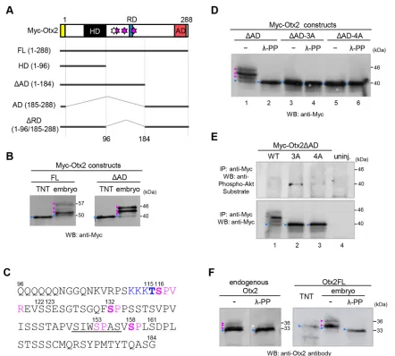
During western blot analyses of exogenous Otx2 in theXenopus laevisembryo (Sudou et al., 2012), we noticed several bands of Otx2 that were migrating more slowly than anin vitrotranslated product. Similarly, several previous studies using western blots showed multiple bands of exogenous mouse Otx2 in transfected cultured cells (Mallamaci et al., 1996) and endogenous Otx2 in the mouse embryo (Boyl et al., 2001), although these have yet to be characterized. To evaluate the modification of Otx2, we constructed N-terminally Myc-tagged full-length Otx2 (Myc-Otx2FL) and its deletion constructs (Myc-Otx2HD,ΔAD, AD andΔRD) (Fig. 1A), and expressed them by mRNA injection intoX. laevisembryos for western blot analysis. The Myc-Otx2FL andΔAD but notΔRD, HD or AD constructs clearly showed several slowly migrating bands (magenta arrowheads in Fig. 1B and Fig. S1A), compared with the in vitro translated product (blue arrowheads in Fig. 1B and Fig. S1A). These data suggest that modification mainly occurs in the region of amino acid positions 96-184 (Fig. 1A).
The amino acid sequence from position 96 to 184 indicated possible phosphorylation sites for Cdk (S*/T*-P or S*/T*-P-X-R/ K; asterisk indicates phosphorylatable residues), mitogen-activated protein kinase (MAPK), casein kinase (S*/T*-X0-2-D/E/Sp1-3; Sp, primary phosphorylatable serine residue), and Akt kinase (R/K-X2 -S*/T*) (Fig. 1C). Mouse Otx2 reportedly has an Akt phosphorylation site at S115 (the corresponding site of X. laevis Otx2 is T115; Fig. S2), according to the database for the prediction of phosphorylation sites (see Materials and Methods). To identify modification sites, we first examined seven serine residues (S116, S122, S123, S132, S153, S158 and S161) and one threonine (T115) by introducing point mutations in Myc-Otx2ΔAD. Replacement of a single serine with alanine revealed that S116A, S132A and S158A constructs eliminated the uppermost band and increased the lower bands compared with the wild type (WT) and other mutants (S122A, S123A, S153A and S161A) (Fig. S1B,C). Furthermore, a triple-alanine mutant of Myc-Otx2ΔAD for S116, S132 and S158 (namedΔAD-3A) showed no slowly migrating bands (Fig. 1D, lane 3). These data suggest that S116, S132 and S158, which have the S-P consensus motif for Cdk and MAS-PK, are responsible for multiple shifted bands (up to seven bands by combinations of three sites) of Myc-Otx2. To examine this suggestion, embryonic lysates expressing Myc-Otx2 constructs were treated with λ-protein phosphatase (λ-PP). λ-PP treatment removed all of the modified
bands of Myc-Otx2ΔAD (Fig. 1D, lanes 1 and 2) and did not change the band ofΔAD-3A itself (Fig. 1D, lanes 3 and 4), suggesting that slowly migrating bands of Myc-Otx2ΔAD were caused by phosphorylation at S116, S132 and S158. The same experiment was performed with Myc-Otx2FL, with similar results, whereby all modified bands of Myc-Otx2FL were removed byλ-PP treatment except for a weak band that is shifted (Fig. S1D). Consistently, Myc-Otx2FL-3A had only a single weak shifted band, which was not removed with λ-PP (Fig. S1D, orange arrowhead), suggesting another distinct modification site around the activation domain.
We next asked whether T115 of Xenopus Otx2 is an Akt phosphorylation site, although we did not detect a shifted band in the Otx2 mutant 3A (ΔAD-3A) using western blotting (Fig. 1D, lanes 3 and 4). In addition, there was no difference in band positions between ΔAD-3A (remaining T115) and ΔAD-4A (alanine mutation at T115 in addition to 3A) regardless ofλ-PP (Fig. 1D, lanes 3-6). However, it was still possible that phosphorylation at T115 either did not cause a band shift in ΔAD-3A in western blotting or was inhibited by alanine mutations at the three sites including S116 next to T115. Therefore, we performed immunoprecipitation (IP)-western blot assays using anti-Phospho-Akt Substrate antibody, which recognizes the R-X-X-Sp/Tp motif (Sp/Tp, phosphorylated residues). Myc-Otx2ΔAD in embryonic lysates was immunoprecipitated with anti-Myc antibody and then subjected to western blotting with anti-Phospho-Akt Substrate antibody. A band was detected with mutantΔAD-3A but not with ΔAD-4A (Fig. 1E, upper panel, lanes 2 and 3), suggesting T115 as an Akt site. Thus, we identified four possible phosphorylation sites for Akt (T115) and Cdk/MAPK (S116, S132 and S158) in Otx2, and these sites are well conserved in vertebrate Otx2 as well as an Otx2 paralog/ohnolog, Otx5/Crx (Fig. S2).
Phosphorylation of endogenous Otx2
Although it is important to determine whether endogenous Otx2 is phosphorylated, no antibodies against phosphorylated sites of Otx2 are available. As far as we know, there is no report on detecting the phosphorylation of TFs expressed region specifically and endogenously in Xenopus embryos, such as Otx2, as evidenced by band shift in western blotting, possibly because of the limited amounts of such TFs in the whole embryo. Therefore, we first estimated the sensitivity of detection using a diluted series ofotx2 mRNA injected into Xenopusembryos. Modified bands of full-length Otx2 without a Myc-tag (Otx2FL) were clearly detected when >200 pg otx2FL mRNA was exogenously injected per embryo (Fig. S3). Based on RNA-seq data from X. tropicalis (Owens et al., 2016), the amount ofotx2mRNA was estimated to be in the order of 1 pg per embryo at the neurula stage. Therefore, we predicted that endogenous Otx2 can be detected by western blotting of samples from more than 200X. tropicalisembryos. As expected, we successfully detected multiple bands of endogenous Otx2 protein immunoprecipitated from lysates of approximately 800X. laevisneurula embryos in total, and the upper bands were reduced by λ-PP treatment (Fig. 1F, left). Similarly, shifted bands of exogenous Otx2FL were detected, and were abolished by λ-PP treatment (Fig. 1F, right). Thus, endogenous Otx2 is phosphorylated, although the intensities of phosphorylated bands of endogenous Otx2 appeared to be lower than those of exogenous Otx2 (see Discussion).
Phosphorylation of Otx2 depends on Cdk activity
Given that S116, S132 and S158 sites have the S-P motif (Fig. 1C), we speculated that MAPK or cyclin/Cdk complexes phosphorylate
DEVEL
O
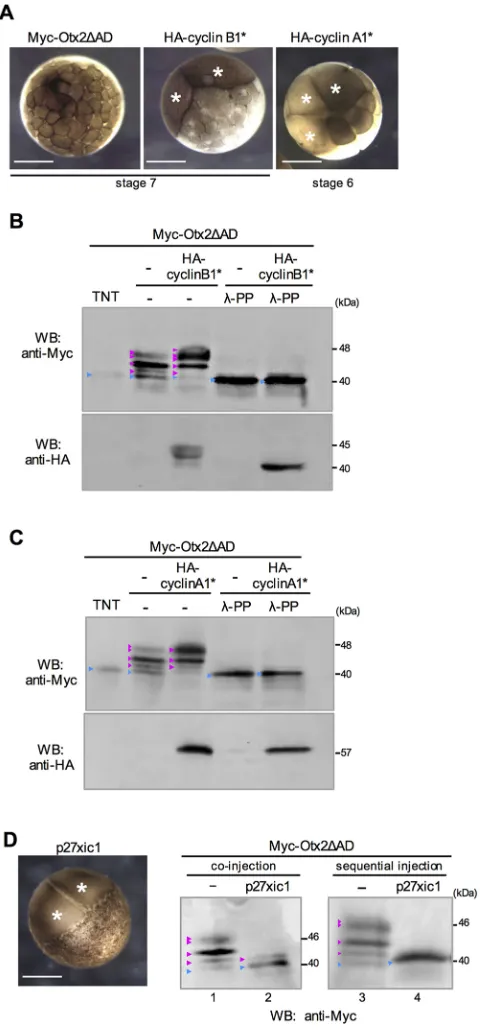
Otx2. We first observed that western blot bands of Otx2 were not affected by either MAPKK (Kosako et al., 1993) or a MAPKK inhibitor (Fig. S4A,B). We then tried to hyperactivate endogenous Cdk activity inXenopusembryos by overexpressing stable mutants of cyclin B1 (cyclin B1* for Cdk1) and cyclin A1 (cyclin A1* for Cdk1 and Cdk2), both of which lack the destruction box (Geley et al., 2001; Yamano et al., 2004). Hyperactivation of Cdks by mRNA injection of HA-cyclin B1* or HA-cyclin A1* was verified by cell-cycle arrest of mRNA-injected blastomeres (Fig. 2A, white asterisks). Under these conditions, modifications of Myc-Otx2ΔAD were dramatically enhanced (Fig. 2B,C), and these
modified bands were almost removed by λ-PP treatment (Fig. 2B,C). These data suggest that Cdk is involved in the phosphorylation of Otx2.
[image:3.612.87.529.54.452.2]If Otx2 is phosphorylated by cyclin/Cdks, then the S116, S132 and S158 sites might prefer either cyclin B/Cdk or cyclin A/Cdks. The data suggest that S116 and S132 preferred both cyclin B/Cdk and cyclin A/Cdks, whereas S158 preferred cyclin A/Cdks more than cyclin B/Cdk (Fig. S5), implying that cyclin B/Cdk and cyclin A/Cdks are differentially involved in the phosphorylation of Otx2. To further examine Cdk-dependent phosphorylation of Otx2, we overexpressed the Cdk inhibitor p27xic1 in Xenopus embryos Fig. 1. Exogenous and endogenous Otx2 is phosphorylated.(A) Schematic structures of Myc-Otx2 and deletion constructs. HD, homeodomain; RD, repression domain; AD, activation domain; yellow box, Myc-tag; white star, Akt site; pink stars, Cdk sites. FL and deletion constructs are indicated by thick lines and positions of amino acid residues. (B) Western blotting of Myc-FL and Myc-ΔAD. TNT,in vitrotranslational products; embryo, lysate fromXenopus
gastrula embryos expressing constructs as indicated. (C) Amino acid sequence of Otx2 from position 96 to 184. Numbers, position of amino acid; underline, repression domain; magenta, putative Cdk site; blue, putative Akt site; bold, possible phosphorylation sites identified in this study. (D) Western blotting of Myc-Otx2ΔAD constructs, WT, 3A and 4A. Lysates were treated with or withoutλ-PP. (E) IP-western assay for phospho-Akt sites. Myc-Otx2ΔAD was
immunoprecipitated with anti-Myc antibody and subjected to western blotting with the anti-Phospho-Akt substrate antibody (upper) or anti-Myc antibody (lower). uninj. indicates uninjected control. The reasons why no clear band was detected in WT (lane 1) might be that: (i) phospho-Akt signals were dispersed in several shifted bands caused by phosphorylation at other sites; and (ii) phosphorylation at T115 was inhibited by phosphorylation at S116 or the other sites. (F) Western blotting of endogenous Otx2 (left) and exogenous Otx2FL (right) with anti-Otx2 antibodies. Lysates were treated with or withoutλ-PP. Apparent molecular masses (kDa) of the nascent and upper-most modified bands are indicated on the right (B,D-F). Calculated and apparent molecular masses of constructs (kDa): Myc-Otx2FL, 43 and 50; Myc-Otx2ΔAD, 32 and 40; Otx2FL, 31 and 33. White arrowhead indicates a band resistant toλ-PP or incomplete digests. 3A, alanine mutant at S116, S132 and S158; 4A, alanine mutant at T115, S116, S132 and S158; blue arrowheads indicate nascent proteins; magenta arrowheads indicate modified proteins. The amount of injected mRNA ( pg/embryo) wasMyc-Otx2FLandMyc-Otx2ΔAD, 500;Otx2FL, 250. Antibodies used for western blotting (WB) or IP were as indicated.
DEVEL
O
(Vernon, 2003). We first verified cleavage arrest byp27xic1mRNA injection (Fig. 2D, left, white asterisks) as reported elsewhere (Ohnuma et al., 1999). When a mixture of p27xic1 and Myc-Otx2ΔAD mRNAs were injected at the two-cell stage, modified bands of Myc-Otx2ΔAD were reduced, but a weak band remained, which might result from phosphorylation occurring prior to the accumulation of p27xic1 protein translated from the injected mRNA (Fig. 2D, lanes 1, 2). Therefore, we first injectedp27xic1mRNA alone at the two-cell stage, followed by injection of Myc-Otx2ΔAD mRNA into the cleavage-arrested blastomeres at the 32-cell-stage equivalent. p27xic1 completely abolished modified bands of Myc-Otx2ΔAD (Fig. 2D, lanes 3 and 4), suggesting that phosphorylation of Otx2 depends on Cdk activity. These data also imply that the reduction of modified Otx2 by p27xic1 expression was not caused by dephosphorylation, because, once Otx2 was phosphorylated at the 2- to 32-cell stages (lane 2), it appeared to be stable until the late blastula stage when western blotting was performed (compare lanes 2 and 4 in Fig. 2D). Given that phosphorylation of Otx2 appears to start early in development, we examined developmental changes in phosphorylation levels using Myc-Otx2ΔAD and Myc-Otx2FL (Fig. S5D,E). The data showed that modification of Otx2ΔAD was already detectable at the 16-cell stage (stage 5), gradually increased until the early gastrula stage (stage 10.5) (Fig. S5D; see boxed areas for WT), and then reached almost a plateau at stage 10.5 onwards (Fig. S5E). The increase in phosphorylation levels until stage 9 correlates well with the cleavage cycle, supporting again the possibility that Otx2 is a substrate of cyclin/Cdks.
Taken together, Otx2 has four possible phosphorylation sites, and we refer to T115 as A-site ( putative Akt site) and S116, S132 and S158 as C-sites ( putative Cdk sites) in the following experiments.
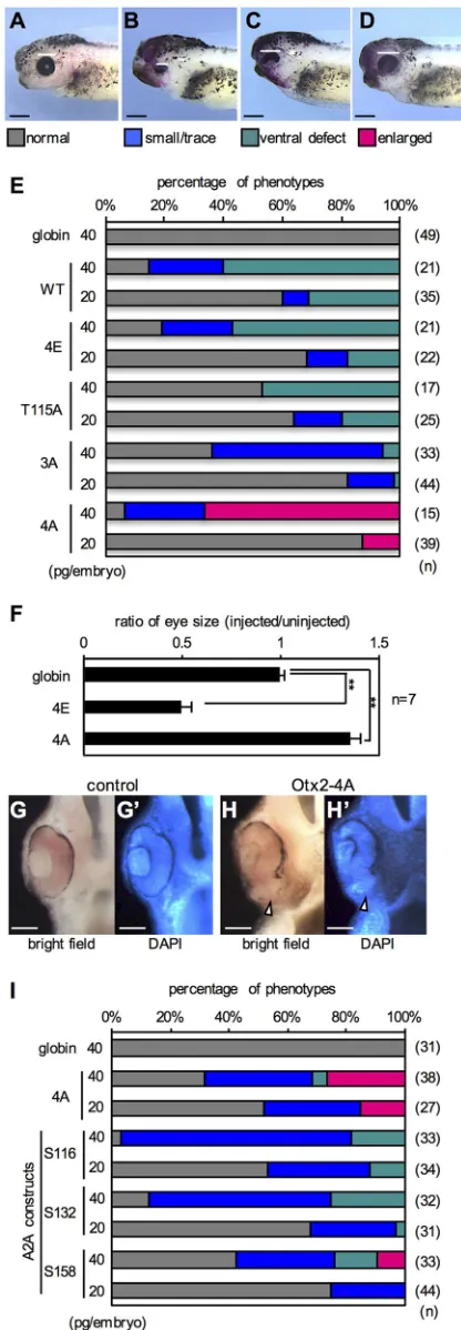
Otx2 mutants 4A and 4E have distinct functions in eye formation
[image:4.612.55.295.57.574.2]To determine whether known Otx2 activities are affected by phosphorylation states, we overexpressed phosphomimetic or nonphosphorylatable Otx2 inXenopus embryos and analyzed the resulting eye phenotypes, which have been well documented for Otx2 functions (Martinez-Morales et al., 2001; Nishihara et al., 2012). A phosphomimetic mutant, named Otx2-4E, was made by replacing A-site (T115) and three C-A-sites (S116, S132 and S158) with glutamate in Otx2FL. Nonphosphorylatable mutants, named Otx2-T115A, -3A and -4A, were similarly made by replacing A-site, C-sites or all four sites with alanine, respectively. Each Otx2 construct was expressed unilaterally in the anterior neuroectoderm, which includes the eye field, and the phenotypes were scored at stages 38-42. We categorized abnormal eye phenotypes into three types compared with the normal-looking phenotype (Fig. 3A-D). Expression of Otx2-WT, -4E -T115A, or -3A caused small/trace eye and ventral defect phenotypes in a dose-dependent manner (Fig. 3E), in which stronger phenotypes appeared to be obtained by expression of Otx2-WT, -4E, -3A and -T115A in order. By contrast, only Otx2-4A caused enlarged eye phenotypes, which have not previously been reported following the expression of any other Otx2 mutants in Xenopus(Andreazzoli et al., 1997; Isaacs et al., 1999). Notably, both Otx2-T115A and -3A reduced rather than enlarged the eyes (Fig. 3E), indicating that both mutations are necessary for the activity of Otx2-4A. Quantitative analysis of the area of the eye vesicle showed that eye size in Otx2-4A-expressing embryos was enlarged by approximately 1.3 times, whereas that in Otx2-4E-expressing embryos was reduced by approximately 0.5 times, compared with control (Fig. 3F). Section examination of the enlarged eye phenotype by Otx2-4A showed that the retinal tissue was expanded ventrally with a reduction in the retinal Fig. 2. Cdk-dependent phosphorylation of Otx2.(A) Cleavage arrest by
HA-cyclin B1* or HA-HA-cyclin A1*. mRNA was injected into one blastomere at the two-cell stage, and embryos were observed at the 32-two-cell (stage 6) or early blastula (stage 7) stage. Animal view. (B,C) Western blotting of Myc-Otx2ΔAD co-expressed with HA-cyclin B1* (B) or HA-cyclin A1* (C). Lysates were treated withλ-PP as indicated. (D) Phenotype of p27xic1-overexpressing embryos (left) and western blotting of Myc-Otx2ΔAD co-expressed with p27xic1 (middle, right). Co-injection (middle):Myc-Otx2ΔADmRNA (500 pg/embryo) was co-injected with (lane 2) or without (lane 1)p27xic1mRNA (2 ng/embryo) into one blastomere at the two-cell stage. Sequential injection (right):p27xic1mRNA (1.5 ng/embryo) was injected into one blastomere at the two-cell stage, then mRNAs forMyc-Otx2ΔAD(500 pg/embryo) andp27xic1(500 pg/embryo) were co-injected into the cleavage-arrested blastomeres at the 32-cell-stage equivalent (lane 4).Myc-Otx2ΔADmRNA was injected at the two-cell stage as control (lane 3). Lysates were prepared from embryos at stage 8 (B), stage 9 (C and D, lanes 1 and 2) or stage 9.5 (D, lanes 3 and 4). White asterisks indicate
cleavage-arrested blastomeres. Scale bars: 500μm in A and D.
DEVEL
O
pigment epithelium (Fig. 3H,H′, arrowheads) compared with the control (Fig. 3G,G′). These results clearly demonstrated that Otx2-4A has different activities in eye formation compared with the other mutants, and that exogenous Otx2-WT functions in a phosphorylated form, given that WT exhibits similar activities to 4E (Fig. 3E).
To further examine whether all C-site mutations are required for the activity of Otx2-4A, we constructed triple-alanine mutants of Otx2 (named A2A constructs), in which only one of the C-sites (S116, S132 or S158) was intact, referred to as A2A-S116, -S132 or -S158, respectively. A2A-S116 and A2A-S132 both caused small/ trace and ventral defect eye phenotypes (Fig. 3I). By contrast, A2A-S158 caused an enlarged eye phenotype in a dose-dependent manner, although the fraction of enlarged eye phenotypes by A2A-S158 was lower than that of Otx2-4A. These data indicate that mutations at T115, S116 and S132 are essential for enlarged eye phenotypes.
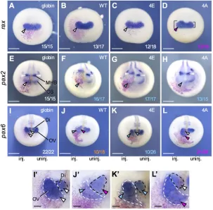
[image:5.612.68.276.57.656.2]We next examined the early effect of Otx2-4A and -4E by focusing on the eye field marker generax. Overexpression of Otx2-4A increased the expression ofraxcompared with the uninjected side (Fig. 4D), whereas Otx2-WT and -4E as well as the globin control did not affectraxexpression (Fig. 4A-C). The expansion of rax expression by Otx2-4A could explain the enlarged-eye phenotypes seen in these embryos. The enlarged eye phenotypes with aberrant ventral eye structures (Fig. 3H,H′) also suggested that Otx2-4A converts the presumptive optic stalk to the retina. Therefore, we examined the expression of pax2and pax6in the late neurula, in whichpax2is expressed in the optic stalk (OS) and MHB (Fig. 4E), whereaspax6is expressed in the optic vesicle (OV) and diencephalon (Di) side by side with different whole-mount in situ hybridization (WISH) intensities (Fig. 4I). Overexpression of Otx2-WT, -4E and -4A all inhibited pax2 expression in the OS (Fig. 4E-H), whereas Otx2-WT and -4E, but not -4A caused a posterior shift in pax2 expression in the MHB (open arrowheads in Fig. 4E-H). The reduction inpax2 in the OS by Otx2-WT, -4E and -4A (Fig. 4F-H) is consistent with the aberrant eye morphology at the tailbud stage on the ventral side where the OS is formed (Fig. 3C,E). Otx2-WT, -4E and -4A differently affected the expression ofpax6. WT reduced OV but expanded Dipax6expression (Fig. 4J,J′; dashed black lines, Di; dashed white lines, OV); and Otx2-4E slightly reducedpax6OV expression similar to WT (Fig. 4K,K′), consistent with the small/ trace eye phenotypes of these embryos (Fig. 3B,E). By contrast, Otx2-4A expanded OVpax6expression (Fig. 4L,L′), consistent with its enlarged eye phenotypes (Fig. 3D,E). These data demonstrated that Otx2-4E and -4A have different activities in eye formation. This raised the next question of whether Otx2-4E and -4A have different activities in cell proliferation and anteroposterior patterning, which have been reported for Otx2 (Pilo et al., 2001; Acampora et al., 2000).
Fig. 3. Eye phenotypes caused by Otx2 mutants.(A-D) Representative eye phenotypes. mRNAs for Otx2 construct andnbgalas a tracer were co-injected into the presumptive eye field of the dorsal-left blastomere at the four-cell stage. Phenotypes were scored at stages 38-42: normal indicates normal-looking eyes; small/trace indicates small or trace eye; ventral defect indicates normal eye width with pigmentation defect on the ventral side; enlarged indicates enlarged eye. Some injected embryos showed gastrulation arrest, which were removed from the total scores. White bars indicate the diameter of the eye. Anterior to the left, dorsal is up. (E) Incidence of eye phenotypes. See A-D for color codes of phenotypes. mRNAs for Otx2 construct or globin (negative control) was injected at 20 or 40 pg/embryo as indicated.nindicates the total number of scored embryos. (F) Quantitative analysis of the eye size. **P<0.01 (Student’st-test); error bars, s.e.m.;nindicates the number of samples. (G-H′) Transverse hemisection across the left eye. (G,G′) Control. (H,H′) Enlarged eye caused by Ox2-4A. (G,H) Bright field. (G′,H′) DAPI staining. Arrowheads indicate pigmentation defect in the ventral retina. (I) Incidence of eye phenotypes caused by Otx2-A2A constructs. In the A2A constructs, the remaining intact serine residue is indicated. Scale bars: 500μm in A-D; 100μm in G,H.
DEVEL
O
Otx2-4E stimulates cell proliferation
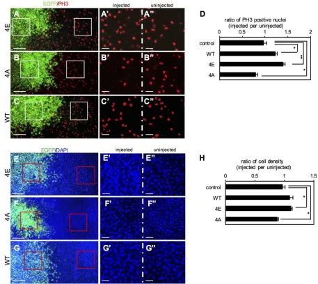
To investigate the effect of Otx2 on cell proliferation, Otx2-4E or -4A was expressed in the dorsoanterior region of the embryo, and immunostaining was then performed for phospho-histone H3 (PH3) as a mitosis marker. The numbers of PH3-positive nuclei were counted in EGFP-positive and negative areas for quantitative analysis (Fig. 5A-C). The data showed that the ratio of the number of PH3-positive nuclei in injected versus uninjected areas was 1.27 ±0.07 (mean±s.e.m.) and 1.38±0.05 in Otx2-WT and -4E-expressing embryos, respectively, whereas it was 0.727±0.062 in Otx2-4A-expressing embryos. These ratios significantly differed from the control (0.944±0.014) (Fig. 5D), suggesting that Otx2-WT and -4E both stimulate cell proliferation, whereas Otx2-4A reduces it. To confirm this, we counted the number of nuclei stained by DAPI (Fig. 5E-G). Quantitative analysis showed that the ratio of cell number in injected versus uninjected areas was 1.08±0.061 and 1.13± 0.02 in Otx2-WT- and -4E-expressing embryos, respectively, whereas it was 0.894±0.019 in Otx2-4A-expressing embryos. The ratios in 4E and 4A embryos differed significantly from the control (1.02± 0.04; P<0.05) (Fig. 5H). Thus, these data suggest that Otx2-4E and -4A have opposite effects on cell proliferation: Otx2-4E stimulates whereas -4A reduces proliferation.
To examine the in vivo relevance of Otx2 function in cell proliferation, we next performed loss-of-function experiments using antisense morpholino oligos (MOs) for otx2 and its functionally redundant paralogotx5, according to our previous report (Yasuoka et al., 2014). The number of PH3-positive nuclei was almost unchanged in the anterior neuroectoderm injected with standard control MO (Fig. S6A-A″,C), whereas it significantly decreased in the region injected with otx2/otx5-MOs (Fig. S6B-B″,C). Consistent with this, the number of nuclei decreased in theotx2/ otx5-MO-injected area (Fig. S6E-E″,F), but not affected in the control MO-injected area (Fig. S6D-D″,F). These data indicate that
Otx2 and Otx5 are required for cell proliferation in the anterior neuroectoderm, and these in vivo data are consistent with the enhancement of cell proliferation by exogenous Otx2-WT and -4E (Fig. 5). This also suggests that the reduction in cell proliferation caused by Otx2-4A results from the dominant-negative action of Otx2-4A against endogenous phosphorylated Otx2, which stimulates cell proliferation, and the unphosphorylated form of Otx2 is unable to take its place.
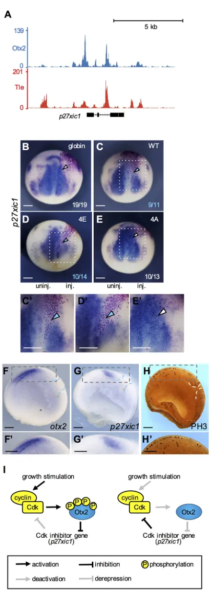
To determine how Otx2 stimulates proliferation, we focused on p27xic1because Otx2 reportedly inhibits thep27gene (Bunt et al., 2012). Analysis of our ChIP-seq data using theX. tropicalisgastrula (Yasuoka et al., 2014) showed the co-occupancy of Otx2 and Tle1 of several potential CRMs nearp27xic1(Fig. 6A), suggesting that p27xic1is a direct target gene of Otx2. Using mRNA injection, Otx2-WT, -4E or -4A with nuclear β-galactosidase (nβ-gal) as a tracer were unilaterally expressed, and the expression ofp27xic1 was examined by WISH. Expression of Otx2-WT and -4E, but not of -4A, downregulated p27xic1, compared with globin-injected negative control and on the uninjected side (Fig. 6B-E′), consistent with the stimulation of cell proliferation by Otx2-4E (Fig. 5D,H).
To assess the gain-of- and loss-of-function data discussed above, the expression domains ofotx2andp27xic1and the distribution of PH3-positive cells in the early neurula were examined using sagittal hemisections. The data showed that theotx2-expressing region did not overlap with that ofp27xic1(Fig. 6F-G′) and had more mitotic cells than the posterior region (Fig. 6H,H′), consistent with the involvement ofotx2in cell proliferation in the anterior neuroectoderm. These data suggest the existence of a positive feedback loop involving Otx2, Cdk and p27xic in the regulation of cell proliferation (Fig. 6I).
[image:6.612.48.351.59.357.2]Activities of Otx2-4E and -4A in anteroposterior patterning In the neural plate, Otx2 is involved in anteroposterior patterning by directly inhibiting posterior genes, which are suggested to include
Fig. 4. Effects of Otx2 mutants on eye marker genes.
Embryos were co-injected on the right side with mRNA for globin, WT, 4E or 4A (20 pg/embryo) andnbgalas described in the legend to Fig. 3, and were subjected to WISH forrax(A-D),
pax2(E-H) andpax6(I-L) at early neurula stages (stages 13-14, A-D) or late neurula stages (stages 18-20, E-L). The fractions indicate the proportion of the presented phenotype per total number. Colors of numbers: white indicates normal expression; magenta indicates upregulation; blue indicates downregulation; orange indicates combined effects of enlarged Di and reduced OV. Square brackets indicate the sizes of the eye field showing the difference between left and right. Open arrowhead indicates the position of MHB. Anterior view with dorsal up. (I′-L′) Enlarged images of I-L. Dashed black and white lines indicate Di and OV, respectively, where
pax6is expressed. Colors of arrowheads: white indicates normal expression; magenta indicates upregulation; blue indicates downregulation (A-L′). Scale bars: 500μm in A-L; 100μm in I′-L′.
DEVEL
O
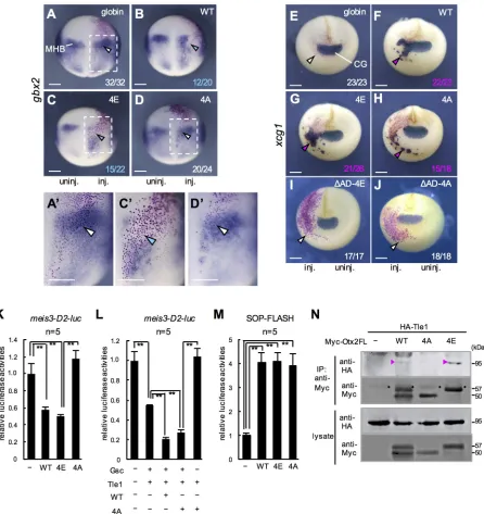
gbx2and meis3(Yasuoka et al., 2014), and by directly activating anterior genes, such as xcg1(Gammill and Sive, 1997) andrax (Danno et al., 2008). We first examined gbx2 by ectopically expressing Otx2-WT, -4E or -4A, in the posterior neuroectoderm. Both Otx2-WT and -4E downregulated the expression of gbx2 (Fig. 7B-C′, arrowheads) compared with the uninjected side and globin-injected control (Fig. 7A,A′). This activity explains the posterior shift inpax2expression in the MHB by Otx2-WT and -4E (Fig. 4F,G). Conversely, Otx2-4A did not downregulate gbx2 expression (Fig. 7D,D′), suggesting that only phosphorylated Otx2 has the ability to inhibit posteriorization.
xcg1is expressed in the cement gland, which is the anterior-most organ (Fig. 7E). Ectopic expression of Otx2-WT, -4E and -4A all upregulatedxcg1(Fig. 7F-H), and this activation was observed in the ventrolateral ectoderm, but not in the neuroectoderm, as reported
elsewhere (Gammill and Sive, 1997). To assess whether the ectopic upregulation of xcg1 by the Otx2 mutants required their transactivation activity, we developed transactivation domain-deleted constructs of 4E (ΔAD-4E) and 4A (ΔAD-4A). Neither ΔAD-4E- norΔAD-4A upregulatedxcg1(Fig. 7I,J), indicating that the transactivation domain of Otx2-4E and -4A is required for their activation of xcg1. These data also suggest that Otx2 has transactivation activity regardless of its phosphorylation state.
[image:7.612.75.532.57.465.2]The above data suggest that the difference in function between Otx2-4E and -4A is repression activity. To further examine whether phosphorylation states affect the repression and activation activity of Otx2, we performed luciferase reporter assays. We first examined the response of the silencer of meis3, a posterior gene, which is expected to be repressed by Otx2. Themeis3-D2 silencer (Yasuoka et al., 2014) was inserted upstream of the SV40 promoter in the Fig. 5. Phosphomimetic Otx2-4E stimulates cell proliferation.(A-C) mRNA for Otx2-4E (A), -4A (B) or -WT (C) together with EGFP as a tracer was injected into one blastomere at the four-cell stage. Embryos were immunostained for PH3 at the late gastrula stage. PH3-positive nuclei are shown in red, Otx2 mutant-expressing cells (mutant-expressing EGFP) are shown in green. White boxes indicate enlarged areas (A′-C″). (D) Quantitative analysis of PH3-positive nuclei. More than 90 PH3-positive nuclei were counted in one embryos, and six to ten embryos were analyzed for calculating the mean ratio (and s.e.m.) of the number of PH3-positive nuclei in injected versus uninjected areas. (E-G) Representative images of DAPI-stained nuclei in Otx2-4E (E), -4A (F) or -WT (G)-expressing gastrula embryos. Nuclei are shown in blue, and Otx2 mutant-expressing cells (expressing EGFP) are shown in green. Red boxes indicate enlarged areas (E′-G″). The dorsoanterior region was observed. (H) Quantitative analysis of cell numbers. More than 500 nuclei in total were counted in one embryo, and five to seven embryos were analyzed for calculating the mean ratio (and s.e.m.) of the number of nuclei in injected versus uninjected areas. **P<0.01, *P<0.05 (Student’st-test); error bars, s.e.m. (D,G). Scale bars: 200μm in A-G; 50μm in A′-G″.
DEVEL
O
reporter construct, and this reporter alone showed a high level of luciferase activity in the animal pole region. Under this condition, reporter activity was significantly repressed by Otx2-WT and -4E, but not by -4A (Fig. 7K). We next tested the combinatorial repressive activity of Otx2 with the interacting partner Gsc and the co-repressor Tle1, which form the repression complex for negative
target genes of Otx2 (Yasuoka et al., 2014). Repression of the meis3-D2reporter by Gsc and Tle1 was enhanced by co-expression of Otx2-WT, and this enhancement was also observed with Otx2-4A (Fig. 7L), suggesting that phosphorylation of Otx2 is not required for, and neither does unphosphorylated Otx2 interfere with, the formation of a ternary repression complex. Given that Otx2-4A and Tle1 did not repress the reporter (Fig. 7L), repression activity by a combination of Otx2-4A, Gsc and Tle1 probably results from recruitment of Tle1 to Gsc. Although Otx2-4A does not interfere with complex formation, Otx2-4A might weaken the repression activity of the complex, as implied in Fig. S7 (compare Otx2-4A with Otx2-4E).
We next examined an activated target gene of Otx2,rax, using the reporter gene SOP-FLASH, which has eight tandemly repeatedrax enhancer elements binding for Otx2 and Sox2 (Danno et al., 2008). The data showed that both Otx2-4E and -4A exerted transactivation activity (Fig. 7M), similarly to activation of thexcg1gene by Otx2-4E and -4A (Fig. 7G,H). Given that Sox2 is expressed in the neuroectoderm (Mizuseki et al., 1998) and has a transactivation domain (Nowling et al., 2000), a combination of Sox2 and Otx2 could function as an activator regardless of the phosphorylation states of Otx2. In addition, because Otx2-4E did not upregulate the endogenousraxgene (Fig. 4C), activation of the raxreporter by Otx2-4E might result from the CRM boosted by eight tandem repeats. Taken together, it is suggested that phosphorylation of Otx2 is required for its repression activity but does not affect the transactivation activity of this protein.
Otx2-4E, but not -4A interacts with Tle1
[image:8.612.66.283.49.657.2]The eh1 repression domain of Otx2 reportedly interacts with TLE/ Groucho (Heimbucher et al., 2007). Therefore, we tested the physical interaction between Otx2 mutants and HA-tagged Tle1 by co-immunoprecipitation (Co-IP) assays (Fig. 7N). A co-immunoprecipitated band of HA-Tle1 was detected with Myc-Otx2-4E as well as with Myc-Otx2-WT (magenta arrowheads in Fig. 7N), but barely with Myc-Otx2-4A, consistent with the data that neither Otx2-4A alone nor co-expression with Tle1 exerted repressive activity on themeis3-D2luciferase reporter (Fig. 7K,L). The reason why the intensity of the co-immunoprecipitated Tle1 band with WT was weaker than that of 4E (Fig. 7N) might be because of the partial phosphorylation of WT. These data suggest that phosphorylation of Otx2 is required for the interaction with Fig. 6. Phosphomimetic Otx2-4E negatively regulatesp27xic1.
(A) Genome browser representation showing occupancies of Otx2 and Tle in
Xenopus tropicalisgastrula embryos around thep27xic1gene. (B-E) WISH analysis ofp27xic1at the neurula stage (stages 13-14). mRNA (30 pg/embryo) for globin (B), WT (C), 4E (D) or 4A (E) together withnbgalmRNA was injected in the presumptive posterior neuroectoderm at the four-cell stage. Dorsal view with anterior up. inj. indicates the injected side; uninj. indicates the uninjected side. Colors of numbers and arrowheads are the same as in Fig. 4. (C′,D′,E′) Enlarged image of C, D and E (dotted white box). (F-H) WISH and immunostaining of sagittal hemisection. WISH forotx2andp27xic1(F,G). Immunostaining of PH3 (H). Dorsal up, anterior to the left. (F′,G′,H′) Enlarged images of F,G, H (dashed gray box). Dashed blue or white lines indicate the position of the blastopore. (I) A schematic model of feedback loops involving Cdk, Otx2 and p27xic in cell-proliferation (left) and non-proliferation states (right). Left, upon growth stimulation, (1) activation of Cdk causes
phosphorylation of Otx2; (2) phosphorylated Otx2 downregulatesp27xic1; and (3) the reduction in p27xic1 increases Cdk activity, thereby enhancing both cell proliferation and a phosphorylation state of Otx2. Right, upon the reduction of growth stimulation, (1) reduced Cdk activity causes the reduction of phosphorylation of Otx2; (2) the reduction in phosphorylated Otx2 derepresses
p27xic1; and (3) increased p27xic1 inhibits Cdk activity, thereby reducing both cell proliferation and a phosphorylation state of Otx2. Scale bars: 300μm in B-H,C′-H′.
DEVEL
O
Tle1, which enables Otx2 to then exert its own repression activity. The phosphorylation-dependent interaction of eh1 domains with TLE has not previously been reported.
DISCUSSION
Repressor activity of Otx2 conferred by post-translational modification
In this study, we showed that Otx2 exists in two distinct states: phosphorylated and unphosphorylated. Our data suggest that
[image:9.612.84.530.56.529.2]phosphorylated Otx2 functions as both a repressor and activator, whereas unphosphorylated Otx2 functions only as an activator. To date, only a few TFs have been reported to be converted from being an activator and a repressor by post-translational modification. The SoxE group of TFs, including Sox9, are converted by SUMOylation from an activator to a repressor by displacing CBP/p300 and recruiting Tle4/Grg4 (Lee et al., 2012). Another case is Pax2, transactivation of which is enhanced through JNK kinase-mediated phosphorylation in the activation domain by blocking TLE/ Fig. 7. Repression and/or activation activities of Otx2-4E and -4A and Tle interaction.(A-D) WISH forgbx2at the late gastrula to early neurula stage (stages 12.5-13). Dorsal view with anterior up. (A′,C′,D′) Enlarged images of A, C and D (dashed white box). (E-J) WISH forxcg1at the late neurula stage (stages 18-20). Anterior view with dorsal up. CG indicates the cement gland. mRNA injection was the same as in Fig. 6. Colors of numbers and arrowheads are the same as in Fig. 4. (K-M) Luciferase reporter assays.meis3-D2-luc reporter DNA was co-injected with mRNA for Otx2-WT, -4E or -4A (50 pg/embryo) (K) or with combinations of mRNAs for Gsc (12.5 pg/embryo), Tle1 (12.5 pg/embryo), Otx2-WT and -4A (25 pg/embryo) as indicated (L). SOP-FLASH reporter DNA was co-injected with mRNA for Otx2-WT, -4E or -4A (50 pg/embryo) (M). **P<0.01 (Student’st-test); error bars, s.e.m.; N.S., not significant;nindicates the number of samples. (N) Physical interaction between Tle1 and Otx2FL mutants as assayed by Co-IP. The amounts of expressed protein were verified by western blotting (lower panels). Apparent molecular masses (kDa) of Myc-Otx2FL-WT, -4A and -4E were 50, 50 and 57, respectively. Magenta arrowheads indicate co-immunoprecipitated bands; circles indicate heavy chains of IgG from the anti-Myc antibody. Scale bars: 300μm in A-J,A′,C′,D′.
DEVEL
O
Groucho (Cai et al., 2003). Thus, we have shown a third case, in which phosphorylation of Otx2 enables it to function as a repressor through its interaction with Tle1 (Fig. 7N), opposite to the case of Pax2.
Otx2 has a novel eh1 domain with a Cdk site (SIWSPASISP; underlined is the possible Cdk site), which is only present in the Otx family proteins (Otx1, Otx2 and Otx5/Crx) as well as a few other TFs (Copley, 2005). The eh1 domain is present in many metazoan TFs (Copley, 2005) and interacts with TLE family proteins through its conserved C-terminal WD40 repeat domain (Heimbucher et al., 2007; Pickles et al., 2002). Given that some WD40 domains of ubiquitin ligases and phosphatases, such as β-Trcp and Cdc4, interact with a phosphorylated serine residue in their substrates (Yaffe and Elia, 2001), it is possible that one of the WD40 repeats of TLE interacts with the eh1 domain with a phosphorylated site in Otx2.
Role of cyclin/Cdk-dependent phosphorylation of TFs
We propose a positive feedback loop model for Otx2 functions in cell proliferation (Fig. 6I). In this model, in a proliferative state, Otx2 is phosphorylated to repress target genes, includingp27xic1as well asgbx2andmeis3, whereas, when the cell cycle is arrested, unphosphorylated Otx2 increases to activate the target genes, rax and pax6. Thus, the phosphorylated and unphosphorylated states of Otx2 can be toggled by the cell cycle. However, it is not known how the phosphorylated state of Otx2 is changed to the unphosphorylated state. There are two possibilities: (1) dephosphorylation of Otx2 by phosphatase; and (2) degradation of phosphorylated Otx2. We did not observe any differences in the instability of the Otx2-4E mutant compared with -4A by western blotting (not shown), which does not support the second possibility, and denies phosphorylation-dependent degradation. A putative PEST sequence, an indicator of rapidly degraded protein, is present in the Xenopus Otx2 protein (Mori et al., 1994; Williams and Holland, 1998), and Otx2 might have a short half-life. Therefore, unphosphorylated Otx2 could be generated through the rapid turnover of Otx2 regardless of its phosphorylation state.
We showed that endogenous Otx2 is phosphorylated, but that its phosphorylation levels were lower than those of exogenous Otx2 (Fig. 1F). This difference might be caused by the timing of protein production and the number of proliferating cells where phosphorylation of Otx2 supposedly occurs. For exogenous Otx2, mRNA was injected into the animal pole region at two- or four-cell stages, and translated in the presumptive ectoderm from early cleavage stages; the translation product was phosphorylated until the blastula stage (Fig. S5D,E) by Cdk1 activity, which oscillates with a period of∼30 min during the cleavage cycle (Hörmanseder et al., 2013). By contrast, in thein vivosituation, the transcription of otx2starts in the dorsal endoderm and mesoderm at the late blastula stage and in the dorsal ectoderm at the early gastrula stage (Sudou et al., 2012); endogenous Otx2 protein then accumulates in the head organizer and the anterior neuroectoderm, and the proliferation rates in these regions dramatically decrease, especially in the head organizer (Fig. 6H). Therefore, even if phosphorylation levels of endogenous Otx2 in proliferating cells are high, the average phosphorylation level of Otx2 in major nonproliferating and minor proliferating cell populations in a gastrula or neurula embryo is lower than those of exogenous Otx2.
We have proposed a positive feedback loop involving Cdk-dependent phosphorylation of Otx2 for cell proliferation (Fig. 6I). Similar feedback loops involving other TFs are also reported, but
the regulatory mode is different from that of Otx2. That is, TFs are switched ‘on’ or ‘off’ by Cdk-dependent phosphorylation to promote cell cycle progression. An example of regulation by an ‘on’switch is Foxm1. Foxm1 is activated by the phosphorylation of the N-terminal inhibitory domain by cyclin/Cdk to upregulate cyclin B and cdc25B, thereby forming a positive feedback loop for cell proliferation (Park et al., 2008; Major et al., 2004). An example for an‘off’switch is Foxo. Foxo upregulates the Cdk inhibitor genes p27kip1andp21WAF1, but, upon phosphorylation by cyclin/Cdk, undergoes nuclear export, which almost switches off the function of Foxo, leading to cell proliferation (Schuff et al., 2010; Liu et al., 2008; Stahl et al., 2002). In the case of Otx2, Cdk-dependent phosphorylation confers repressor activity to Otx2 for repressing p27xic; otherwise, it functions as an activator on its own, as mentioned above, which differs from simple switch ‘on’or‘off’ regulation.
Regarding tissue specification and differentiation, Cdk-dependent phosphorylation generally negatively regulates the activity of TFs that directly activate differentiation genes. For example, the phosphorylation of the myogenic TF MyoD by cyclin/ Cdk leads to its degradation to prevent muscle differentiation (Kitzmann et al., 1999). During neurogenesis, the Cdk-dependent phosphorylation of neurogenin 2 (Ngn2, or Neurog2) inhibits its neural differentiation activity (Ali et al., 2011; Hindley et al., 2012). In these cases, Cdk-dependent phosphorylation of the TFs switches off their functions. Taken together, phosphorylation of TFs by cyclin/Cdks either promotes cell cycle progression (e.g. Foxm1 and Foxo) or inhibits tissue differentiation (e.g. MyoD and Ngn2). By contrast, Cdk-dependent phosphorylation not only inhibits the function of Otx2 in differentiation, but also converts it to be able to function in proliferation. Thus, this study has revealed a new category of TFs, in which post-translational modification of a single TF converts it from having a differentiation- to a proliferation-orientated role.
How does Otx2 coordinate its activator and repressor functions?
How Otx2 properly functions as an activator or a repressor for its target genes, and coordinates proliferation, tissue patterning and differentiation during embryogenesis, has been unclear. One answer to this question was reported in our previous paper (Yasuoka et al., 2014), showing the combinatorial regulation of target genes with Lim1 (activator) or Gsc (repressor). In the current study, we showed the post-translational regulation of Otx2 activity to be another answer, in which phosphorylation of Otx2 confers repressor activity at least partly through its interaction with Tle1. However, how the combinatorial and post-translational regulations work together remains elusive.
During eye formation, it was reported that expression of a repressor form of Otx2 (Otx2-EnR) by mRNA injection inXenopus embryos results in a small eye or eyeless phenotype (Isaacs et al., 1999), and that expression of another type of repressor form, EnR-Otx2, in chick eyes by electroporation caused pigmentation defects of the retinal pigment epithelium and a reduction ofpax6(Nishihara et al., 2012). These activities of repressor forms of Otx2 are similar to that of Otx2-4E in Xenopus embryos (Fig. 3E, Fig. 4J,K), consistent with the repressor activity of Otx2-4E (Fig. 7). In contrast with Otx2-4E, Otx2-4A expanded the expression ofraxat early neurula stages (Fig. 4D) without stimulating cell proliferation (Fig. 5D,H); at later stages, Otx2-4A expanded the expression of pax6(retina) but inhibitedpax2(OS) on the ventral side, leading to ventral expansion of the retina. These data suggest that Otx2-4A
DEVEL
O
expands the eye field, and subsequently alters patterning of the neural retina and OS. In other words, enlarged eye phenotypes caused by Otx2-4A result from changes in patterning, not proliferation. Our data are reminiscent of a previous report on the function of Otx2 forXenopusretina formation, in which an activator type of Otx2 (Otx2-VP16) promotes the bipolar cell fate without stimulating retinal proliferation (Viczian et al., 2003), supporting the possibility that unphosphorylated Otx2 acts mainly as an activator in retina formation.
Several heterozygousOTX2mutations in human were reportedly linked with severe ocular malformation. For example, point mutations of P133T, P134A and P134R, which occur near one of the four putative phosphorylation sites, S132, found in our study (Fig. S2), are associated with microphthalmia, anophthalmia, sclerocornea and retinal detachment (Beby and Lamonerie, 2013). It is not certain whether these mutations affect the phosphorylation of Otx2 or the interaction with putative binding partners of Otx2, but it is possible that ocular defects in humans are caused by the alteration of phosphorylation states of Otx2.
In summary, we demonstrated that Otx2 undergoes phosphorylation in vivo, and that phosphomimetic and nonphosphorylatable mutants of Otx2 exhibit distinct activities in cell proliferation, patterning and differentiation in Xenopus embryos. Post-translational regulation of Otx2 repressor activity (shown here) in addition to combinatorial regulations with partner TFs (Yasuoka et al., 2014) could enable Otx2 to have versatile roles during development.In vivoanalysis of the phosphorylation sites, such as a mutated gene knock-in approach, is now awaited to explore the role of phosphorylated Otx2 in embryonic development.
MATERIALS AND METHODS cDNA cloning and plasmid constructs
Point mutants and deletion constructs of Otx2 were made by using PCR methods. PCR fragments of Otx2 mutants were cloned into the pCSf107mT (Mii and Taira, 2009), pCSf107_MTmT (for Myc tagging) and pCSf107_4HAmT (for HA tagging) vectors (Shibano et al., 2015), which
contain SP6 terminator sequences. pGEX-GST-ΔN106cyclin B1 (Iwabuchi
et al., 2002) was recloned into pCSf107_HAmT. pGEM-Δcyclin A (deletion
of 55 amino acids from the N terminus of the protein) was gifted by Dr N. Furuno (Hiroshima University) before publication, and recloned into pCSf107_HAmT. All constructs made in this study are listed in Table S1.
Xenopusembryo and microinjection
Xenopus laevis and X. tropicalis embryos were obtained by artificial
fertilization, dejellied, incubated in 0.1×Steinberg’s solution (Peng, 1991),
and staged according to Nieuwkoop and Faber (1967). Microinjection experiments were conducted as previously described (Yasuoka et al., 2014). For mRNA synthesis, pCSf107mT constructs with 4×SP6 terminators were transcribed with SP6 polymerase (mMESSAGE mMACHINE SP6 kit, Ambion). mRNAs were injected into one blastomere at the two- or four-cell
stage. Nuclear β-galactosidase (nbgal) mRNA (50-100 pg/embryo) or
EGFP mRNA (250 pg/embryo) was co-injected for lineage tracing.
Antisense or standard control morpholino oligos (MOs) (Gene Tools) were injected into a dorsal blastomere at the four-cell stage. FITC-dextran (5 ng/embryo) was co-injected for lineage tracing. Sequences and
specificities of antisense MOs for X. tropicalis otx2and otx5 were as
described previously (Yasuoka et al., 2014).
Western blotting and protein phosphatase treatment
Lysates were prepared from embryos (stages 10-10.5 unless mentioned otherwise) that had been injected with mRNA (500 pg/embryo) into both of
blastomeres in the animal pole region at the two-cell stage. In vitro
translation was performed using the TNT SP6 Quick-Coupled
Transcription/Translation System (Promega) as previously described
(Yamamoto et al., 2003). λ-PP treatment of lysates was carried out as
previously described (Shibano et al., 2015). SDS-PAGE was carried out with 12-12.5% polyacrylamide gels. Western blotting was performed essentially as described elsewhere (Shibata et al., 2005), using anti-Myc (9E10), Otx2 (Sudou et al., 2012), p44/42 MAP kinase and anti-phospho-p44/42 MAPK (CST) and anti-HA (12CA5) antibodies. Apparent molecular masses (kDa) of bands were estimated based on the standard curve of size markers (BioDynamics Laboratory).
IP assays for putative phosphorylated Akt sites and the detection of endogenous Otx2 protein
Possible Akt sites of Otx2 were searched for using the database
PhosphoSitePlus, (www.phosphosite.org/). IP-western assays were
performed essentially as described elsewhere (Shibano et al., 2015). Immunoprecipitates with anti-Myc antibody were boiled in 2×SDS sample buffer and analyzed by western blotting with anti-Phospho-Akt Substrate antibody (CST). To detect the endogenous Otx2, we modified the preparation
of the embryonic lysates.Xenopusneurula embryos were homogenized with
lysis buffer A (50 mM Tris-Cl, pH 8.0, 10% glycerol, 0.1%NP-40, 40μg/ml
aprotinin, 20μg/ml leupeptin, 1 mM PMSF) plus 400 mM NaCl, and
homogenates were centrifuged at 4°C for 1 h at 13,200 rpm (16,000 g).
Supernatants were diluted four times with buffer A and centrifuged at 4°C for 1 h at 13,200 rpm. Equivalent amounts of lysate were incubated with the
anti-Otx2 antibody (Sudou et al., 2012) and 25μl protein A-agarose beads (Roche).
The following wash steps of beads for IP-western assays were performed essentially as described elsewhere (Shibano et al., 2015). Immunoprecipitates with anti-Otx2 antibody were boiled in 2×SDS sample buffer and analyzed by SDS-PAGE with a 15% polyacrylamide gel and western blotting with anti-Otx2 antibody (Abcam, ab21990).
Quantitation of the eye size in tailbud-stage embryos
Embryos injected for phenotypic analysis were reared until stages 38-42 and fixed with MEMFA for 1 h. Experiments were carried out with at least three clutches. The lengths of semi-major and semi-minor axes of the eye vesicle were measured, and the eye size were calculated by using the formula for the area of an ellipse.
Immunostaining and whole-mountin situhybridization
Immunostaining with anti-phospho-Histone H3 (Ser10) antibody (anti-PH3, 06-570, Merck), DAPI staining of nuclei, and counting of PH3-positive and DAPI-stained nuclei were performed as described elsewhere (Shibano et al., 2015). mRNA or MO with EGFP mRNA or FITC dextran, respectively, as a lineage tracer was injected into one blastomere at the four-cell stage. Embryos with a lineage tracer in the anterior neuroectoderm were
selected for counting PH3- or DAPI-positive nuclei in an area (0.44 mm2) in
the anterior neuroectoderm on the injected or uninjected side. The effect was scored by dividing the number of nuclei on the injected side with that on the
uninjected side. The statistical significance (Pvalue) was calculated using
Student’st-test after a one-way analysis of variance (ANOVA). Experiments
were carried out with either four or three clutches for the mRNA or MO injection experiments, respectively. WISH was performed according to Harland (1991) with BM Purple (Roche) as a substrate. Antisense RNA probes were transcribed from linearized plasmids as described in Table S2.
Experiments for WISH were carried out with two clutches forrax,pax2and
p27xic1, and with three clutches forpax6, gbx2andxcg1. Hemisection of fixed embryos was performed as described elsewhere (Sudou et al., 2012).
Luciferase reporter assays
Embryos were co-injected with pGL4p-meis3-D2-luc reporter DNA and mRNA into two animal blastomeres at the two-cell stage, or co-injected with SOP-FLASH reporter (Danno et al., 2008) and mRNA into two dorsal animal blastomeres at the four-cell stage. Five pools of three injected embryos were assayed for luciferase activity at stage 10.5 for pGL4p-meis3-D2-luc reporter, and at stage 12 for SOP-FLASH reporter using the
luciferase assay system (Promega) according to the manufacturer’s protocol
and as described elsewhere (Mii and Taira, 2009). Experiments were carried out at least three times; when similar results were obtained, a representative
result is presented.
DEVEL
O
Co-IP assay
mRNA for HA-Tle1 (700 pg/embryo) was co-injected with or without mRNA for Myc-Otx2-WT, Myc-Otx2-4A or Myc-Otx2-4E (500 pg/ embryo) into the animal pole region of both blastomeres at the two-cell stage. Embryonic lysates were prepared at stage 10.5, and subjected to Co-IP as described elsewhere (Shibano et al., 2015). Myc-Otx2 protein was co-immunoprecipitated with anti-Myc antibody and subjected to SDS-PAGE with a 10% polyacrylamide gel, followed by western blotting with anti-HA and anti-Myc antibodies.
Acknowledgments
We thank Keita Ohsumi for pGEX-GST-ΔN106cyclin B1 (a stable mutant of cyclin B1), Nobuaki Furuno for pGEM-Δcyclin A (a stable mutant of cyclin A1), Yukiko Gotoh for pSP64T-MAPKK*, and Hidetoshi Saiga and Mariko Kondo for critical reading of the manuscript. Male and female frogs ofX. tropicaliswere obtained from the National Bioresource Project (Amphibian Research Center, Hiroshima University).
Competing interests
The authors declare no competing or financial interests.
Author contributions
Conceptualization: Y.S.; Methodology: Y.S.; Validation: Y.S.; Investigation: K.M., E.H., H.O., Y.Y., T.S.; Writing - original draft: Y.S., M.T.; Writing - review & editing: Y.S., Y.Y., T.S., T.T., M.T.; Visualization: Y.S.; Supervision: M.T.; Project administration: Y.S.; Funding acquisition: M.T.
Funding
This work was supported by the Japan Society for the Promotion of Science KAKENHI grant numbers 24657147 and 25251026 (M.T.) and 17KT0114 and 16K21559 (Y.Y.).
Supplementary information
Supplementary information available online at
http://dev.biologists.org/lookup/doi/10.1242/dev.159640.supplemental
References
Acampora, D., Postiglione, M. P. I. A., Avantaggiato, V., Bonito, M. D. I. and Simeone, A. (2000). The role of Otx and Otp genes in brain development.
Int. J. Dev. Biol.44, 669-677.
Agoston, Z. and Schulte, D.(2009). Meis2 competes with the Groucho co-repressor Tle4 for binding to Otx2 and specifies tectal fate without induction of a secondary midbrain-hindbrain boundary organizer. Development 136, 3311-3322.
Ali, F., Hindley, C., McDowell, G., Deibler, R., Jones, A., Kirschner, M., Guillemot, F. and Philpott, A. (2011). Cell cycle-regulated multi-site phosphorylation of Neurogenin 2 coordinates cell cycling with differentiation during neurogenesis.Development138, 4267-4277.
Andreazzoli, M., Pannese, M. and Boncinelli, E.(1997). Activating and repressing signals in head development: the role of Xotx1 and Xotx2.Development124, 1733-1743.
Andreazzoli, M., Gestri, G., Angeloni, D., Menna, E. and Barsacchi, G.(1999). Role of Xrx1 in Xenopus eye and anterior brain development.Development126, 2451-2460.
Beby, F. and Lamonerie, T.(2013). The homeobox gene Otx2 in development and disease.Exp. Eye Res.111, 9-16.
Blitz, I. L. and Cho, K. W.(1995). Anterior neurectoderm is progressively induced during gastrulation: the role of the Xenopus homeobox gene orthodenticle.
Development121, 993-1004.
Boyl, P. P., Signore, M., Acampora, D., Martinez-Barbera, J. P., Ilengo, C., Annino, A., Corte, G. and Simeone, A. (2001). Forebrain and midbrain development requires epiblast-restricted Otx2 translational control mediated by its 3′UTR.Development128, 2989-3000.
Brzezinski, J. A. and Reh, T. A.(2015). Photoreceptor cell fate specification in vertebrates.Development142, 3263-3273.
Bunt, J., Hasselt, N. E., Zwijnenburg, D. A., Hamdi, M., Koster, J., Versteeg, R. and Kool, M. (2012). OTX2 directly activates cell cycle genes and inhibits differentiation in medulloblastoma cells.Int. J. Cancer131, E21-E32.
Cai, Y., Brophy, P. D., Levitan, I., Stifani, S. and Dressler, G. R.(2003). Groucho suppresses Pax2 transactivation by inhibition of JNK-mediated phosphorylation.
EMBO J.22, 5522-5529.
Copley, R. R. (2005). The EH1 motif in metazoan transcription factors.BMC Genomics6, 169.
Danno, H., Michiue, T., Hitachi, K., Yukita, A., Ishiura, S. and Asashima, M.
(2008). Molecular links among the causative genes for ocular malformation: Otx2 and Sox2 coregulate Rax expression.Proc. Natl. Acad. Sci. USA105, 5408-5413.
Gammill, L. S. and Sive, H. (1997). Identification of otx2 target genes and restrictions in ectodermal competence during Xenopus cement gland formation.
Development124, 471-481.
Gammill, L. S. and Sive, H.(2001). otx2 expression in the ectoderm activates anterior neural determination and is required for Xenopus cement gland formation.
Dev. Biol.240, 223-236.
Geley, S., Kramer, E., Gieffers, C., Gannon, J., Peters, J.-M. and Hunt, T.(2001). Anaphase-promoting complex/cyclosome-dependent proteolysis of human cyclin A starts at the beginning of mitosis and is not subject to the spindle assembly checkpoint.J. Cell Biol.153, 137-148.
Harland, R. M.(1991). Appendix G: in situ hybridization: an improved whole-mount method for Xenopus embryos.Methods Cell Biol.36, 685-695.
Heimbucher, T., Murko, C., Bajoghli, B., Aghaallaei, N., Huber, A., Stebegg, R., Eberhard, D., Fink, M., Simeone, A. and Czerny, T.(2007). Gbx2 and Otx2 interact with the WD40 domain of Groucho/Tle corepressors.Mol. Cell. Biol.27, 340-351.
Hindley, C., Ali, F., McDowell, G., Cheng, K., Jones, A., Guillemot, F. and Philpott, A.(2012). Post-translational modification of Ngn2 differentially affects transcription of distinct targets to regulate the balance between progenitor maintenance and differentiation.Development139, 1718-1723.
Holland, L. Z., Carvalho, J. E., Escriva, H., Laudet, V., Schubert, M., Shimeld, S. M. and Yu, J.-K.(2013). Evolution of bilaterian central nervous systems: a single origin?Evodevo4, 27.
Hörmanseder, E., Tischer, T. and Mayer, T. U.(2013). Modulation of cell cycle control during oocyte-to-embryo transitions.EMBO J.32, 2191-2203.
Isaacs, H. V., Andreazzoli, M. and Slack, J. M. W. (1999). Anteroposterior patterning by mutual repression of orthodenticle and caudal-type transcription factors.Evol. Dev.1, 143-152.
Iwabuchi, M., Ohsumi, K., Yamamoto, T. M. and Kishimoto, T. (2002). Coordinated regulation of M phase exit and S phase entry by the Cdc2 activity level in the early embryonic cell cycle.Dev. Biol.243, 34-43.
Katahira, T., Sato, T., Sugiyama, S., Okafuji, T., Araki, I., Funahashi, J.-I. and Nakamura, H.(2000). Interaction between Otx2 and Gbx2 defines the organizing center for the optic tectum.Mech. Dev.91, 43-52.
Kitzmann, M., Vandromme, M., Schaeffer, V., Carnac, G., Labbé, J.-C., Lamb, N. and Fernandez, A.(1999). cdk1- and cdk2-mediated phosphorylation of MyoD Ser200 in growing C2 myoblasts: role in modulating MyoD half-life and myogenic activity.Mol. Cell. Biol.19, 3167-3176.
Kosako, H., Nishida, E. and Gotoh, Y.(1993). cDNA cloning of MAP kinase kinase reveals kinase cascade pathways in yeasts to vertebrates.EMBO J.12, 787-794.
Lee, P.-C., Taylor-Jaffe, K. M., Nordin, K. M., Prasad, M. S., Lander, R. M. and LaBonne, C.(2012). SUMOylated SoxE factors recruit Grg4 and function as transcriptional repressors in the neural crest.J. Cell Biol.198, 799-813.
Liu, P., Kao, T. P. and Huang, H.(2008). CDK1 promotes cell proliferation and survival via phosphorylation and inhibition of FOXO1 transcription factor.
Oncogene27, 4733-4744.
Major, M. L., Lepe, R. and Costa, R. H.(2004). Forkhead box M1B transcriptional activity requires binding of Cdk-cyclin complexes for phosphorylation-dependent recruitment of p300/CBP coactivators.Mol. Cell. Biol.24, 2649-2661.
Mallamaci, A., Di Blas, E., Briata, P., Boncinelli, E. and Corte, G.(1996). OTX2 homeoprotein in the developing central nervous system and migratory cells of the olfactory area.Mech. Dev.58, 165-178.
Martinez-Morales, J. R., Signore, M., Acampora, D., Simeone, A. and Bovolenta, P.(2001). Otx genes are required for tissue specification in the developing eye.Development128, 2019-2030.
Matsuo, I., Kuratani, S., Kimura, C., Takeda, N. and Aizawa, S.(1995). Mouse Otx2 functions in the formation and patterning of rostral head.Genes Dev.9, 2646-2658.
Mii, Y. and Taira, M.(2009). Secreted Frizzled-related proteins enhance the diffusion of Wnt ligands and expand their signalling range.Development136, 4083-4088.
Mizuseki, K., Kishi, M., Matsui, M., Nakanishi, S. and Sasai, Y.(1998). Xenopus Zic-related-1 and Sox-2, two factors induced by chordin, have distinct activities in the initiation of neural induction.Development125, 579-587.
Mori, H., Miyazaki, Y., Morita, T., Nitta, H. and Mishina, M.(1994). Different spatio-temporal expressions of three otx homeoprotein transcripts during zebrafish embryogenesis.Mol. Brain Res.27, 221-231.
Nakamura, H., Katahira, T., Matsunaga, E. and Sato, T.(2005). Isthmus organizer for midbrain and hindbrain development.Brain Res. Rev.49, 120-126.
Nieuwkoop, P. D. and Faber, J.(1967).Normal Table of Xenopus Laevis (Daudin). Garland Publishing.
Nishihara, D., Yajima, I., Tabata, H., Nakai, M., Tsukiji, N., Katahira, T., Takeda, K., Shibahara, S., Nakamura, H. and Yamamoto, H.(2012). Otx2 is involved in the regional specification of the developing retinal pigment epithelium by preventing the expression of Sox2 and Fgf8, factors that induce neural retina differentiation.PLoS ONE7, e48879.
DEVEL
O
Nowling, T. K., Johnson, L. R., Wiebe, M. S. and Rizzino, A.(2000). Identification of the transactivation domain of the transcription factor Sox-2 and an associated co-activator.J. Biol. Chem.275, 3810-3818.
Ohnuma, S.-I., Philpott, A., Wang, K., Holt, C. E. and Harris, W. A.(1999). p27Xic1, a Cdk inhibitor, promotes the determination of glial cells in Xenopus retina.Cell99, 499-510.
Omodei, D., Acampora, D., Mancuso, P., Prakash, N., Di Giovannantonio, L. G., Wurst, W. and Simeone, A.(2008). Anterior-posterior graded response to Otx2 controls proliferation and differentiation of dopaminergic progenitors in the ventral mesencephalon.Development135, 3459-3470.
Owens, N. D. L., Blitz, I. L., Lane, M. A., Patrushev, I., Overton, J. D., Gilchrist, M. J., Cho, K. W. Y. and Khokha, M. K.(2016). Measuring absolute RNA copy numbers at high temporal resolution reveals transcriptome kinetics in development.Cell Rep.14, 632-647.
Pannese, M., Polo, C., Andreazzoli, M., Vignali, R., Kablar, B., Barsacchi, G. and Boncinelli, E.(1995). The Xenopus homologue of Otx2 is a maternal homeobox gene that demarcates and specifies anterior body regions.Development121, 707-720.
Park, H. J., Wang, Z., Costa, R. H., Tyner, A., Lau, L. F. and Raychaudhuri, P.
(2008). An N-terminal inhibitory domain modulates activity of FoxM1 during cell cycle.Oncogene27, 1696-1704.
Peng, H. B.(1991). Xenopus laevis: pratical uses in cell and molecular biology. Solutions and protocols.Methods Cell Biol.36, 657-662.
Pickles, L. M., Roe, S. M., Hemingway, E. J., Stifani, S. and Pearl, L. H.(2002). Crystal structure of the C-terminal WD40 repeat domain of the human Groucho/ TLE1 transcriptional corepressor.Structure10, 751-761.
Pilo, P., Signore, M., Annino, A., Pedro, J., Barbera, M., Acampora, D. and Simeone, A.(2001). Otx genes in the development and evolution of the vertebrate brain.Int. J. Neurosci.19, 353-363.
Schuff, M., Siegel, D., Bardine, N., Oswald, F., Donow, C. and Knöchel, W.
(2010). FoxO genes are dispensable during gastrulation but required for late embryogenesis in Xenopus laevis.Dev. Biol.337, 259-273.
Shibano, T., Mamada, H., Hakuno, F., Takahashi, S.-I. and Taira, M.(2015). The inner nuclear membrane protein Nemp1 is a new type of RanGTP-binding protein in eukaryotes.PLoS ONE10, 1-21.
Shibata, M., Itoh, M., Hikasa, H., Taira, S. and Taira, M.(2005). Role of crescent in convergent extension movements by modulating Wnt signaling in early Xenopus embryogenesis.Mech. Dev.122, 1322-1339.
Spitz, F. and Furlong, E. E. M.(2012). Transcription factors: from enhancer binding to developmental control.Nat. Rev. Genet.13, 613-626.
Stahl, M., Dijkers, P. F., Kops, G. J. P. L., Lens, S. M. A., Coffer, P. J., Burgering, B. M. T. and Medema, R. H.(2002). The Forkhead transcription factor FoxO regulates transcription of p27 Kip1 and Bim in response to IL-2.J. Immunol.168, 5024-5031.
Suda, Y., Kurokawa, D., Takeuchi, M., Kajikawa, E., Kuratani, S., Amemiya, C. and Aizawa, S.(2009). Evolution of Otx paralogue usages in early patterning of the vertebrate head.Dev. Biol.325, 282-295.
Sudou, N., Yamamoto, S., Ogino, H. and Taira, M.(2012). Dynamic in vivo binding of transcription factors to cis-regulatory modules of cer and gsc in the stepwise formation of the Spemann-Mangold organizer.Development139, 1651-1661.
Vernay, B., Koch, M., Vaccarino, F., Briscoe, J., Simeone, A., Kageyama, R. and Ang, S.-L.(2005). Otx2 regulates subtype specification and neurogenesis in the midbrain.J. Neurosci.25, 4856-4867.
Vernon, A. E.(2003). A single cdk inhibitor, p27Xic1, functions beyond cell cycle regulation to promote muscle differentiation in Xenopus.Development130, 71-83.
Viczian, A. S., Vignali, R., Zuber, M. E., Barsacchi, G. and Harris, W. A.(2003). XOtx5b and XOtx2 regulate photoreceptor and bipolar fates in the Xenopus retina.
Development130, 1281-1294.
Williams, N. A. and Holland, P. W. H.(1998). Gene and domain duplication in the chordate Otx gene family: insights from amphioxus Otx.Mol. Biol. Evol.15, 600-607.
Wortham, M., Jin, G., Sun, J. L., Bigner, D. D., He, Y. and Yan, H.(2012). Aberrant Otx2 expression enhances migration and induces ectopic proliferation of hindbrain neuronal progenitor cells.PLoS ONE7, e36211.
Yaffe, M. B. and Elia, A. E. H.(2001). Phosphoserine/threonine-binding domains.
Curr. Opin. Cell Biol.13, 131-138.
Yamamoto, S., Hikasa, H., Ono, H. and Taira, M.(2003). Molecular link in the sequential induction of the Spemann organizer: direct activation of the cerberus gene by Xlim-1, Xotx2, Mix.1, and Siamois, immediately downstream from Nodal and Wnt signaling.Dev. Biol.257, 190-204.
Yamano, H., Gannon, J., Mahbubani, H. and Hunt, T.(2004). Cell cycle-regulated recognition of the destruction box of Cyclin B by the APC/C in Xenopus egg extracts.Mol. Cell13, 137-147.
Yasuoka, Y., Suzuki, Y., Takahashi, S., Someya, H., Sudou, N., Haramoto, Y., Cho, K. W., Asashima, M., Sugano, S. and Taira, M.(2014). Occupancy of tissue-specific cis-regulatory modules by Otx2 and TLE/Groucho for embryonic head specification.Nat. Commun.5, 4322.
Zuber, M. E.(2010). Eye field specification in Xenopus laevis.Curr. Top. Dev. Biol.
93, 29-60.