INTRODUCTION
The normal function of the pancreas to maintain blood sugar homeostasis relies on appropriate numbers of functional hormone-secreting cells within the pancreatic islets. The islets are the endocrine component of the pancreas and are composed primarily of insulin-producing beta cells, glucagon-producing alpha cells and somatostatin-producing delta cells. In all vertebrates, both the endocrine and exocrine components of the pancreas develop from cells of the endoderm germ layer (Ober et al., 2003; Stainier, 2005). Pancreas progenitors can already be recognized in the zebrafish at 15 hours post-fertilization (hpf) by expression of pdx1 in bilateral endoderm domains. Expression of pdx1 and of the endocrine progenitor marker isl1 precede expression of the endocrine hormone genes (Argenton et al., 1999; Biemar et al., 2001). As development proceeds, pancreas progenitor cells merge in the midline to form the dorsal pancreatic bud adjacent to somites 3-4 by 24 hpf (reviewed by Kinkel and Prince, 2009). As in other vertebrates, the complete zebrafish pancreas forms from both dorsal and ventral buds. The ventral bud, which is the sole source of exocrine cells, arises later in development at 40 hpf and subsequently the buds merge to produce the complete organ (Field et al., 2003). The
development of differentiated pancreatic cell types in appropriate numbers relies on both extrinsic secreted signals and intrinsic transcription factors (Kinkel and Prince, 2009).
A variety of secreted signaling molecules have been shown to play important roles in regionalization of the foregut endoderm and in the specification of pancreatic progenitors. These include Shh, Nodals, BMPs and FGFs (DiIorio et al., 2002; Tiso et al., 2002; Poulain et al., 2006; Shin et al., 2007; Chung and Stainier, 2008). We and others have shown that retinoic acid (RA) signaling also plays a central role in specifying pancreatic cell types in a variety of vertebrate models (Stafford and Prince, 2002; Chen et al., 2004; Stafford et al., 2004; Martín et al., 2005; Molotkov et al., 2005; Stafford et al., 2006; Ostrom et al., 2008). In zebrafish, disruptions of RA signaling cause a complete absence of all pancreatic cell types, whereas exogenous RA causes anterior endoderm to take on pancreatic fates (Stafford and Prince, 2002). Using cell transplantation we have confirmed that mesoderm-derived RA signals are received and transduced in endodermal cells, where they ultimately lead to differentiation of pancreatic cell types (Stafford et al., 2006). We have made good progress in understanding the regulation of RA signaling, recently establishing that the anterior limit of the pancreatic field is restricted by RA-degrading Cyp26 enzymes (Kinkel et al., 2009). By contrast, the gene-regulatory network that functions downstream of RA signaling to specify endocrine pancreas is not well characterized.
To better understand how RA regulates pancreas development we used a microarray approach to identify RA-regulated genes in the developing zebrafish endoderm (Kinkel et al., 2009). Using this strategy, we identified zebrafish motor neuron and pancreas homeobox 1 (mnx1) as an RA-regulated gene that is expressed in early endoderm and have confirmed that mnx1regulation by RA is conserved between zebrafish and mouse (Martín et al., 2005). mnx1encodes a homeodomain transcription factor, previously Development 138, 4597-4608 (2011) doi:10.1242/dev.067736
© 2011. Published by The Company of Biologists Ltd
1Department of Organismal Biology and Anatomy, University of Chicago, Chicago, IL 60637, USA. 2Department of Biology, Adelphi University, Garden City, NY 11530, USA. 3Department of Neuroscience and Center for Molecular Neurobiology, The Ohio State University, Columbus, OH 43210, USA. 4Department of Cell and Developmental Biology, Oregon Health and Science University, School of Medicine, Portland, OR 97239, USA.
*Author for correspondence (vprince@uchicago.edu)
Accepted 24 August 2011
SUMMARY
The vertebrate endocrine pancreas has the crucial function of maintaining blood sugar homeostasis. This role is dependent upon the development and maintenance of pancreatic islets comprising appropriate ratios of hormone-producing cells. In all vertebrate models studied, an initial precursor population of Pdx1-expressing endoderm cells gives rise to separate endocrine and exocrine cell lineages. Within the endocrine progenitor pool a variety of transcription factors influence cell fate decisions, such that hormone-producing differentiated cell types ultimately arise, including the insulin-producing beta cells and the antagonistically acting glucagon-producing alpha cells. In previous work, we established that the development of all pancreatic lineages requires retinoic acid (RA) signaling. We have used the zebrafish to uncover genes that function downstream of RA signaling, and here we identify mnx1 (hb9) as an RA-regulated endoderm transcription factor-encoding gene. By combining manipulation of gene function, cell transplantation approaches and transgenic reporter analysis we establish that Mnx1 functions downstream of RA within the endoderm to control cell fate decisions in the endocrine pancreas progenitor lineage. We confirm that Mnx1-deficient zebrafish lack beta cells, and, importantly, we make the novel observation that they concomitantly gain alpha cells. In Mnx1-deficient embryos, precursor cells that are normally destined to differentiate as beta cells instead take on an alpha cell fate. Our findings suggest that Mnx1 functions to promote beta and suppress alpha cell fates.
KEY WORDS: Zebrafish, Pancreas, mnx1,hb9, Retinoic acid
Zebrafish
mnx1
controls cell fate choice in the developing
endocrine pancreas
Gokhan Dalgin1, Andrea B. Ward2, Le T. Hao3, Christine E. Beattie3, Alexei Nechiporuk4and
Victoria E. Prince1,*
D
E
V
E
LO
P
M
E
N
termed Hb9 (Harrison et al., 1994). In mouse, Mnx1is expressed in both dorsal and ventral gut endoderm by embryonic day 8, preceding expression of Pdx1in the dorsal pancreatic primordium (Harrison et al., 1999; Li et al., 1999). However, expression of Mnx1in the developing pancreatic buds is transient, and at later stages Mnx1 expression is limited to the murine beta cells. Similarly, expression of zebrafish mnx1is also transient in the early endoderm and precedes pdx1expression: by 16 hpf, mnx1 expression is restricted to the endocrine pancreas domain and by 20 hpf mnx1is expressed exclusively in beta cells (Wendik et al., 2004). Mnx1mutant mice display abnormal islet formation with a very reduced population of insulin-expressing beta cells (Harrison et al., 1999; Li et al., 1999). Furthermore, overexpression of Mnx1under the control of the Pdx1promoter causes the development of intestinal cell types at the expense of pancreatic epithelium (Li and Edlund, 2001). Previous reports of Mnx1 knockdown in zebrafish have similarly shown a reduction in the number of beta cells (Wendik et al., 2004).
In this study, we show that RA signaling regulates the expression of zebrafish mnx1 in the endoderm and establish that mnx1 functions downstream of RA signaling to promote beta cell fate. Using cell transplantation we confirm that mnx1 function is required directly within the endoderm for normal beta cell development. Remarkably, when Mnx1 function is disrupted, we find not only a reduction in beta cells, but also an increase in alpha cells. Using a new transgenic zebrafish line we demonstrate that, in Mnx1-deficient specimens, the beta cell precursors fail to activate insulin expression and instead go on to express markers of alpha cell fate, indicating a switch in cell fate choice. Together, our findings suggest that mnx1 plays a crucial role in endocrine pancreas development, functioning downstream of RA signaling to promote the differentiation of progenitors to the beta cell fate and to ensure an appropriate balance of alpha and beta cells.
MATERIALS AND METHODS Zebrafish husbandry
Zebrafish (Danio rerio) were maintained as described (Westerfield, 1995). Zebrafish embryos were obtained from wild type AB, Tg(–5.0sox17:EGFP)zf99 (Mizoguchi et al., 2008), TgBAC(neurod:EGFP)nl1 (Obholzer et al., 2008) or Tg(mnx1:0.6hsp70:GFP)os26lines and staged as described (Kimmel et al., 1995).
Generation of the Tg(mnx:0.6hsp70:GFP)os26line
The zebrafish mnx1promoter from mnx1:GFP in the pBluescript SK plasmid (Flanagan-Steet et al., 2005) was digested with BamHI and EcoRI and cloned into a pBluescript vector that contained two SceI sites (Grabher et al., 2004) to generate the mnx1/pBluescript SceI plasmid. The 3⬘0.6 kb region (0.6hsp70) of the zebrafish heat shock 70promoter, which can drive expression after heat shock treatment (Halloran et al., 2000), was amplified together with the GFPsequence from the pHSP70/4 EGFP-1 plasmid using forward primer HS-Kpn-F (5⬘TAGGTACCAACTGGAG -GCCTTCCAGAACAG-3⬘) and reverse primer GFP-Kpn-R (5⬘ -GAGGTACCCTTAAGATACATTGATGAGTTTG-3⬘). The PCR product was cloned into mnx1/pBluescript SceI to produce mnx:0.6hsp70:GFP/ pBluescript SceI, which was injected into one-cell stage embryos. At 1 day post-fertilization (dpf), embryos were heat shocked (30 minutes at 37°C) and those with the strongest transient expression were grown to adulthood (~30 embryos). These F0 fish were outcrossed and the embryos examined at 1 dpf for GFP expression in motoneurons (see Flanagan-Steet et al., 2005). F1 fish with GFP expression were used to establish transgenic lines and line Tg(mnx1:0.6hsp70:GFP)os26was used in this study. These fish express GFP in motoneurons and beta cells and can be heat shocked to express GFP ubiquitously. We found that introduction of the 0.6 kb hsp70 promoter enhances expression from the mnx1 promoter.
Microinjection of morpholino antisense oligonucleotides
Morpholino (MO) antisense oligonucleotides for mnx1 [Mnx1 MO1, 5⬘-AACAGATTAACGCCTCGTTCGTAAC-3⬘; Mnx1 MO2, 5⬘ -TTTTTAGATTTCTCCATCTGGCCCA-3⬘(Wendik et al., 2004)] and a standard control MO were purchased from Gene Tools (Philomath, OR, USA). Embryos were microinjected at the one- to two-cell stage with 1-2 ng MO and raised at 28°C until harvested.
Whole-mount in situ hybridization, immunohistochemistry and imaging
Whole-mount in situ hybridization was performed as described (Prince et al., 1998) using published probes (Korzh et al., 1998; Stafford and Prince, 2002). Fluorescent in situ hybridization (FISH) was performed as described (Elsen et al., 2009) and can be found at ZFIN protocols (https:// wiki.zfin.org/display/prot/wholemount+flourescence+in+situ+hybridization +and+fluorescein+tyramide+conjugation). Fluorescent images of chromogenic in situ hybridizations were taken as described (Jékely and Arendt, 2007; Trinh et al., 2007). For whole-mount immunohistochemistry, embryos were fixed with 4% paraformaldehyde for 2 hours at room temperature or overnight at 4°C. Embryos were manually deyolked and blocked for 1 hour in PBS containing 2 mg/ml BSA, 0.2% Triton X-100 (Sigma), 1% DMSO and 2% goat serum.
The following antibodies were used: polyclonal rabbit anti-active caspase 3 (1:100; Millipore AB3623), monoclonal mouse anti-GFP (1:500; Clontech 632381), polyclonal rabbit anti-GFP (1:500; Invitrogen A6455), monoclonal mouse anti-glucagon (1:200; Sigma G2654), polyclonal rabbit anti-phospho-histone H3 (pH3) (1:100; Millipore 06-570), polyclonal guinea pig anti-insulin (1:100; Dako A0564), monoclonal mouse anti-Islet1 [1:50; Developmental Studies Hybridoma Bank (DSHB), clone 39.4D5], polyclonal rabbit anti-somatostatin (1:200; MP Biomedicals 11180) and monoclonal mouse anti-myosin (1:100; DSHB, clone A4.1025).
Specimens were incubated overnight at 4°C with the primary and appropriate Alexa Fluor-conjugated secondary antibodies (Invitrogen). For vibratome sections, embryos were embedded in 4% low-melting-point agarose and sectioned at 100 m on a Vibratome TPI Series 1000 Plus. To obtain fluorescent images, embryos were flat-mounted and imaged using a Zeiss LSM 510 confocal microscope with 25⫻or 40⫻objectives. To obtain bright-field images, embryos were deyolked, flat-mounted, and photographed under a Zeiss Axioskop microscope. Cell counting was also performed under the microscope; to enable visualization of single cells, chromogenic samples were monitored to avoid overstaining. Counts for fluorescent specimens were performed using the Zeiss LSM 510 confocal microscope. z-stacks of optical sections (2-4 m) were taken for each specimen and analyzed using ImageJ software. Embryos were counterstained with the nuclear marker TO-PRO-3 (Invitrogen S33025) when appropriate.
Cell transplantation
Transplantation was performed as previously described (Ho and Kane, 1990; Stafford et al., 2006). Host embryos were injected with Sox32 MO (Sakaguchi et al., 2001) at the one-cell stage. Donor embryos were injected at the one-cell stage with synthetic capped sox32 mRNA (Ambion Megascript Kit) or Mnx1 MO2, together with 40 kDa fixable fluorescein dextran (Molecular Probes D1845). At 4 hpf, ~40 cells from a donor embryo were transplanted into the blastoderm margin of each host embryo. Transplanted embryos with a fully reconstituted endoderm of normal morphology were assayed by in situ hybridization for insulin (ins) (Argenton et al., 1999) at 28 hpf; the somites were visualized using anti-myosin antibody.
Proliferation and cell death assay
Tg(–5.0sox17:EGFP)zf99 or TgBAC(neurod:EGFP)nl1 embryos were assayed for proliferating cells. Embryos were immunostained with anti-pH3 antibody and counterstained with TO-PRO-3. No significant differences were observed between controls and mnx1morphants.
Tg(–5.0sox17:EGFP)zf99 embryos were assayed for cell death using either anti-active caspase 3 antibody or the In Situ Cell Death Detection Kit, TMR Red (Roche 12 156 792 910) according to manufacturer’s instructions. No cell death was observed in the endoderm using either assay.
D
E
V
E
LO
P
M
E
N
RESULTS
Endoderm expression of zebrafish mnx1is
positively regulated by RA signaling
To gain insights into how RA signaling promotes zebrafish pancreas development we previously performed a microarray analysis designed to identify endoderm genes regulated by RA (Kinkel et al., 2009). Our strategy was to compare the transcriptional profile of endoderm from 10 hpf wild-type specimens with that of equivalently staged deficient and RA-treated specimens (as outlined in Table 1). To facilitate this analysis, we used the Tg(–5.0sox17:EGFP)zf99 [hereafter Tg(sox17:EGFP)] transgenic line (Mizoguchi et al., 2008) to enable FACS isolation of GFP-expressing endoderm cells; further experimental details are described elsewhere (Kinkel et al., 2009). Our microarray data identified zebrafish mnx1as subject to positive regulation by RA signaling: RA treatment upregulated mnx1 expression in the 10 hpf endoderm by an average of 4.6-fold, whereas expression was reduced by 1.3-fold in RA-deficient specimens. mnx1 is one of many candidates identified by our microarray analysis as subject to RA regulation in the endoderm. The candidate genes encode proteins from a variety of classes, including those involved in RA metabolism (such as Cyp26a1) (Kinkel et al., 2009) and several transcription factors. In our analysis of genes upregulated by RA, mnx1has the twenty-fourth highest fold change.
To follow up on our microarray results we analyzed the expression of mnx1in the developing endoderm. Consistent with previous reports (Wendik et al., 2004), at 11-12 hpf we detected mnx1 transcripts in a sparse punctate pattern typical of the endodermal monolayer, as well as in the axial mesoderm (notochord; Fig. 1A; see Fig. S1A⬘in the supplementary material). We again utilized the Tg(sox17:EGFP) line, which expresses GFP throughout the endoderm, to help visualize endodermal cells. Following in situ hybridization, whole-mount immunolabeling was performed to detect GFP expression in endoderm cells. Our confocal analysis confirmed colocalization of mnx1 transcripts with a subset of GFP-expressing endoderm cells (see Fig. S1B-D in the supplementary material). We further confirmed that this mnx1 expression is within pre-pancreatic endoderm by analyzing transverse sections taken from the anteroposterior level of the first somite (see Fig. S1E-G in the supplementary material), which we have previously shown corresponds to the location of prospective endocrine pancreas (Ward et al., 2007). In addition, and again consistent with previous reports, we detected mnx1expression at later stages in motoneurons and in the pancreatic region (Fig. 1B; data not shown).
Previous analysis has established that by 20 hpf pancreatic expression of mnx1 is confined to insulin-expressing beta cells (Wendik et al., 2004). Our data, taken together with the work of Wendik et al. (Wendik et al., 2004), indicate that broad expression of zebrafish mnx1in the endoderm is transient, that early mnx1 expression precedes that of pdx1, and that later mnx1 expression is restricted to beta cells. This basic expression profile is similar to that described in the mouse (Harrison et al., 1999; Li et al., 1999).
Our microarray data suggest that mnx1 expression in the endoderm is regulated by RA signaling. To confirm this, we examined whether manipulating RA signaling in the embryo alters the expression of mnx1. The pharmacological reagents diethylaminobenzaldehyde (DEAB) and all-trans RA were used to block and activate RA signaling, respectively (Stafford and Prince, 2002) (summarized in Table 1). As predicted by our microarray data, when RA signaling was blocked using DEAB, endodermal mnx1 expression was downregulated at 11 hpf and we found a significant reduction in the number of endodermal cells expressing mnx1 (compare Fig. 1A with 1C). By contrast, the number of mnx1-expressing endodermal cells was expanded at 11 hpf in response to RA treatment (Fig. 1E). Notochord expression of mnx1 was not affected by either treatment. At 24 hpf, DEAB-treated embryos showed little or no expression of mnx1in the pancreatic region when compared with untreated controls, similar to our previous findings with other beta cell markers, whereas motoneuron expression was unaffected (compare Fig. 1B with 1D). We have also previously shown that RA treatment causes an anterior expansion of pancreatic cells (Stafford and Prince, 2002), and, consistent with this finding, the 24 hpf pancreatic expression of mnx1expanded towards the anterior following RA treatment (Fig. 1F).
Previously, we have shown that blocking the embryonic function of RA-degrading Cyp26 enzymes mimics the pancreatic phenotype caused by treatment with exogenous RA (Kinkel et al., 2009). To confirm that elevating RA concentrations within the physiological range expands mnx1 expression, we blocked Cyp26 enzyme function with the pharmacological reagent R115866. As expected, treatment with R115866 expanded mnx1 expression towards the anterior in a similar fashion to direct RA treatment (see Fig. S2B,C in the supplementary material). Furthermore, we analyzed whether RA induces mnx1 transcription directly or whether a round of protein synthesis is required. As schematized in Fig. S2A in the supplementary material, at 8 hpf embryos were incubated in the presence of RA alone, of the protein synthesis blocker cycloheximide (CHX) (Poulain and Lepage, 2002) alone, or of CHX and RA together; mnx1transcription was then analyzed by in situ hybridization at 11 hpf. Compared with controls (see Fig. S2D in the supplementary material), CHX treatment alone decreased the expression of mnx1transcripts (see Fig. S2F in the supplementary material); however, mnx1transcripts were elevated above control levels when embryos were co-treated with CHX and RA (see Fig. S2G in the supplementary material). This finding is consistent with the model that RA directly regulates mnx1transcription without any requirement for the synthesis of intermediate transcription factor(s). Consistent with this model, we identified two consensus retinoic acid-response elements (RAREs) in the zebrafish mnx1 non-coding sequence (Ensembl ENSDARG00000035984) located ~7.5 kb upstream (GGTTCAaaacgGGGTCA) and ~4.6 kb downstream (AGTTCAaacccGGGTCA) of the translation start site. Taken together, our data are consistent with a model in which RA directly regulates endodermal mnx1expression via interaction with receptors bound at one or both RAREs. This model is
Table 1. Strategies to modulate RA signaling
Strategy Microarray Molecular analysis
Zebrafish line Tg(sox17:EGFP) Wild type or Tg(sox17:EGFP)
RA induction Incubate embryos in 1 M RA for 1 hour at 8 hpf Incubate embryos in 1 M RA for 1 hour at 8 hpf RA deficiency Inject Aldh1a2 MO at the one-cell stage Incubate embryos in 10 M DEAB for 1 hour at 8 hpf Embryo collection At 10 hpf, FACS isolation of GFP-positive cells Fix between 11 and 30 hpf
D
E
V
E
LO
P
M
E
N
supported by the rapid transcriptional response of mnx1 characterized in our microarray experiment and by our in situ hybridization analysis.
mnx1-deficient embryos have defects in endocrine
pancreas development
To investigate the function of mnx1we used morpholino (MO) knockdown, employing MOs targeted to the 5⬘UTR (Mnx1 MO1) and to the translational start site (Mnx1 MO2) of mnx1 mRNA (Wendik et al., 2004). A standard control MO (CoMO) was used as an injection control. We found that both controls and mnx1MO-injected specimens (morphants) showed normal gross morphology (data not shown). To analyze endocrine pancreas development we examined the expression of the beta cell marker insulin and the pan-endocrine progenitor cell marker isl1using whole-mount double-fluorescent in situ hybridization (FISH). In Mnx1 MO1-injected embryos, the numbers of ins-expressing and isl1-expressing cells were significantly reduced at 24 hpf in comparison to controls (Fig. 2A-G). We observed similar results in response to Mnx1 MO2 injection (data not shown). The reduction in beta cell numbers is consistent with previous reports of a drastic reduction in insexpression at 3.5 dpf (Wendik et al., 2004).
A possible explanation for the decrease in hormone-producing cells is that mnx1 morphants have early defects that lead to a general disruption of gut development. To determine whether gut morphogenesis is properly completed, we examined the expression of the general endodermal markers foxa1,foxa3and prox1 at 30 hpf and confirmed that mnx1 morphants displayed normal gut epithelium and formed a dorsal pancreatic bud (see Fig. S3 in the
supplementary material). These data suggest that the defects caused by mnx1deficiency are not a consequence of more general gut defects.
[image:4.612.53.512.59.286.2]Another possible explanation for the decrease in isl1-expressing and ins-expressing cells in mnx1 morphants is that endocrine progenitors are disrupted or missing. To explore this possibility we used neurodas a marker of early endocrine progenitors (Naya et al., 1997; Chao et al., 2007). We have found that GFP expression in the TgBAC(neurod:EGFP)nl1 line (Obholzer et al., 2008) [hereafter Tg(neurod:EGFP)] recapitulates endogenous neurod mRNA expression (see Fig. S4A-F in the supplementary material), with GFP-positive cells emerging in the pre-pancreatic endoderm as early as 14 hpf (data not shown). As expected for an endocrine progenitor marker, expression of Tg(neurod:EGFP)colocalizes with isl1at 18 hpf (see Fig. S4G-I in the supplementary material) and, by 30 hpf, all Tg(neurod:EGFP)cells in the dorsal pancreatic bud co-express Islet1 (see Fig. S5A-C in the supplementary material). In addition, owing to the perduring expression of stable GFP protein we can follow Tg(neurod:EGFP)cells until stages when hormone markers are expressed: GFP colabels all hormone-expressing cells at 72 hpf (see Fig. S5D-L in the supplementary material), indicating that neurodis a reliable marker of zebrafish endocrine progenitors. In 18 hpf Tg(neurod:EGFP) embryos, GFP-expressing endocrine progenitor cells lie in endoderm adjacent to somites 1-5 (as determined by colabeling for myosin; Fig. 3A,B). In mnx1 morphants, neither the location nor the average number of GFP-expressing progenitor cells was altered (Fig. 3C,D,I); however, GFP-positive cells were observed more frequently at the level of somite 1 in comparison to controls. In situ hybridization to detect neurod transcripts (Fig. 3E-I) Fig. 1. Endoderm expression of mnx1is regulated by retinoic acid. (A-F)Control zebrafish embryos were treated with DMSO carrier (A,A⬘) or untreated (B). Retinoic acid (RA) signaling was blocked by DEAB treatment (C-D) or elevated by RA treatment (E-F). (A,C,E) In situ hybridization for
mnx1at 11 hpf shows punctate expression of mnx1transcripts (blue) in the endoderm and high levels of expression in notochord. In situ
hybridization for the mesodermal marker myod1(myoD, red) marks the adaxial mesoderm at this stage up to the first somite (S1). (A⬘,C⬘,E⬘) Higher magnification views of A,C,E. At 24 hpf, mnx1transcripts are localized to presumptive endocrine pancreas (B). DEAB treatment reduces the endodermal expression of mnx1at 11 hpf (C,C⬘) and abolishes it at 24 hpf (arrowhead), whereas RA treatment increases the number of cells expressing mnx1at 11 hpf (E,E⬘) and expands the mnx1-expressing pancreatic domain (arrowhead) towards the anterior (black line). Note that neither the notochord (A,C,E) nor the spinal cord motoneurons (B,D,F) show altered mnx1expression in response to modulation of RA signaling. n, notochord.
D
E
V
E
LO
P
M
E
N
confirmed that the number of early endocrine progenitors was unaffected by Mnx1 knockdown. Finally, our analysis of pdx1 confirmed previous reports (Wendik et al., 2004) that mnx1 morphants have similar expression levels of this pan-pancreas progenitor marker to controls (data not shown).
Together, these results suggest that mnx1morphants specify the appropriate number of endocrine pancreas precursors, but that further endocrine pancreas development is disrupted. We tested whether this disruption is a consequence of altered cell proliferation or cell death in the pancreatic region, but found no significant changes in either between 18 and 72 hpf (data not shown).
Alpha cell number is increased in mnx1
morphants
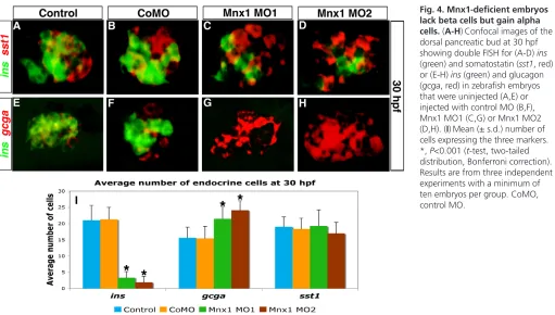
Although mnx1morphants possess the normal complement of endocrine precursors, they fail to produce an appropriate number of ins-expressing beta cells. To investigate when and how this disruption in endocrine pancreas development occurs, we analyzed whether mnx1 morphants showed changes in other hormone-producing cell types. We used FISH to examine 30 hpf mnx1 morphants for expression of the alpha cell marker glucagon (gcga), the delta cell marker somatostatin (sst1), and the epsilon cell marker ghrelin (ghrl), as well as the beta cell marker insulin (ins). As expected, ins expression was significantly decreased in mnx1 morphants (Fig. 4A-I); however, expression of sst1(Fig. 4A-D,I) and ghrl (data not shown) was unaltered. By contrast, gcgashowed increased expression in mnx1 morphants (Fig. 4E-I).Quantitative analysis of each cell type confirmed that mnx1morphants maintained normal delta cell numbers, failed to form beta cells, and produced supernumerary alpha cells (Fig. 4I). Consistent with these data, in mnx1morphants we found more cells expressing the alpha cell marker arxthan in controls, at 24 hpf (see Fig. S6 in the supplementary material) and 30 hpf (data not shown).
To investigate whether additional alpha cells were maintained at later stages of mnx1 morphant pancreas development, Tg(neurod:EGFP)embryos were injected with control MO or Mnx1 MO and fixed at 72 hpf for immunohistochemical analysis. Insulin and glucagon proteins were detected to analyze the fate of beta and alpha cells, respectively. Consistent with our results at 30 hpf, at 72 hpf mnx1 morphant embryos showed a decrease in the number of beta cells, an increase in the number of alpha cells (Fig. 5A-D), and no change in the number of delta cells (not shown) in comparison to controls. Equivalent results were obtained using wild-type non-transgenic (AB) embryos (see Fig. S7 in the supplementary material). These results indicate that mnx1-deficient embryos maintain their altered balance of beta to alpha cells as development continues. In addition, we found that the 72 hpf Tg(neurod:EGFP) mnx1 morphants had equivalent numbers of GFP-expressing cells to control embryos (Fig. 5E-H), indicating that there is no unusual loss of endocrine progenitor cells in mnx1 morphants. We did note some minor alterations in the morphology of the primary islet of mnx1 morphants, which appeared disorganized in comparison to the spherical islet of controls.
These observations are consistent with our finding that mnx1 morphants produce normal numbers of endocrine progenitors and do not undergo aberrant cell proliferation or cell death. In summary, we find that mnx1-deficient specimens initially specify the normal number of endocrine progenitor cells, but that the balance of further differentiation into beta and alpha cells is altered, such that these embryos lose beta cells and gain alpha cells. These findings suggest that the normal role of mnx1is to promote beta cell differentiation, possibly at the expense of alpha cell differentiation.
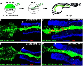
[image:5.612.50.355.60.351.2]mnx1is required in the endoderm for beta cell fate Our data suggest that mnx1functions directly within the endoderm to promote the beta cell fate. To test this hypothesis we used cell transplantation to generate chimeric embryos in which mnx1
Fig. 2. Mnx1-deficient embryos have endocrine pancreas defects. (A-F)Confocal images of the dorsal pancreatic bud at 24 hpf. ins(green) and isl1(red) transcripts were detected by double-fluorescent in situ hybridization (FISH) in control (A-C) or Mnx1 MO1-injected (D-F) zebrafish embryos. Anterior to left. (G)Mean (± s.d.) number of cells positive for insulin (ins) or isl1(red) in control and mnx1morphants. *,
P<0.001 (t-test, two-tailed distribution, Bonferroni correction); control, n35; Mnx1 MO1, n50.
D
E
V
E
LO
P
M
E
N
function is manipulated exclusively within the endoderm. Briefly, we injected donor embryos with sox32 mRNA, causing all mesendodermal cells to take on an endoderm fate, and then transplanted donor endoderm to host embryos rendered endoderm deficient by Sox32 knockdown (schematized in Fig. 6A). As we previously reported (Stafford et al., 2006), in the majority of control transplants in which Sox32-overexpressing donor-derived endoderm cells are transplanted into sox32morphant hosts, the endoderm is fully reconstituted, shows normal morphology (green cells in Fig. 6B) and develops ins-expressing beta cells (Fig. 6B,B⬘). By contrast, when we performed transplants using endoderm cells from donors overexpressing mnx1(mnx1mRNA injected), the donor-derived cells did not survive and thus failed to reconstitute the endoderm lineage (data not shown). Consistent with this observation, mnx1-overexpressing donor embryos become dysmorphic and die early in gastrulation (data not shown) (Wendik et al., 2004).
When we performed transplants using cells from donor embryos depleted of Mnx1 (mnx1 morphants), the chimeric embryos again developed a fully reconstituted endoderm with normal morphology (Fig. 6C). However, these chimeric embryos developed much reduced numbers of beta cells (Fig. 6C,C⬘), similar to embryos injected with mnx1MOs at the one-cell stage. The lack of beta cells in embryos with normal mesoderm and ectoderm but mnx1-deficient endoderm confirms that mnx1 function is required in the endoderm for endocrine progenitors to take on a beta cell fate.
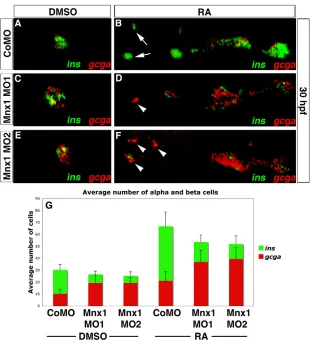
Mnx1 functions downstream of RA to promote the beta cell fate
[image:6.612.52.553.57.350.2]Our microarray results suggest that mnx1 functions downstream of RA signaling in pancreatic endoderm. To explore this finding in more detail we tested the hypothesis that knockdown of Mnx1 might block the expansion of endocrine cells caused by treatment with exogenous RA. We injected wild-type embryos with control or mnx1MOs and treated them with RA (see Table 1). Embryos were fixed at 30 hpf for FISH to detect insulinand glucagon transcripts. Results with control morphants were identical to those with uninjected control embryos (see Fig. S8 in the supplementary material). In RA-treated controls, the number of pancreatic endocrine cells was increased and the domain occupied by these cells expanded dramatically towards the anterior (Fig. 7A,B), consistent with our previous findings (Stafford and Prince, 2002). RA-treated mnx1 morphants showed a similar overall expansion in endocrine cells, indicating that Mnx1 knockdown does not block the consequences of RA treatment. However, in comparison to RA-treated controls, the RA-treated mnx1morphants displayed a significant decrease of beta cells but an increase of alpha cells (Fig. 7C-F). Interestingly, in the RA-treated mnx1 morphants, alpha cells were expanded significantly further towards the anterior of the embryos than in RA-treated controls (Fig. 7D,F, arrowheads), into a location typically populated only by beta cells (Fig. 7B, arrow). Whether embryos were RA treated or not, the ratio of alpha to beta cells in control morphants was 1:2, as compared with ~3:1 in mnx1 Fig. 3. Endocrine progenitors are unaffected by Mnx1 knockdown. (A-D)Confocal images of Tg(neurod:EGFP) zebrafish embryos at 18 hpf. Whole-mount immunolabeling for GFP (green) and for myosin to label somites (blue); nuclear staining with TO-PRO-3 (red). Somites are numbered from anterior to posterior (S1-S6). (E-H)In situ hybridization for neurodat 18 hpf. (A,E)Uninjected controls; (B,F) control morphants; (C,G) Mnx1 MO1-injected embryos; (D,H) Mnx1 MO2-injected embryos. (I)Mean (± s.d.) neurod-expressing cells from two independent experiments and from a minimum of 17 embryos per group.
D
E
V
E
LO
P
M
E
N
morphants (Fig. 7G). We also analyzed other hormone-producing cell types by in situ hybridization for sst1and ghrl. RA treatment expanded delta and epsilon cells as expected, but knockdown of Mnx1 did not alter their numbers (data not shown).
We also tested the possibility that knockdown of Mnx1 might cause premature alpha cell differentiation. We analyzed the numbers of alpha and beta cells at 24 hpf (a stage at which we do not typically detect gcga transcripts) by double in situ hybridization for gcgaand ins. Whereas the number of beta cells was significantly decreased in mnx1morphants at this stage, no alpha cells were detected in either controls or mnx1morphants, whether RA treated or not, suggesting that the increase in alpha cell numbers is not a consequence of premature differentiation (data not shown). Together, our data confirm that mnx1functions
downstream of RA to promote beta cell fates, and provide support for the hypothesis that, in the absence of Mnx1 function, beta cell precursors instead differentiate as alpha cells.
Beta cell progenitors differentiate as alpha cells in mnx1 morphants
[image:7.612.53.564.57.347.2]Next, we tested the hypothesis that reduction of Mnx1 function causes progenitor cells that are normally fated to produce beta cells to instead differentiate as alpha cells. To enable recognition of cells that would normally differentiate as beta cells, we developed a new Tg(mnx1:0.6hsp70:GFP)os26line [hereafter Tg(mnx1:GFP)], in which mnx1regulatory elements drive GFP expression in the beta cells. In agreement with our mnx1expression analysis (Fig. 1), we find that pancreatic expression of Tg(mnx1:GFP) colocalizes Fig. 4. Mnx1-deficient embryos lack beta cells but gain alpha cells. (A-H)Confocal images of the dorsal pancreatic bud at 30 hpf showing double FISH for (A-D) ins
(green) and somatostatin (sst1, red) or (E-H) ins(green) and glucagon (gcga, red) in zebrafish embryos that were uninjected (A,E) or injected with control MO (B,F), Mnx1 MO1 (C,G) or Mnx1 MO2 (D,H). (I)Mean (± s.d.) number of cells expressing the three markers. *, P<0.001 (t-test, two-tailed distribution, Bonferroni correction). Results are from three independent experiments with a minimum of ten embryos per group. CoMO, control MO.
Fig. 5. The altered balance of alpha and beta cells in Mnx1-deficient embryos is maintained after ventral bud development.
Confocal images of whole-mount 72 hpf
Tg(neurod:EGFP)zebrafish
embryos that were (A,E) uninjected, or injected with (B,F) control MO, (C,G) Mnx1 MO1 or (D,H) Mnx1 MO2, immunolabeled for glucagon (red), insulin (blue) and GFP (green) as indicated. Data are representative of three independent experiments.
D
E
V
E
LO
P
M
E
N
[image:7.612.52.449.553.739.2]exclusively with insulin-expressing beta cells (Fig. 8A-D,I-L; see Figs S9 and S10 in the supplementary material). GFP-positive cells are observed in the pancreatic endoderm as early as 16 hpf (see Fig. S9 in the supplementary material), and, by 24 hpf, these cells start to co-express ins(see Fig. S9M,N in the supplementary material); we also observed GFP-positive cells in 9 dpf larvae and in the dissected adult pancreas (data not shown). Importantly, this transgene does not recapitulate the earlier, broad endodermal expression of mnx1that can be detected by in situ hybridization at 10-14 hpf (Fig. 1; data not shown).
Mnx1 MO1 targets the 5⬘UTR of mnx1 mRNA and also recognizes the 5⬘UTR of the Tg(mnx1:GFP) transcript. As expected, Mnx1 MO1 significantly depletes GFP expression in Tg(mnx1:GFP)embryos (see Fig. S9C,G,K,O in the supplementary material), attesting to the specificity of this MO. By contrast, Mnx1 MO2 targets the mnx1start site, which is largely missing from the Tg(mnx1:GFP) transgene. As expected, Mnx1 MO2 does not initially affect GFP expression from the transgene (see Fig. S9D,H in the supplementary material), although we did note a slight reduction in GFP levels by 24 hpf (see Fig. S9L in the supplementary material), suggesting that an autoregulatory mechanism might function to maintain high levels of Mnx1 in beta cells. Furthermore, and consistent with our finding that Mnx1 knockdown does not decrease the number of endocrine precursors, the number of Tg(mnx1:GFP)cells does not decrease in Mnx1 MO2 morphants (compare Fig. S9M with S9N and S9P in the supplementary material). We were therefore able to use Mnx1 MO2 to block Mnx1 protein expression, while at the same time tracing the fates of cells expressing Tg(mnx1:GFP).
Tg(mnx1:GFP) embryos were injected with control MO or Mnx1 MO2 and the fate of Mnx1-GFP cells analyzed at 72 hpf using whole-mount immunohistochemistry. In control MO-injected embryos the Tg(mnx1:GFP) cells give rise exclusively to beta cells (Fig. 8A-D). By contrast, when Mnx1 is knocked down ins
expression is depleted, yet there is no loss of Tg(mnx1:GFP) cells (Fig. 8E-H), consistent with the hypothesis that these cells lose their normal beta cell fate. In accordance with this observation, we did not detect any change in apoptosis levels between controls and mnx1 morphants (data not shown). Analysis of GFP-positive cells in control morphants confirmed that these cells do not express the alpha cell marker glucagon, consistent with the fact that Mnx1-expressing cells do not normally differentiate to an alpha cell fate (Fig. 8I-L). By contrast, in mnx1morphants (Fig. 8M-P) the alpha cell marker glucagon colocalizes with GFP (Fig. 8P, arrows), indicating a cell fate change, such that beta cell precursors give rise to alpha cells. The fate change is limited to beta cell precursors taking on alpha cell fates, as Mnx1 knockdown does not cause Tg(mnx1:GFP) cells to colabel with the delta cell marker somatostatin (data not shown). In addition, we have never detected dual hormone-expressing cells in either wild-type or mnx1 morphant embryos. However, we did note that some GFP-positive cells in the mnx1morphants did not express glucagon (Fig. 8P), or any other endocrine hormone marker, suggesting that some cells retain progenitor properties at least at the stage of our analysis. This finding explains why the observed expansion in alpha cell numbers does not fully account for the reduction in beta cell numbers and, additionally, explains why we see a reduction in isl1-expressing endocrine cell numbers at 24 hpf (Fig. 2).
Together, our data suggest that, in mnx1 morphants, a normal number of endocrine precursors forms, but a large proportion of beta cell precursors fail to follow their usual developmental pathway, with many of these precursors instead taking on the alpha cell fate.
DISCUSSION
[image:8.612.53.402.58.338.2]In this study we have shown that RA signaling regulates the expression of mnx1 in the endoderm, and that the Mnx1 transcription factor functions within the endoderm to regulate the Fig. 6. Mnx1 functions directly in the endoderm to promote beta cell fate.
(A)The cell transplantation approach. Wild-type zebrafish embryos were injected with fluorescein dextran (green) together with either sox32mRNA or
sox32 mRNA and Mnx1 MO. Control or
morphant donor cells were transplanted into sox32morphant hosts then raised until 28 hpf. (B-C⬘) Confocal images of representative 28 hpf control (B,B⬘) and Mnx1 MO (C,C⬘) transplants, at low (B,C) and higher (B⬘,C⬘) magnification, showing whole-mount immunostaining for myosin (blue) and in situ hybridization for insulin
(red). S1, first somite. Anterior to left. Control, n12; Mnx1 MO, n13.
D
E
V
E
LO
P
M
E
N
fate of beta cell precursors downstream of RA signaling. Importantly, we have also found that zebrafish Mnx1 plays a crucial role in establishing the appropriate balance of alpha and beta cells. When Mnx1 is knocked down, beta cell precursors fail to follow their normal differentiation program and instead take on an alpha cell fate. The production of endocrine cell types in appropriate relative proportions is crucial to the development of fully functional islets.
Our in situ analysis confirms that there is a biphasic expression of zebrafish mnx1in the endoderm. There is a transient initial phase of expression in a broad subset of endoderm cells with an anterior limit at approximately the level of the pancreatic anlagen (Ward et al., 2007). We find that in zebrafish, as in the mouse dorsal pancreatic bud (Li et al., 1999), this early broad expression is rapidly downregulated before the onset of pdx1expression. In the second expression phase, mnx1 is localized to the endocrine pancreas anlagen and, as early as insulin can be detected, is restricted to the beta cells (Wendik et al., 2004). The new Tg(mnx1:GFP)line that we have described in this study recapitulates only the second phase of expression, suggesting that the enhancer elements required for early endoderm expression are absent from the transgene. Phenotypes associated with MNX1mutations in humans, as well as studies in mice, have revealed roles for Mnx1 in both pancreatic and posterior gut endoderm (Ross et al., 1998; Hagan et al., 2000; Li and Edlund, 2001; Cretolle et al., 2008), suggesting conserved expression across vertebrates.
Our microarray and expression analyses have revealed that zebrafish mnx1 expression is subject to RA regulation from its onset. The rapid response of mnx1transcription to RA treatment
suggests that it might be a direct target of RA signaling, and, consistent with this model, we find two RAREs in the mnx1 non-coding sequences. Further, we find that CHX treatment to block protein translation does not prevent mnx1 expression from expanding in response to RA treatment. Our previous work (Stafford and Prince, 2002) has shown that all pancreas cell types are expanded towards the anterior in response to a brief treatment with RA at the end of gastrulation. Interestingly, the results we present in this study indicate that although RA enlarges the endocrine progenitor pool, the relative ratios of the different endocrine cell types remain unchanged. In both control and treated specimens the alpha to beta cell ratio is 1:2. Although RA-treated embryos have significantly more pancreatic cells in total, Mnx1 depletion changes the ratio between alpha and beta cells in precisely the same manner in RA-treated versus untreated specimens. In both control and RA-treated specimens, Mnx1 knockdown alters the alpha to beta cell ratio to ~3:1. We propose that at late gastrulation stages, RA signaling directs endoderm cells toward the endocrine pancreas fate, and those progenitors that maintain or initiate mnx1expression at ~15 hpf go on to become beta cells as opposed to alpha cells. Our observation of maintained Tg(mnx1:GFP)expression in the beta cells is consistent with this model. Together, our results suggest that the primary function of mnx1is to establish the correct number of beta cells from the endocrine pancreas progenitor pool.
[image:9.612.52.363.58.400.2]Our results suggest that, in addition to promoting beta cell fate, mnx1 might actively suppress alpha cell fate. Previous analysis in mouse has revealed that Arxfunction is necessary for alpha cell differentiation (Collombat et al., 2003) and, importantly, that Arx
Fig. 7. Mnx1 knockdown alters the alpha:beta cell ratio in a similar fashion in control and RA-treated embryos. (A-F)Confocal images of DMSO carrier-treated (A,C,E) and RA-treated (B,D,F) 30 hpf zebrafish embryos that were injected with control MO (A,B), Mnx1 MO1 (C,D) or Mnx1 MO2 (E,F), showing double FISH for ins(green) and gcga (red). Arrows indicate beta cells and arrowheads indicate alpha cells. (G)Mean (± s.d.) number of alpha and beta cells from four independent experiments with a minimum of 45 embryos per group. Anterior to left.
D
E
V
E
LO
P
M
E
N
misexpression in endocrine progenitors is sufficient to drive them to an alpha cell fate (Collombat et al., 2007). In response to Mnx1 knockdown in zebrafish we observed a significant increase in the number of pancreatic progenitors expressing arx. As we have found that Mnx1 knockdown increases the number of alpha cells, this finding is entirely consistent with the model of an instructive role for Arx in alpha cell differentiation. Indeed, remarkably, misexpression of Arx in mature murine beta cells using an insulin-Cre driver is sufficient to cell-autonomously convert beta cells to the alpha cell fate (Collombat et al., 2007). Together, our findings and published studies suggest a model in which the loss of repression of arxin zebrafish beta cell progenitors is likely to be sufficient to activate the alpha cell differentiation program. In turn, we suggest that one of the normal roles of Mnx1 is to directly or indirectly repress arxexpression in beta cell progenitors to block the alpha cell differentiation program and hence promote the beta cell differentiation program.
Recent work in the zebrafish has suggested that the initial set of dorsal bud-derived beta cells do not expand further and are perhaps less important for blood sugar homeostasis than the later-developing ventral bud-derived beta cells (Hesselson et al., 2009). We find that the imbalance in alpha:beta cell ratio caused by Mnx1 knockdown at 30 hpf is maintained through to at least 72 hpf, by which stage ventral bud-derived cells have begun to contribute to the pancreas. This suggests that Mnx1 is important for beta cell fates in both dorsal bud- and ventral bud-derived beta cells, although the transient nature of MO knockdown does not allow us to extend our experiments to later larval stages. Notably, Harrison and colleagues (Harrison et al., 1999) found that Mnx1 and insulin
continue to be colocalized even in adult mice. Mouse Mnx1null mutants show complete agenesis of the dorsal bud (Li et al., 1999; Harrison et al., 1999), a more drastic phenotype than we observe in our zebrafish morphants, which form a dorsal bud of approximately normal size although with somewhat disorganized morphology. Interestingly, in mouse Mnx1mutants the ventral bud does form but is missing a subset of the beta cells; although alpha cell numbers are unaffected, delta cell number is increased in the Mnx1mutant ventral buds (Li et al., 1999). This alteration in the balance of endocrine cell types is reminiscent of our results in the zebrafish, although in the murine case beta cells might be replaced by delta cells rather than alpha cells. Notably, in both rodents and humans polyhormonal cells have been observed during the primary transition stage of endocrine pancreas development (De Krijger et al., 1992; Teitelman et al., 1993; Polak et al., 2000), but we have not detected polyhormonal cells in either wild-type or Mnx1-deficient zebrafish embryos. The differences between our mnx1 zebrafish morphants and Mnx1mutant mice might reflect species-specific differences in Mnx1gene function, although we cannot rule out the possibility that they reflect the incomplete nature of MO knockdown. Nevertheless, in both species Mnx1 is crucial for normal beta cell development and also plays a role in controlling the balance of endocrine cell types.
[image:10.612.49.419.57.400.2]As zebrafish mnx1is expressed both early, in a broad population of endoderm during late gastrulation, and later, in a much more restricted set of endocrine cells, the question arises as to precisely when in development Mnx1 functions to promote beta cell fates. Our MO knockdown approach depletes Mnx1 function for the duration of at least the first day of development, and therefore does Fig. 8. Beta cell progenitors take on an alpha cell fate in Mnx1-deficient embryos. (A-P)Confocal images of 72 hpf Tg(mnx1:GFP)
zebrafish embryos injected with control MO (A-D,I-L) or Mnx1 MO2 (E-H,M-P). (A-H)Whole-mount immunolabeling for GFP (green) and insulin (red), with nuclear staining with TO-PRO-3 (blue). (I-P)Whole-mount immunostaining for GFP (green) and glucagon (red), with nuclear staining with TO-PRO-3 (blue). Arrowheads indicate colocalization of glucagon and GFP. Data are representative of three independent experiments with a minimum of 20 embryos per group.
D
E
V
E
LO
P
M
E
N
not allow us to distinguish whether Mnx1 function is required at late gastrulation, subsequently in the beta cell progenitors, or, perhaps most likely, at both stages. Nevertheless, the capacity of Mnx1 to regulate arxexpression suggests that Mnx1 is likely to act relatively high up in the transcriptional hierarchy leading to beta cell fate.
Many additional transcription factors have been shown to play important roles during endocrine cell specification in both mice and zebrafish; these include sox4(Mavropoulos et al., 2005; Wilson et al., 2005), nkx2.2 (Sussel et al., 1998; Pauls et al., 2007), neurod (Naya et al., 1997; Chao et al., 2007), nkx6.1 andnkx6.2(Sander et al., 2000; Henseleit et al., 2005; Binot et al., 2010). The details of the gene-regulatory cascade leading to beta cell specification are still not fully understood, but it is likely that complex gene-regulatory networks act to establish the multiple endocrine cell fates (Anderson et al., 2009). In future experiments it will be valuable to investigate how Mnx1 fits into these networks by exploring epistatic relationships with other regulatory genes. In addition, mutation of the murine gene that encodes connective tissue growth factor (Ctgf) leads to a depletion of beta cells and to an increase in alpha cells in the developing pancreas (Crawford et al., 2009). This secreted protein is known to modulate several signaling pathways and it will be of interest to explore whether Ctgf and Mnx1 function in a common pathway. Finally, in view of the important roles that we have uncovered for zebrafish mnx1, we suggest that the role of human MNX1 during the in vitro differentiation of beta cells warrants further exploration.
Acknowledgements
We thank members of the V.E.P. laboratory for helpful advice and discussion; Sean Burns for expert fish care; Drs Didier Stainier and Dirk Meyer for the
prox1 and mnx1 (hb9) constructs; and Mary Kinkel, Stefani Eames Nalle and Devorah Goldman for helpful comments on the manuscript.
Funding
This work was supported by the National Institutes of Health [grant DK064973 to V.E.P.]; a Juvenile Diabetes Research Foundation (JDRF) fellowship to G.D.; an Intellectual and Developmental Disabilities Research Centers (IDDRC) grant to the J.P. Kennedy Mental Retardation and Developmental Disabilities Center [NIH 5POHD054275]; the Digestive Disease Research Core Center of the University of Chicago [DK42086]; a National Center for Research Resources (NCRR) grant to the University of Chicago Imaging Institute; and a Clinical and Translational Science Award (CTSA) [UL1RR024999]. Deposited in PMC for release after 12 months.
Competing interests statement
The authors declare no competing financial interests.
Supplementary material
Supplementary material for this article is available at
http://dev.biologists.org/lookup/suppl/doi:10.1242/dev.067736/-/DC1
References
Anderson, K. R., Torres, C. A., Solomon, K., Becker, T. C., Newgard, C. B., Wright, C. V., Hagman, J. and Sussel, L.(2009). Cooperative transcriptional regulation of the essential pancreatic islet gene NeuroD1 (beta2) by Nkx2.2 and neurogenin 3. J. Biol. Chem. 284, 31236-31248.
Argenton, F., Zecchin, E. and Bortolussi, M.(1999). Early appearance of pancreatic hormone-expressing cells in the zebrafish embryo. Mech. Dev. 87, 217-221.
Biemar, F., Argenton, F., Schmidtke, R., Epperlein, S., Peers, B. and Driever, W.(2001). Pancreas development in zebrafish: early dispersed appearance of endocrine hormone expressing cells and their convergence to form the definitive islet. Dev. Biol. 230, 189-203.
Binot, A.-C., Manfroid, I., Flasse, L., Winandy, M., Motte, P., Martial, J. A., Peers, B. and Voz, M. L.(2010). Nkx6.1 and nkx6.2 regulate alpha- and beta-cell formation in zebrafish by acting on pancreatic endocrine progenitor beta-cells.
Dev. Biol. 340, 397-407.
Chao, C. S., Loomis, Z. L., Lee, J. E. and Sussel, L.(2007). Genetic identification of a novel NeuroD1 function in the early differentiation of islet alpha, PP and epsilon cells. Dev. Biol. 312, 523-532.
Chen, Y., Pan, F., Brandes, N., Afelik, S., Solter, M. and Pieler, T.(2004). Retinoic acid signaling is essential for pancreas development and promotes endocrine at the expense of exocrine cell differentiation in. Dev. Biol. 271, 144-160.
Chung, W. and Stainier, D.(2008). Intra-endodermal interactions are required for pancreatic cell induction. Dev. Cell14, 582-593.
Collombat, P., Mansouri, A., Hecksher-Sorensen, J., Serup, P., Krull, J., Gradwohl, G. and Gruss, P.(2003). Opposing actions of Arx and Pax4 in endocrine pancreas development. Genes Dev. 17, 2591-2603.
Collombat, P., Hecksher-Sørensen, J., Krull, J., Berger, J., Riedel, D., Herrera, P. L., Serup, P. and Mansouri, A.(2007). Embryonic endocrine pancreas and mature beta cells acquire alpha and PP cell phenotypes upon Arx misexpression.
J. Clin. Invest. 117, 961-970.
Crawford, L. A., Guney, M. A., Oh, Y. A., Deyoung, R. A., Valenzuela, D. M., Murphy, A. J., Yancopoulos, G. D., Lyons, K. M., Brigstock, D. R., Economides, A. et al.(2009). Connective tissue growth factor (CTGF) inactivation leads to defects in islet cell lineage allocation and b-cell proliferation during embryogenesis. Mol. Endocrinol. 23, 324-336.
Cretolle, C., Pelet, A., Sanlaville, D., Zerah, M., Amiel, J., Jaubert, F., Revillon, Y., Baala, L., Munnich, A., Nihoul-Fekete, C. et al.(2008). Spectrum of HLXB9 gene mutations in Currarino syndrome and genotype-phenotype correlation. Hum. Mutat.29, 903-910.
De Krijger, R. R., Aanstoot, H. J., Kranenburg, G., Reinhard, M., Visser, W. J. and Bruining, G. J.(1992). The midgestational human fetal pancreas contains cells coexpressing islet hormones. Dev. Biol. 153, 368-375.
DiIorio, P. J., Moss, J. B., Sbrogna, J. L., Karlstrom, R. O. and Moss, L. G. (2002). Sonic hedgehog is required early in pancreatic islet development. Dev. Biol. 244, 75-84.
Elsen, G. E., Choi, L. Y., Prince, V. E. and Ho, R. K.(2009). The autism susceptibility gene met regulates zebrafish cerebellar development and facial motor neuron migration. Dev. Biol. 335, 78-92.
Field, H. A., Dong, P. D. S., Beis, D. and Stainier, D. Y. R.(2003). Formation of the digestive system in zebrafish. II. Pancreas morphogenesis. Dev. Biol. 261, 197-208.
Flanagan-Steet, H., Fox, M. A., Meyer, D. and Sanes, J. R.(2005). Neuromuscular synapses can form in vivo by incorporation of initially aneural postsynaptic specializations. Development132, 4471-4481.
Grabher, C., Joly, J. S. and Wittbrodt, J.(2004). Highly efficient zebrafish transgenesis mediated by the meganuclease I-SceI. Methods Cell Biol. 77, 381-401.
Hagan, D. M., Ross, A. J., Strachan, T., Lynch, S. A., Ruiz-Perez, V., Wang, Y. M., Scambler, P., Custard, E., Reardon, W., Hassan, S. et al.(2000). Mutation analysis and embryonic expression of the HLXB9 Currarino syndrome gene. Am. J. Hum. Genet. 66, 1504-1515.
Halloran, M. C., Sato-Maeda, M., Warren, J. T., Su, F., Lele, Z., Krone, P. H., Kuwada, J. Y. and Shoji, W.(2000). Laser-induced gene expression in specific cells of transgenic zebrafish. Development127, 1953-1960.
Harrison, K. A., Druey, K. M., Deguchi, Y., Tuscano, J. M. and Kehrl, J. H. (1994). A novel human homeobox gene distantly related to proboscipedia is expressed in lymphoid and pancreatic tissues. J. Biol. Chem.31, 19968-19975. Harrison, K. A., Thaler, J., Pfaff, S. L., Gu, H. and Kehrl, J. H.(1999). Pancreas dorsal lobe agenesis and abnormal islets of Langerhans in Hlxb9-deficient mice.
Nat. Genet. 23, 71-75.
Henseleit, K. D., Nelson, S. B., Kuhlbrodt, K., Hennings, J. C., Ericson, J. and Sander, M.(2005). NKX6 transcription factor activity is required for alpha- and beta-cell development in the pancreas. Development132, 3139-3149. Hesselson, D., Anderson, R. M., Beinat, M. and Stainier, D. Y. R.(2009).
Distinct populations of quiescent and proliferative pancreatic beta-cells identified by HOTcre mediated labeling. Proc. Natl. Acad. Sci. USA106, 14896-14901. Ho, R. K. and Kane, D. A.(1990). Cell-autonomous action of zebrafish spt-1
mutation in specific mesodermal precursors. Nature348, 728-730. Jékely, G. and Arendt, D.(2007). Cellular resolution expression profiling using
confocal detection of NBT/BCIP precipitate by reflection microscopy.
BioTechniques42, 751-755.
Kimmel, C. B., Ballard, W. W., Kimmel, S. R., Ullmann, B. and Schilling, T. F. (1995). Stages of embryonic development of the zebrafish. Dev. Dyn. 203, 253-310.
Kinkel, M. D. and Prince, V. E.(2009). On the diabetic menu: zebrafish as a model for pancreas development and function. BioEssays 31, 139-152. Kinkel, M. D., Sefton, E. M., Kikuchi, Y., Mizoguchi, T., Ward, A. B. and
Prince, V. E.(2009). Cyp26 enzymes function in endoderm to regulate pancreatic field size. Proc. Natl. Acad. Sci. USA106, 7864-7869. Korzh, V., Sleptsova, I., Liao, J., He, J. and Gong, Z.(1998). Expression of
zebrafish bHLH genes ngn1 and nrd defines distinct stages of neural differentiation. Dev. Dyn. 213, 92-104.
Li, H. and Edlund, H.(2001). Persistent expression of Hlxb9 in the pancreatic epithelium impairs pancreatic development. Dev. Biol. 240, 247-253. Li, H., Arber, S. and Edlund, T. M. J. H.(1999). Selective agenesis of the dorsal
pancreas in mice lacking homeobox gene Hlxb9. Nature23, 67-70.
D
Martín, M., Gallego-Llamas, J., Ribes, V., Kedinger, M., Niederreither, K., Chambon, P., Dollé, P. and Gradwohl, G.(2005). Dorsal pancreas agenesis in retinoic acid-deficient Raldh2 mutant mice. Dev. Biol. 284, 399-411.
Mavropoulos, A., Devos, N., Biemar, F., Zecchin, E., Argenton, F., Edlund, H., Motte, P., Martial, J. A. and Peers, B.(2005). sox4b is a key player of pancreatic alpha cell differentiation in zebrafish. Dev. Biol. 285, 211-223. Mizoguchi, T., Verkade, H., Heath, J., Kuroiwa, A. and Kikuchi, Y.(2008).
Sdf1/Cxcr4 signaling controls the dorsal migration of endodermal cells during zebrafish gastrulation. Development135, 2521-2529.
Molotkov, A., Molotkova, N. and Duester, G.(2005). Retinoic acid generated by Raldh2 in mesoderm is required for mouse dorsal endodermal pancreas development. Dev. Dyn. 232, 950-957.
Naya, F. J., Huang, H. P., Qiu, Y., Mutoh, H., DeMayo, F. J., Leiter, A. B. and Tsai, M. J.(1997). Diabetes, defective pancreatic morphogenesis, and abnormal enteroendocrine differentiation in BETA2/neuroD-deficient mice. Genes Dev. 11, 2323-2334.
Ober, E. A., Field, H. A. and Stainier, D. Y. R.(2003). From endoderm formation to liver and pancreas development in zebrafish. Mech. Dev. 120, 5-18. Obholzer, N., Wolfson, S., Trapani, J. G., Mo, W., Nechiporuk, A.,
Busch-Nentwich, E., Seiler, C., Sidi, S., Söllner, C., Duncan, R. N. et al.(2008). Vesicular glutamate transporter 3 is required for synaptic transmission in zebrafish hair cells. J. Neurosci. 28, 2110-2118.
Ostrom, M., Loffler, K. A., Edfalk, S., Selander, L., Dahl, U., Ricordi, C., Jeon, J., Correa-Medina, M., Diez, J. and Edlund, H.(2008). Retinoic acid promotes the generation of pancreatic endocrine progenitor cells and their further differentiation into beta-cells. PLoS ONE3, e2841.
Pauls, S., Zecchin, E., Tiso, N., Bortolussi, M. and Argenton, F.(2007). Function and regulation of zebrafish nkx2.2a during development of pancreatic islet and ducts. Dev. Biol. 304, 875-890.
Polak, M., Bouchareb-Banaei, L., Scharfmann, R. and Czernichow, P.(2000). Early pattern of differentiation in the human pancreas. Diabetes49, 225-232. Poulain, M. and Lepage, T.(2002). Mezzo, a paired-like homeobox protein is an
immediate target of Nodal signalling and regulates endoderm specification in zebrafish. Development129, 4901-4914.
Poulain, M., Fürthauer, M., Thisse, B., Thisse, C. and Lepage, T.(2006). Zebrafish endoderm formation is regulated by combinatorial Nodal, FGF and BMP signalling. Development133, 2189-2200.
Prince, V. E., Moens, C. B., Kimmel, C. B. and Ho, R. K.(1998). Zebrafish hox genes: expression in the hindbrain region of wild-type and mutants of the segmentation gene, valentino. Development125, 393-406.
Ross, A. J., Ruiz-Perez, V., Wang, Y., Hagan, D.-M., Scherer, S., Lynch, S. A., Lindsay, S., Custard, E., Belloni, E., Wilson, D. I. et al.(1998). A homeobox gene, HLXB9, is the major locus for dominantly inherited sacral agenesis. Nature
20, 358-361.
Sakaguchi, T., Kuroiwa, A. and Takeda, H.(2001). A novel sox gene, 226D7, acts downstream of Nodal signaling to specify endoderm precursors in zebrafish.
Mech. Dev. 107, 25-38.
Sander, M., Sussel, L., Conners, J., Scheel, D., Kalamaras, J., Dela Cruz, F., Schwitzgebel, V., Hayes-Jordan, A. and German, M.(2000). Homeobox gene Nkx6.1 lies downstream of Nkx2.2 in the major pathway of beta-cell formation in the pancreas. Development127, 5533-5540.
Shin, D., Shin, C. H., Tucker, J., Ober, E. A., Rentzsch, F., Poss, K. D., Hammerschmidt, M., Mullins, M. C. and Stainier, D. Y. R.(2007). Bmp and Fgf signaling are essential for liver specification in zebrafish. Development134, 2041-2050.
Stafford, D. and Prince, V. E.(2002). Retinoic acid signaling is required for a critical early step in zebrafish pancreatic development. Curr. Biol. 12, 1215-1220. Stafford, D., Hornbruch, A., Mueller, P. R. and Prince, V. E.(2004). A
conserved role for retinoid signaling in vertebrate pancreas development. Dev. Genes Evol.214, 432-441.
Stafford, D., White, R. J., Kinkel, M. D., Linville, A., Schilling, T. F. and Prince, V. E.(2006). Retinoids signal directly to zebrafish endoderm to specify insulin-expressing beta-cells. Development133, 949-956.
Stainier, D. Y. R.(2005). No organ left behind: tales of gut development and evolution. Science307, 1902-1904.
Sussel, L., Kalamaras, J., Hartigan-O’Connor, D. J., Meneses, J. J., Pedersen, R. A., Rubenstein, J. L. and German, M. S.(1998). Mice lacking the homeodomain transcription factor Nkx2.2 have diabetes due to arrested differentiation of pancreatic beta cells. Development125, 2213-2221. Teitelman, G., Alpert, S., Polak, J. M., Martinez, A. and Hanahan, D.(1993).
Precursor cells of mouse endocrine pancreas coexpress insulin, glucagon and the neuronal proteins tyrosine hydroxylase and neuropeptide Y, but not pancreatic polypeptide. Development118, 1031-1039.
Tiso, N., Filippi, A., Pauls, S., Bortolussi, M. and Argenton, F.(2002). BMP signalling regulates anteroposterior endoderm patterning in zebrafish. Mech. Dev. 118, 29-37.
Trinh, L. A., McCutchen, M. D., Bonner-Fraser, M., Fraser, S. E., Bumm, L. A. and McCauley, D. W.(2007). Fluorescent in situ hybridization employing the conventional NBT/BCIP chromogenic stain. BioTechniques42, 756-759. Ward, A. B., Warga, R. M. and Prince, V. E.(2007). Origin of the zebrafish
endocrine and exocrine pancreas. Dev. Dyn. 236, 1558-1569.
Wendik, B., Maier, E. and Meyer, D.(2004). Zebrafish mnx genes in endocrine and exocrine pancreas formation. Dev. Biol. 268, 372-383.
Westerfield, M.(1995). The Zebrafish Book: A Guide for the Laboratory Use of Zebrafish(Danio rerio). Eugene: University of Oregon Press.
Wilson, M. E., Yang, K. Y., Kalousova, A., Lau, J., Kosaka, Y., Lynn, F. C., Wang, J., Mrejen, C., Episkopou, V., Clevers, H. C. et al.(2005). The HMG box transcription factor Sox4 contributes to the development of the endocrine pancreas. Diabetes54, 3402-3409.