ORIGINAL RESEARCH ARTICLE
STATISTICAL APPROACH OF THE PHYSICALCHEMICAL INVESTIGATION OF
CHITOSAN/COLLAGEN BIOMATRIX CROSSLINKED WITH GRADIENT OF GENIPIN FOR
TISSUE ENGINEERING
1,2,3
Idiberto José Zotarelli Filho,
1Maria Christiane Valéria Braga Braile-Sternieri,
2Durval Ribas Filho,
2Victor Rodrigues Ribeiro Ferreira,
2Eliana Migliorini Mustafa,
1
Sofia Braile Sabino,
2Giovanni Braile Sternieri,
1Cibele Olegário Vianna Queiroz,
1
Bethina Canaroli Sbardellini,
1Lúcia Angélica Buffulin de Faria,
1Domingo Marcolino Braile
and
3Marinônio Lopes Cornélio
1
Domingo Braile Institute of Sao Jose do Rio Preto (SP), Rua Luíz Vaz de Camões, 3111 - Vila Redentora, São
José do Rio Preto SP Brazil 15015-750
2
Associação Brasileira de Nutrologia (ABRAN) / Brazilian Association of Nutrology, Catanduva/SP, Rua Belo
Horizonte, 909 - Centro, Catanduva SP Brazil 15801-150
3
State University of São Paulo - IBILCE-UNESP, Department of Physics (Molecular Biophysics), Rua Cristovão
Colombo 2265, São José do Rio Preto SP Brazil 15054-000
ARTICLE INFO ABSTRACT
Introduction: More than 60 million people in the United States have had an increase in their life expectancy in recent years because of artificial tissue and organ therapy, with every five people over 65 years old. In Brazil, this figure was higher. Thus, tissue engineering contemplates numerous advantages that meet the needs of the injured tissue or organ for the regeneration process.
Objective: to investigate the physicochemical properties of chitosan/collagen cross linked with a gradient of genipin for tissue engineering.
Methods: Chitosan/collagen biomatrices were prepared and crosslinked with a gradient of genipin for swelling, degradation and cross linking degree investigations, with statistical approach.
Results: 0.75 % v/v genomic composite biomatrix indicated to present the best physicochemical characteristics for future clinical studies.
Conclusion: With the increase of the degree of interlocking with genipin, the biomatrices became more rigid, reducing the degree of swelling and of free amino groups. Thus, through the present investigation, the chitosan/collagen biomatrix with 0.75 % v/v genipin provided the best physicochemical properties for future cell and clinical studies.
Copyright © 2018,Idiberto José Zotarelli Filho et al. This is an open access article distributed under the Creative Commons Attribution License, which permits unrestricted use, distribution, and reproduction in any medium, provided the original work is properly cited.
INTRODUCTION
More than 60 million people in the United States have had an increase in their life expectancy due to the therapy of artificial
*Corresponding author: Idiberto José Zotarelli Filho,
Domingo Braile Institute of Sao Jose do Rio Preto (SP), Rua Luíz Vaz de Camões, 3111 - Vila Redentora, São José do Rio Preto SP Brazil 15015-750.
tissues and organs, and every five people over 65 years of age have benefited from tissues and organs generated "in vitro"
(Muzzarelli et al., 2016;Varoni et al., 2017; Abbott, 2012). In
Brazil, this figure was higher (Zotarelli Filho et al., 2015).
Thus, it is imperative to develop new strategies to meet the demand with the development of biomaterials, whose main purpose is the regeneration of tissues and organs and the
amortization of costs (Muzzarelli et al., 2016; Alsarra, 2009;
ISSN: 2230-9926
International Journal of Development Research
Vol. 08, Issue, 05, pp.20215-20224, May,2018
Article History:
Received 08th February, 2018
Received in revised form 20th March, 2018
Accepted 17th April, 2018
Published online 28th May, 2018
Available online at http://www.journalijdr.com
Key Words:
Biomatrix, Chitosan,
Collagen, Physico chemical properties. Tissue engineering.
Citation: Idiberto José Zotarelli Filho, Maria Christiane Valéria Braga Braile-Sternieri, Durval Ribas Filho et al., 2018. “Statistical approach of the
physicalchemical investigation of chitosan/collagen biomatrix crosslinked with gradient of genipin for tissue engineering”, International Journal of
Development Research, 8, (05), 20215-20224.
Baldwin and Kiick, 2010). Thus, tissue engineering contemplates numerous advantages that meet the needs of
injured tissue or organ for the regeneration process (Yang et
al., 2017; Beppu et al., 1999). For this, the understanding of
chemical, physical and biological processes is necessary both biological material and the biological niche of the host (Varoni
et al., 2017; Ko et al., 2010). The cross-referencing of
compatible information between microenvironments allows cell recognition and signaling cascades for neovascularization
(Boccafoschi et al., 2005). Another advantage is the minimally
invasive surgical intervention, which allows the use of surgical techniques that are faster and cause less risk to the patient (Langer and Vacanti, 1999). For this, in the present study, chitosan-collagen-genipin biomatrices were used, because chitosan presents low molar mass with degrees of deacetylation greater than 0.4 are easily soluble in acidic
solvents (Yang et al., 2017; Alsarra, 2009). The
physico-chemical behavior of chitosan in aqueous solutions is highly dependent on pH and degree of deacetylation and has been the
target of a large number of studies (Yang et al., 2017; Alsarra,
2009). Thus, the degree of deacetylation (GD) is an important criterion in the activity of chitosan in the process of tissue regeneration. This was analyzed with the in vitro growth of cultures of human keratinocytes and fibroblasts treated with chitosan solutions with different degrees of deacetylation (Chan and King, 2009). On the other hand, collagen is an important biopolymer in tissue engineering and is present in several commercial products used in dermatology and
aesthetics (Bet et al., 1997). Thus, because of its
physicochemical and biological properties, collagen can be processed in different geometric forms without losing its
intrinsic properties (Panopoulos et al., 2012).
It can undergo chemical changes by cross-linking and hydrolysis for use as matrices both in their pure form, as in collagen gels, or in the form of blends with chitosan and other biopolymers. The mixture of the two biopolymers reconciles the biocompatibility of collagen with the adhesion forces of chitosan (Domard and Taravel, 1995). Collagen is bioadhesive by specific arginine-glycine-aspartate (RGD) sites (Zotarelli
Filho et al., 2013). The RGD group promotes cell adhesion
through binding to integrin receptors, thus promoting cell
growth and differentiation (Zotarelli Filho et al., 2013). In
addition, genipin is a hydrophilic organic compound that had its structure discovered in 1960 and is extracted from
geniposide (origin: Gardenia fruit) (Jin et al., 2004). Although
biocompatibility has not been tested in humans, it has been shown that genipin is non-cytotoxic "in vitro" and that it is
biocompatible in rats Sung et al. (1999). verified that genipin
was 10,000 times less cytotoxic than glutaraldehyde, which justifies its use as a cross-linking agent in biomaterials without causing chronic inflammatory problems. Therefore, in this scenario of technological advances in regenerative medicine and human tissue engineering, the present study aimed to investigate statistically the physicochemical properties of chitosan / collagen crosslinked biomatrix with genipin gradient for tissue engineering.
MATERIALS AND METHODS
Materials
Chitosan, collagen and genipin were purchased from Sigma-Aldrich (St. Louis, MO, USA). Collagen type 1 bovine from Sigma-Aldrich (St. Louis, MO, USA). Acetic acid and other
reagents also were purchased from Sigma-Aldrich (St. Louis, MO, USA). Device Microplate Spectrophotometer (BioTek® Instruments, USA).
Protocols
Preparation of the chitosan-collagen-genipin biomatrix:
Based on the method described by Baldwin; Kiick (2010), the previously characterized chitosan (Mw = 115 kDa and degree of deacetylation of 85.25 %) was dissolved in 10.0 mL of 2.5 % v/v acetic acid solution for 24 hours at room temperature. The collagen was dissolved. The chitosan-collagen biomatrix was prepared by mixing the two solutions under stirring for 96 h. The solutions were transferred to a 96-well plate, with a volume of 170.0 μL, in a ratio of 1: 1 v/v. Finally, the biomatrix was crosslinked obeying the concentration in ascending order of genipin of 0.10 %, 0.25 %, 0.50 %, 0.75 % and 1.00 % v/v. The mixture was then frozen in liquid nitrogen (-196.0 ° C) and then lyophilized.
Determination of the degree of swelling (DS) of the biomatrizes: The matrices were weighed when dry and swollen with alpha MEM cell culture medium (pH = 7.4) to determine the degree of swelling, with n = 10 and the measured data were studied and analyzed in the Minitab 17 statistical program. The amount of blood in absorbed alpha MEM was calculated by equation 1:
DS (%) = (mw – md) / mw (x 100) (Equation 1)
Where : mw = wet mass
md = dry mass
Study of the "in vitro" degradation of the biomatrices with blood sample in ALFA MEM culture medium: For the determination of the degradation index (DI) the samples in
replicates with initial mass, Mi, were oven dried at 40 ± 2oC
and weighed after weight stabilization (Mid). After weighing
the samples were placed in containers with blood solution in ALFA MEM culture medium obeying the relation between the surface area and the volume of solution = 0,1 cm -1 (27,28).
Samples were maintained at (37 ± 1 0C) in a water bath. Then,
they were removed from the container and dried in an oven at
(40 ± 2 0C), after reaching weight stabilization it was recorded.
The DI was obtained according to equation 1, where Mid is the
initial dry mass and Mfd is the mass of the sample after the
final drying. Equation 2:
The evaluation was performed for the times of: 0, 10, 30, 60, 90, 120, 180, 240 and 300 minutes, with n = 10.
0.50 %, 0.75 % and 1.00 % v/v by crosslinking and free amino groups in the biomatrices. The amino acid L-arginine was used as standard in different concentrations (1.0, 2.0, 3.0, 4.0, 5.0,
6.0, 7.0 and 8.0 mg mL-1) for the construction of the analytical
equation. Ten measurements were performed for each biomatrix group (n = 10). The amount of free amine groups in the biomatrices was determined by the optical absorbance of the solution at 570 nm, with "white" subtraction in a
Microplate Spectrophotometer apparatus (BioTek®
Instruments, USA).
Statistical analysis
Statistical analysis of the data was performed and interpreted by the author of the present study. For data analysis a database was built in the Microsoft Excel spreadsheet which was exported to the Minitab 17 statistical program. A common descriptive statistical analysis and Anderson-Darling normality test were performed for all variables and controls, with reference p> 0.10 as "normal". As there were continuous and categorical predictors (chitosan / collagen biomaterials crosslinked with increasing genipin concentrations of 0.10 %, 0.25 %, 0.50 %, 0.75 % and 1.00 % v/v) and the response predictors (chitosan, collagen and chitosan / collagen biomatrizes), linear regression and residual Durbin-Watson analysis were applied. For all linear regression tests, alpha level lower than 0.05 was adopted as significant. For Durbin-Watson residue analysis, the reference significance level was 0.05, adopting as acceptable range of independence 0.95 <dw <1.54 (according to the Durbin-Watson standard table, dU <dw <4-dU ), with two explanatory variables for sample size of n = 10.
RESULTS
Study of Normality (Anderson-Darling)
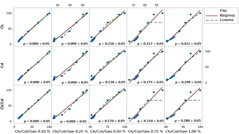
In all analyzes of the present study, according to figures 1, 5 and 8, it was observed that all the samples presented normal distribution, with p> 0.10, followed by parametric statistical analysis.
Degree of Swelling
Figure 2 shows that the chitosan bio matrix, due to its higher number of hydrophilic groups, showed a higher degree of swelling, followed by collagen, chitosan-collagen and collagen chitosan-collagen bio matrices with a gradient of 0.10 % to 1.00 %. Due to the increase in the rigidity of these biomaterials with the increase of genipin concentration, the 0.75 % bio matrix presented moderate stiffness and swelling degree around 80.0 %, pointing to be chosen for future studies. Statistical regression analysis between continuous and categorical predictors (chitosan / collagen biomaterials crosslinked with increasing genipine concentrations of 0.10 %, 0.25 %, 0.50 %, 0.75 % and 1.00 % v/v) and the predictors re sponse (chitosan, collagen and chitosan / collagen biomatizes) showed that there was no statistical significance between the continuous and categorical predictors in relation to the biomarkers response chitosan and collagen, evidencing that there was no difference in the degree of swelling, with p> 0.05, however, already in relation to the predictor bio matrix response of chitosan/collagen was statistically significant, with p <0.05, as shown in figure 3. In the Durbin-Watson residual analysis, all correlation results between the degree of swelling
of the bio matrices were within the acceptable range of independence 0.95 <dw <1.54 (according to the Durbin-Watson standard table, dU <dw <4-dU), with two explanatory variables and a sample size of n = 10. Therefore, there was no relationship of dependence (significance) between the data analyzed. Thus, the results are confirmed by figure 4, where the residues appear to follow a straight line. There is no evidence of discrepant points or unidentified variables; the residues appear to be randomly scattered around zero. There is no evidence of non-constant variance, absent terms, discrepant points or influential points; the histogram does not follow a normal curve; the residues appear to be randomly scattered around zero. There is no evidence that the error terms are correlated with each other.
Biomatrix Degradation Index
Figure 6 shows the rate of weight loss of the matrices during immersion in a solution of human blood. Matrices with increasing gradient of genipin content of 0.10 % to 1.00 % present in this sequence, a tendency of reduction in the degradation rate since the biomatrix that has lower levels of crosslinker tends to increase the degradation rate with time. Statistical regression analysis between continuous and categorical predictors (chitosan / collagen biomatrices crosslinked with increasing concentrations of genipin 0.10 %, 0.25 %, 0.50 %, 0.75 % and 1.00 % v/v) and the response predictors (chitosan, collagen and chitosan / collagen biomatrices) showed that there was no statistical significance between the continuous and categorical predictors in relation to the biomarkers response chitosan and collagen up to the concentration of 0.25 % v/v genipin, showing that there was no difference in the degree of degradation, with p <0.05. However, since 0.50 % v/v of genipin, there was a statistical difference, with p> 0.05, as shown in figure 7. In the Durbin-Watson residual analysis, all correlation results between the degree of swelling of the biomatrices were within the acceptable range of independence 0.95 <dw <1.54 (according to the Durbin-Watson standard table, dU <dw <4-dU ), with two explanatory variables and a sample size of n = 10. Therefore, there was no relationship of dependence (significance) between the data analyzed. In this way, the waste appears to follow a straight line. There is no evidence of discrepant points or unidentified variables; the residues appear to be randomly scattered around zero. There is no evidence of non-constant variance, absent terms, discrepant points or influential points; the histogram does not follow a normal curve; the residues appear to be randomly scattered around zero. There is no evidence that the error terms are correlated with each other (Figure 4).
Degree of cross-linking of bio matrix with genipin gradient
Before measurements of the degree of cross linking with genipin, the standard curve (analytical equation) was made with the amino acid L-arginine with a concentration gradient
of 1.0 to 8.0 mg mL-1. Figure 9 shows the degree of
0.75 % and 1.00 % v/v) and the predictors response (chitosan, collagen and chitosan/collagen bio matrices) showed that there was no statistical significance between the continuous and categorical predictors in relation to the biomarkers response chitosan and collagen, evidencing that there was no difference
[image:4.595.140.462.49.232.2]in the degree of swelling, with p> 0.05. However, in relation to the biomaterial response of chitosan / collagen, there was a statistical significance, with p <0.05, as shown in Figure 10. In the Durbin-Watson residual analysis, all correlation results between the degree of swelling of the bio matrices were within
[image:4.595.141.462.263.463.2]Figure 1. Normality test for the swelling test, with p> 0.10 as significant
Figure 2. Box-Plot model showing statistical values of mean, standard deviation and decline of the swelling curve with increasing genipin concentration
[image:4.595.125.467.507.701.2]the acceptable range of independence 0.95 <dw <1.54 (according to the Durbin-Watson standard table, dU <dw <4-dU ), with two explanatory variables and a sample size of n = 10. Therefore, there was no relationship of dependence (significance) between the data analyzed. Thus, the results are confirmed by Figure 4, where the residues appear to follow a straight line. There is no evidence of discrepant points or unidentified variables; the residues appear to be randomly scattered around zero.
[image:5.595.114.481.68.256.2]There is no evidence of non-constant variance, absent terms, discrepant points or influential points; the histogram does not follow a normal curve; the residues appear to be randomly scattered around zero. There is no evidence that the error terms are correlated with each other (figure 4). These results demonstrated that genipin is favorable cross linking reagent for collagen and chitosan, as it can efficiently crosslink amino groups, even at low concentration.
[image:5.595.127.473.296.459.2]Figure 04. Graphs representative of the three trials of the present study, showing in a generic way the results of the residual analysis
Figure 05. Normality test for the degradation index test, with p> 0.10 as significant
[image:5.595.139.455.493.667.2]Figure 7. Graph Matrix-Plot model showing the results of the regression analysis between the continuous / categorical predictors and the response in the degree of degradation assay
Figure 8. Normality test for the cross linking grade assay, as p> 0.10 as significant
[image:6.595.123.487.567.750.2]In addition, as the concentration of genipin increased, more amino groups were cross-linked. The region highlighted in blue in Table 1 shows t he bio matrix with 0.75 % v/v of genipin that was the one that presented the best physicochemical characteristics in the present study and was therefore the bio matrix selected for the biological studies of other works.
DISCUSSION
Biomatrix in tissue engineering
Tissue engineering is a tool that enables the creation of a suitable biological niche for the construction and regeneration
of any tissues and organs (Muzzarelli et al., 2016; Varoni et
al., 2017; Bonfield, 2006; Breyner et al., 2010). For this,
autogenous or allogeneic graft pathways are used, with and
without the use of cells (Irioda et al., 2016). For example,
biomaterials such as chitosan, collagen and
polyhydroxybutyrate, which are biodegradable, biocompatible and non-toxic, can act in the controlled release of drugs, gene transfection and tissue regeneration (Friedenstein, 1976). According to the Conference of the National Institute for the
Development of Health Consensus in 1982, biomaterials are beneficial organic compounds, or a combination thereof, that can be used for a period of time, wholly or partially as part of a system that treats, replace any tissue, organ or function of the
human body (Planat Bernard et al., 2004). The great challenge
is to understand that the science of biomaterials is multidisciplinary and its application requires adequate processing, sterilization and structural modifications that favor interaction with the tissue of interest. There are several biomaterial manufacturing models. These models follow geometric representations that must be in agreement with the type of tissue or organ of interest. To satisfy the diversity of tissues and organs, biomaterials can be manufactured in the form of porous bio matrices, thin superimposed layers, beads
and in the form of elongated yarns (Breyner et al., 2010). The
biomaterial type also directs the type of cell line it can store and stimulate proliferation or differentiation from stem cells
(Zazakowny et al., 2016; Zotarelli Filho et al., 2015, Chan et
al., 2009), as presented with the use of chitosan-collagen
[image:7.595.74.521.53.284.2]genipin in the present study. Thus, bioengineering and cell therapy work together for regenerative medicine, favoring and improving biological conditions to accelerate repair and tissue regeneration, and thus maintain tissue homeostasis naturally.
Figure 10. Graph Matrix-Plot model showing the results of the regression analysis between the continuous / categorical predictors and response in the crosslinking degree test
Table 1. Values as a percentage of the decrease in the degree of crosslinking with increasing genipin concentration
Biomatrices Degree of crosslinking%
L-arginina (standard) 100,0
Chitosan 80,0
Collagen 34,1
Chitosan/Collagen 45,9
Chitosan/Collagen/Genipin (0.10 % v/v) 39,0
Chitosan/Collagen/Genipin (0.25 % v/v) 32,4
Chitosan/Collagen/Genipin (0.50 % v/v) 23,0
Biomatrices Degree of crosslinking%
L-arginina (standard) 100,0
Chitosan 80,0
Collagen 34,1
Chitosan/Collagen 45,9
Chitosan/Collagen/Genipin (0.10 % v/v) 39,0
Chitosan/Collagen/Genipin (0.25 % v/v) 32,4
Chitosan/Collagen/Genipin (0.50 % v/v) 23,0
Chitoan/Collagen/Genipin (0.75 % v/v 12,9
[image:7.595.123.481.347.521.2]This condition is maintained because the required cellular elements, cell proliferation and differentiation factors, and supramolecular structures are provided which guarantee the functional stereochemical organization of the generated tissues
and their systemic integration (Chen et al., 2006).
Physical chemical Studies of Bio matrices
Degradation of chitosan-collagen-genipin matrices in the presence of human blood in culture media decreased as the degree of cross-linking of the bio matrix with genipin increased. This is because of the greater energy required for depolymerization of the polymer chains crosslink (crosslink covalent) than that showed low level of cross linking agent. The results below show the preferred cross linking via NH2 (chitosan) instead of hydroxyl (collagen) therefore is expected in a mixture degradation kinetics for the blends, the collagen that is physically crosslinked undergoes rapid solvation and chitosan/collagen are covalently crosslinked via deploy merization slower degradation. In comparison with the studies
by Chiono et al. (2008), matrix solubility increased as the
degree of cross linking with genipin was also increased, due to the increased vulnerability to dissolve the matrices by increasing the degree of dissociation of chitosan and collagen with protonation of the groups aminos. This revealed that the best concentration of genipin for the manufacture of the bio matrices was 0.75 % (v/v), similar to the results reported by Baldwin; Kiick (2010). Thus, when the degree of crosslinking with genipine was less than 0.50 % v/v biomaterial was very fragile, with low resistance, and when the genipin concentration was greater than 1.00 % v/v, the biomaterial ruptured and pieces of " flakes "left the bio matrices. The morphology of the matrix structure as well as its porosity were determined by scanning electron microscopy, showing the presence of spaces (pores) for the passage of nutrients, gases and cellular metabolites, as well as cell adhesion and
proliferation (Ko et al., 2010; Langer and Vacanti, 1999).
The biocompatibility of the matrices was due to the adhesion, proliferation and modulation of the cellular activities of CTMA and macrophages by the activity of alkaline phosphatase and nitrogen oxide, respectively, as a similar work
done by Planat Bernard et al. (2004). The miscibility of
chitosan with collagen was due to electrostatic interactions in ion-ion, dipole-dipole and dipole-dipole interactions, Van der Waals interactions, π-electrons and charge transfer complexes, forming ionic bonds of hydrogen and covalent bonds between the polymeric components, as well as free Gibbs free energy (ΔG <0), according to the Domard literature; Taravel (1995) and Israelachvili (2011)( Israelachvili, 1973, Israelachvili, 2011), in the mixture among other polymers, although these are of high molecular weight. The hydroxyproline (OH-) groups of collagen can form hydrogen bonds between the chains and interactions with other side groups are important in the formation of collagen fibers. These side groups may also
form hydrogen bonds with -OH and NH2 of chitosan. In
addition, the terminal groups -COOH and -NH2 may form
hydrogen bonds with -OH and –NH2 of the chitosan. This
mixture of polyactions (chitosan) and polyanions (collagen) may have led to spontaneous aggregation and release of counterions, causing an entropy gain (ΔS> 0) (Israelachvili, 2011). The presence of the counterions, as well as the presence of the cations and anions of the individual polymer chains can stimulate the cellular activities of human tissues and thus favor
tissue regeneration (Zhan et al., 2012).
Action of the intermolecular forces in the constitution of the biomatrix
Chitosan reacts easily by nucleophilic attack of its amino groups with carbonyl compounds, forming a covalent bond with some collagen and genipin clusters (Antonov and
Moldenaers, 2012; Arof et al., 2010). The miscibility of
chitosan is due to electrostatic interactions in the ion-ion, dipole-ion, and dipole-dipole interactions, Van der Waals interactions, π-electrons and charge transfer complexes, forming ionic, hydrogen and covalent bonds between the polymer components, producing a negative Gibbs (ΔG <0) free energy in the mixture, despite the high molecular weight of the polymers (Alsarra, 2009; Domard and Taravel, 1195; Drago, 1977; Israelachvili, 2011). The hydroxyproline (-OH) groups of collagen make hydrogen bonds between the chains and the interactions with the other side groups of the collagen are important in the formation of fibers. These side groups also
form hydrogen bonds with -OH and NH2 of chitosan. In
addition, the collagen-COOH and -NH2 end groups of the
collagen may also form hydrogen bonds with -OH and –NH2
of chitosan (Zhan et al., 2012; Hill, 1963). The two polymer
chains of chitosan-collagen can intertwine, forming a complex with higher viscosity (Hill, 1963). Mixtures of polyacites (chitosan) and polyanions (collagen) lead to spontaneous aggregation and the release of counterions, leading to an entropy gain (ΔS> 0) (Israelachvili, 1973).
Conclusion
As the degree of interlocking with genipin increased, the biomatrices became more rigid, reducing the degree of swelling and free amino groups. Thus, through the present investigation, the chitosan/collagen biomatrix with 0.75 % v/v genipin provided the best physico-chemical properties for future cell and clinical studies.
Declaration of Potential Conflict of Interest
The authors declare no conflict of interest.
Acknowledgment
Thank you very much Duke University – Medical Center (North Caroline, USA) for all teaching and training in scientific writing and statistical analysis of the present study. We also strongly appreciate the support of the ABRAN (Associação Brasileira de Nutrologia, Catanduva/SP). We also strongly appreciate the support of the hospital Beneficência Portuguesa de São José do Rio Preto/SP and Mitosis Lab.
REFERENCES
Abbott, A. 2012. Cell rewind wins medicine nobel. Nature,
490:151-152, 2012.
Alsarra, IA. 2009.Chitosan topical gel formulation in the
management of burn wounds. International Journal of
Biological Macromolecules, Guildford; 5 :16-21,.
Antonov, YA. and Moldenaers, P. 2012. Strong polyelectrolyte – Induced mixing in concentrated biopolymer aqueous
emulsions. Food Hydrocolloid., Oxford, 28, 1 : 213 223.
Arof, AK., Buraidah, MH., Teo, IP., Majid, SR., Yahya, R. and Taha, RM. 2010. Characterizations of Chitosan Based
Polymer Electrolyte Photovoltaic Cells. Int. J.
Baldwin, AD. and Kiick, KL. 2010. Polysaccharide-modified
synthetic polymeric biomaterials. biopolymers; 94 :128–
140.
Beppu, MM., Arruda, E. and Santana, CC. 1999. Synthesis and characterization of porous structures of dense and
chitosan. Polymers: Science and Technology,9 (4)
:163-169.
Bet, MR., Goissis, G. and Plepis, AMG. 1997. Compósitos colágeno aniômico: fosfato de cálcio. preparação e
caracterização, Química Nova; 20 : 475-477.
Boccafoschi, F., Habermehl, J., Vesentini, S. and Mantovani, D. 2005. Biological performances of collagen-based
scaffolds for vascular tissue engineering. Biomaterials; 26
(35) : 7410-7417.
Bonfield ,W. 2006.Designing porous scaffold for tissue
engineering. Phil Trans R Soc., A364 : 227-232.
Breyner, NM., Hell, RC. and Carvalho LR. 2010. Effect of three dimensional chitosan porous scaffold on the
differentiation of mesenchymal stem cells into
chondrocytes. CTO., 191 : 119-28.
Chan, BP. and So, KF. 2005. Photochemical crosslink improves the physicochemical properties of collagen
scaffolds. Journal Biomedical Materials Research, part a;
75 (3): 689-701.
Chan, YL. and King, NM. 2009.Use of focused íon bean milling for investigating the mechanical properties of biological tissues: a study of human primary molars.
Journal of the mechanical behavior of biomedical
materials, 2 (4): 375-383.
Chen, H., Ouyang, W., Jones, M., Martoni, C., Haque, T., Cohen, R. and Prakash, S. 2006. Genipin cross-linked
alginate-chitosan microcapsules: membrane
characterization and optimization of crosslinking reaction.
Biomacromolecules, 7: 2091-2098.
Chiono, V., Pulieri, E., Vozzi, G., Ciardelli, G., Ahluwalia, A. and Giusti, P. 2008. Genipin-crosslinked chitosan/gelatin
blends for biomedical applications. J Mater SCI Mater
Med., 19: 889–898.
Domard, A. and Taravel, MN.1995.Collagen and its
interaction with chitosan: ii. influence of the
physicochemical characteristics of collagen. Biomaterials.,
16(11):865–71.
Drago, RS. 1977. Physical methods in chemistry. Saunders and Co., London.
Friedenstein, AJ. 1976. Precursor cells of mechanocytes. Int
Rev Cytol, 1976, 47:327-59.
Hill, TL. 1963. Thermodynamics of small systems. Benjamin, New York: Dover Pubns.
Irioda, AC. et al. 2016. Human adipose-derived mesenchymal
stem cells cryopreservation and thawing decrease α
4-integrin expression. Stem Cells International, v. 2016, p. 1–
9.
Israelachvili, JN. 2011. Measurements of hydration forces
between macroscopic surfaces. Chemica Scripta, 25(1), 7–
14.
Israelachvili, JN. 2011. Van der Waals forces in biological
systems. Quarterly Reviews of Biophysics, 2011, 6(4), 341–
387.
Israelachvili, JN. 1973. Van der Waals dispersion force contributions to works of adhesion and contact angles on
the basis of macroscopic theory. Journal of the Chemical
Society, Faraday Transactions, 2(69), 1729–1738.
Jacob, JLB., Zotarelli Filho, IJ., Greco, OT. and Kassis, EN. 2015. Albuquerque MC, Sinibaldi FT and Cornélio ML. Significant improvement in ejection fraction and functional
class after two years of cells transplantation. Cell Biol., res
ther, 4:2.
Jayakumar, R., Menon, D., Manzoor, K., Nair, SV. and Tamura, H. 2010. Biomedical applications of chitin and
chitosan nanomaterials — a short review. Carbohydr
polym, 82 : 227–32.
Jeon, ES., Moon, HJ., Lee, MJ., Song, HY., Kim, YM., Bae YC., Jung, JS. and Kim, JH. 2006. Sphingo sylphosphorylcholine induces differentiation of human mesenchymal stem cells into smooth-muscle-like cells
through a tgf-beta-dependent mechanism. Journal of cell
science, part 23. v. 119, p. 4994-5005.
Jin, J., Song, M. and Hourston, DJ. 2004. novel chitosan-based films cross-linked by genipin with improved physical
properties. Biomacromolecules, 5 : 162-168.
Jui-Yang, I. 2012. Biocompatibility of genipin and glutaraldehyde cross-linked chitosan materials in the
anterior chamber of the eye. Int. J. Mol. Sci., 13.
Kassis, EN., Zotarelli Filho, IJ., Cornélio, ML., Cicarelli, AJ., Frascino, LF. and Araújo, TSB. 2015. Potential use of mesenchymal stem cells and bio-oss® in surgery
skull-maxillofacial: a review. International journal of
development research, vol. 05, issue, 03, pp. 3821-3827.
Khan, F., Tare, RS., Oreffo, ROC. and Bradley, M. 2009. Versatile biocompatible polymer hydrogels: scaffolds for
cell growth, angew. Chem. Int., 48 : 978 – 982.
Ko, HF., Sfeir, C. and Kumta, PN. 2010. Novel synthesis strategies for natural polymer and composite biomaterials
as potential scaffolds for tissue engineering. Phil. Trans. R.
Soc. A., 368: 1981-1997.
Langer, R. and Vacanti, JP. 1999. Tissue engineering: the design and fabrication of living replacement devices for
surgical reconstruction and transplantation. Lancet, 354:
32–34.
Muzzarelli, R.A., El Mehtedi, M, Bottegoni, C. and Gigante A.
2016. Physical properties imparted by genipin
to chitosan for tissue regeneration with human stem cells:
a review. Int J Biol Macromol., dec;93(pt b):1366-1381.
Naoum, FA., Espósito, BP. and IJ. 2017. Impact of labile plasma iron and iron chelation on the viability of cultured mononuclear cells from patients undergoing autologous
hematopoietic stem cell transplantation. Blood Res.,
jun;52(2):135-136.
Panopoulos, AD., Yanes, O. and Ruiz S. The metabolome of induced pluripotent stem cells reveals metabolic changes
occurring in somatic cell reprogramming. Cell Res, 2012,
22:168-177.
Planat Bernard, V., Silvestre, JS. and Cousin, B. 2004. Plasticity of human adipose lineage cells towards
endothelial cells: physiological and therapeutic
perspectives. Circulation, 109 : 656 -63.
Strauer, B., Yousef, M. and Schannwell, CM. 2010. The acute and long-term effects of intracoronary stem cell transplantation in 191 patients with chronic heart failure:
the star-heart study. European journal of heart failure, 12 :
721-729.
Sung, HW., Huang, RN., Huang, LL. and Tsai CC. 1999. In
vitro evaluation of cytotoxicity of a naturally occurring
cross-linking reagent for biological tissue fixation.
Biomater sci polym ed, 10:63–78.
Varoni, E.M., Vijayakumar, S., Canciani, E., Cochis, A., De Nardo, L., Lodi, G., Rimondini, L. and Cerruti, M. 2017. Chitosan-based trilayer scaffold for multitissue periodontal
Yang, J.P., Anderson, A.E., Mccartney, A., Ory, X., Garret, MA., Pappalardo, E., Bader, J. and Elisseeff, JH. 2017. Metabolically. Active three-dimensional brown adipose tissue engineered from white adipose-derived stem cells. Tissue engineering: part a volume 00, number 00, mary ann liebert, inc. doi: 10.1089/ten.tea.2016.0399.
Zazakowny, K., Lewandowska – łańcucka, J., Mastalska-popławska, J., Kamiński, K., Kusior, A., Radecka, M. and Nowakowska, M. 2016. Biopolymeric hydrogels - nanostructured tio2 hybrid materials as potential injectable scaffolds for bone regeneration. Colloids surf b biointer faces. dec 1;148:607-614.
Zhan, R., Thomasson, E., Guillaume Gentil, O., Voros, J. and Zambelli, T. 2012. Ion induced cell sheet detachment from standard cell culture surfaces coated with
polyelectrolytes. Biomaterials, Oxford, 33, 12: 3421-3427.
Zotarelli Filho, IJ, Greco, OT., Bilaqui, A., Kassis, EN.,
Araújo, JD., Jacob, JLB., Borralho, CMT et al. 2013.
Measurement and feasibility of hematopoietic stem cell
was greater for equipment in closed system. International
Journal of Development Research, vol. 3, issue, 12,
pp.033-039.
Zotarelli Filho, IJ., Bilaqui, A., Kassis, EN., Cornélio, ML. and Frascino, IF. 2015.Chronic obstructive lung disease,
stem cells and telocytes: review of therapeutic. Cell Biol:
Res Ther, 3:1 4:1.
Zotarelli Filho, IJ., Frascino, LF., Greco, OT., Araujo, JDD., Bilaqui, A., Kassis, EN., Ardito, RV. and Bonilla-Rodriguez, GO. 2013. Chitosan-collagen scaffolds can regulate the biological activities of adipose mesenchymal
stem cells for tissue engineering. J Regen Med Tissue Eng.,
2013, 2:12.
Zuk, PA., Zhu, M. and Ashjian, P. 2002. Human adipose
tissue is a source of multipotent stem cells. Mol Biol Cell.,
13:4279–4295.