THE C-DI-GMP REGULATORY NETWORK IN CLOSTRIDIOIDES DIFFICILE AND ITS ROLE IN MODULATING SURFACE ADHERENCE AND PERSISTENCE IN THE
MAMMALIAN GUT
Robert Woodrow McKee
A dissertation submitted to the faculty at the University of North Carolina at Chapel Hill in partial fulfillment of the requirements for the degree of Doctor in Philosophy in the Department
of Microbiology and Immunology.
Chapel Hill 2018 Approved by: Peggy A Cotter Jonathan J. Hansen Virginia L. Miller Rita Tamayo Mathew C Wolfgang
© 2018
Robert Woodrow McKee ALL RIGHTS RESERVED
ABSTRACT
Robert Woodrow McKee: The c-di-GMP regulatory network in Clostridioides difficile and its role in modulating surface adherence and persistence in the mammalian gut
(Under the direction of Rita Tamayo)
Clostridioides difficile (Clostridium difficile) is a spore-forming bacterial pathogen responsible for hundreds of thousands of infections each year in the United States. C. difficile outbreaks are common in hospitals because C. difficile spores can persist for months on surfaces and are resistant to many disinfectants. Despite the significant disease burden that C. difficile represents, we know surprisingly little about the factors necessary for C. difficile to colonize and persist in the mammalian intestine. Previous work demonstrated that the signaling molecule cyclic diguanylate (c-di-GMP) regulates a variety of processes in C. difficile including production of the toxins that are required for disease symptoms. Using monolayers of human intestinal epithelial cells, we demonstrate that c-di-GMP promotes attachment of C. difficile to intestinal epithelial cells. We also demonstrate that regulation of type IV pili (TFP) by c-di-GMP promotes prolonged adherence of C. difficile to epithelial cells in vitro. C. difficile mutants lacking TFP were cleared more quickly than the parental strain during single strain mouse
infections and were outcompeted by the parental strain during in vivo competition experiments in mice. Thus, our data provides evidence that TFP promote persistence of C. difficile in the
intestine. To determine what other genes c-di-GMP regulates in C. difficile, we performed RNA-sequencing comparing the transcriptome of C. difficile with elevated c-di-GMP to that of C. difficile with basal levels of c-di-GMP. We demonstrate that c-di-GMP regulates the expression
of 166 genes greatly expanding the known members of the c-di-GMP regulon. We demonstrate that c-di-GMP regulation of several transcripts in C. difficile is dependent on c-di-GMP sensing riboswitches present in the 5’ untranslated regions of these transcripts. Our results also show that c-di-GMP regulates a number of cell envelope proteins in addition to TFP and flagella. These data suggest a broader role for c-di-GMP in remodeling the C. difficile cell surface.
ACKNOWLEDGEMENTS
I would like to thank my advisor, Rita Tamayo, for taking me under her wing and showing me how to be a scientist. I would not be where I am today without her outstanding mentorship and guidance. I would also like to thank the many members of the Tamayo lab who have provided helpful feedback and advice throughout my years in the lab. I would especially like to thank Naira Aleksanyan and Liz Garrett for their contributions to Chapter 2, and Carissa Harvest for her contributions to Chapter 3. I am also grateful for the outstanding support that the UNC-CH Microbiology and Immunology program gives to its graduate students and the work the faculty and staff do to make the department an outstanding place to do science. I want to thank my friends and family who have supported me during my graduate career. Lastly, I want to thank my partner Mike, whose loving support has been critical to my success both in and beyond the lab.
TABLE OF CONTENTS
LIST OF TABLES ... viii
LIST OF FIGURES ... ix
LIST OF ABBREVIATIONS ... xi
CHAPTER 1: INTRODUCTION ... 1
DISCOVERY OF CLOSTRIDIOIDES DIFFICILE ... 1
THE C. DIFFICILE LIFECYCLE AND TRANSMISSION ... 3
RISK FACTORS FOR CDI ... 5
PATHOGENESIS OF CDI ... 7
REGULATION OF TOXIN PRODUCTION ... 8
COLONIZATION FACTORS OF C. DIFFICILE ... 10
C-DI-GMP SIGNALING ... 12
C-DI-GMP SIGNALING IN C. DIFFICILE ... 15
FIGURES ... 18
REFERENCES ... 20
CHAPTER 2: TYPE IV PILI PROMOTE CLOSTRIDIUM DIFFICILE ADHERENCE AND PERSISTENCE IN A MOUSE MODEL OF INFECTION ... 33
SUMMARY ... 33
INTRODUCTION ... 34
MATERIALS AND METHODS ... 37
DISCUSSION ... 48
FIGURES ... 51
REFERENCES ... 64
CHAPTER 3: TRANSCRIPTIONAL REGULATION IN C. DIFFICILE BY THE SECOND MESSENGER C-DI-GMP ... 73
SUMMARY ... 73
INTRODUCTION ... 73
RESULTS ... 77
DISCUSSION ... 82
MATERIALS AND METHODS ... 86
FIGURES ... 92
REFERENCES ... 99
CHAPTER 4: DISCUSSION ... 104
SUMMARY OF RESULTS AND SIGNFICANCE ... 104
IMPORTANCE ... 111
CONCLUSIONS AND FUTURE DIRECTIONS ... 114
REFERENCES ... 115
LIST OF TABLES
Table 2.1. Primers used in this study. ... 62
Table 2.2. Strains and plasmids used in this study. ... 63
Table 3.1. Putative cell envelope proteins whose expression is regulated by c-di-GMP. ... 97
Table 3.2. Changes in transcript abundance for c-di-GMP riboswitches and the downstream genes. ... 97
Table 3.3 Fold changes in transcripts for genes controlled by class II c-di-GMP riboswitches ... 98
Table 3.4. Fold changes in transcripts for genes controlled by GEMM riboswitches. ... 98
Table A1.1 Genes regulated by c-di-GMP in C. difficile ... 121
Table A1.2 Plasmids and Strains used in this study ... 126
LIST OF FIGURES
Figure 1.1. c-di-GMP signaling in C. difficile. ... 18
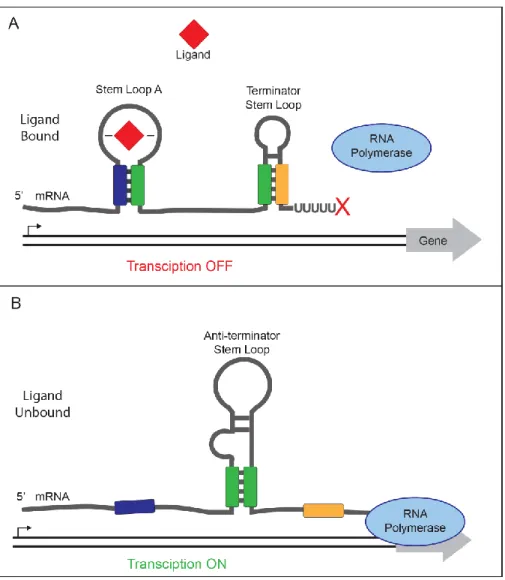
Figure 1.2 Transcriptional termination controlled by an “off” riboswitch. ... 19
Figure 2.1. Viability of epithelial cell lines under anaerobic conditions. ... 51
Figure 2.2. c-di-GMP promotes C. difficile attachment to epithelial cell monolayers in vitro. ... 52
Figure 2.3. Aflagellate C. difficile adhere better to HT-29 cell monolayers. ... 53
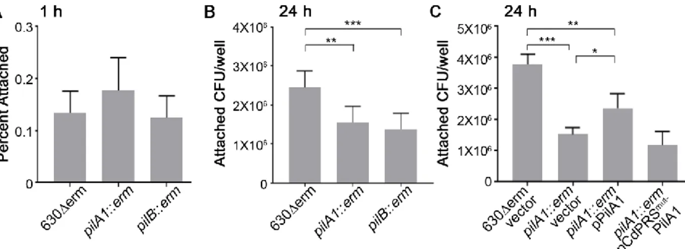
Figure 2.4. Attachment to HT-29 cell monolayers at 1 hour is not dependent on type IV pili. ... 54
Figure 2.5. Type IV pili promote adherence to MDCK cell monolayers at 24 hrs. ... 55
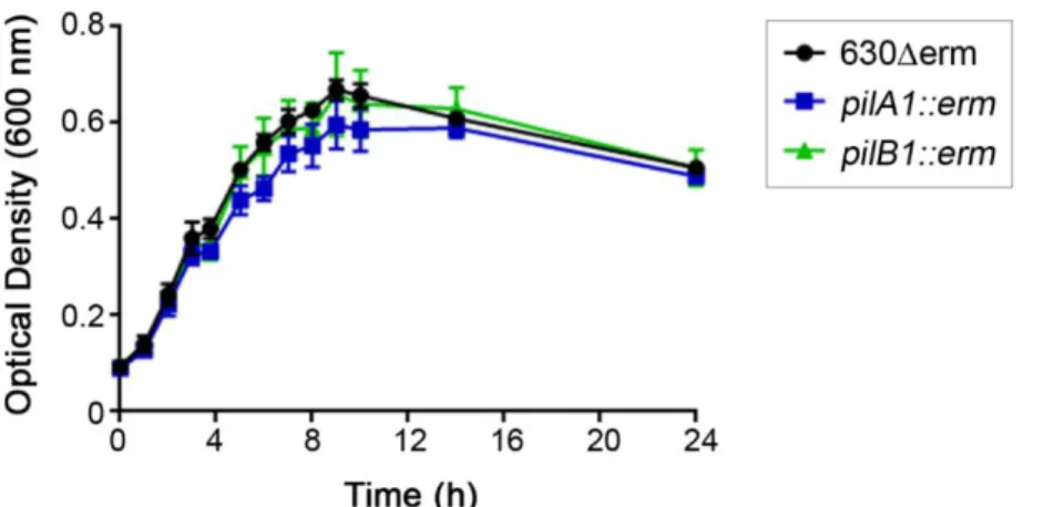
Figure 2.6. Growth of C. difficile strains in DMEM culture medium. ... 56
Figure 2.7. Swimming motility of 630Δerm and TFP mutants. ... 57
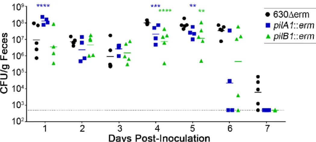
Figure 2.8. Single strain mouse infections of 630Δerm, pilA1 mutant and pilB1 mutant. ... 58
Figure 2.9. C. difficile pilus mutants are outcompeted by the parental strain in murine co-infections. ... 59
Figure 2.10. Competition of R20291 and R20291 pilB1::ermB strains in the mouse model. ... 60
Figure 2.11. TFP promote association of C. difficile with the cecal epithelium. ... 61
Figure 3.1. C. difficile genes regulated by c-di-GMP grouped by Riley classification of predicted gene products. ... 92
Figure 3.2. Biofilm formation is promoted by ectopic expression of genes encoding cell envelope proteins. ... 93
Figure 3.3. Transcript levels of genes downstream of GEMM riboswitches in C. difficile with increasing c-di-GMP. ... 95
Figure 3.4. Transcript regulation of genes downstream of class II c-di-GMP riboswitches in C. difficile with increasing c-di-GMP... 95
Figure 3.5. Alkaline phosphatase reporter assays of riboswitch-adjacent gene promoters. ... 96
Figure A1.2 RNA-sequencing reads for the region surrounding Cdi-1-2. ... 128
Figure A1.3 RNA-sequencing reads for the region surrounding Cdi-1-3. ... 129
Figure A1.4 RNA-sequencing reads for the region surrounding Cdi-1-4. ... 130
Figure A1.5 RNA-sequencing reads for the region surrounding Cdi-1-5. ... 130
Figure A1.6 RNA-sequencing reads for the region surrounding Cdi-1-6. ... 131
Figure A1.7 RNA-sequencing reads for the region surrounding Cdi-1-7. ... 131
Figure A1.8 RNA-sequencing reads for the region surrounding Cdi-1-8. ... 132
Figure A1.9 RNA-sequencing reads for the region surrounding Cdi-1-9. ... 132
Figure A1.10 RNA-sequencing reads for the region surrounding Cdi-1-10. ... 133
Figure A1.11 RNA-sequencing reads for the region surrounding Cdi-1-11. ... 133
Figure A1.12 RNA-sequencing reads for the region surrounding Cdi-1-12. ... 134
Figure A1.13 RNA-sequencing reads for the region surrounding Cdi-2-1. ... 134
Figure A1.14 RNA-sequencing reads for the region surrounding Cdi-2-2. ... 135
Figure A1.15 RNA-sequencing reads for the region surrounding Cdi-2-3. ... 135
Figure A1.16 RNA-sequencing reads for the region surrounding Cdi-2-4. ... 136
Figure A1.17 Sequence analysis of the GEMM riboswitches in C. difficile. ... 137
LIST OF ABBREVIATIONS
ANOVA Analysis of variation
BHIS Brain heart infusion supplemented with yeast extract CDI Clostridiodes difficile infection
c-di-GMP Cyclic diguanylate CFU Colony forming unit
CI Competitive Index
CROP Combined repetitive oligopeptide DGC Diguanylate cyclase
DMEM Dulbecco’s modified Eagle’s medium DNA Deoxyribonucleic acid
DPBS Dulbecco’s modified phosphate buffered saline DSS Dextran sodium sulfate
Erm Erythromycin
FBS Fetal bovine serum GTP Guanosine triphosphate IBD Inflammatory bowel disease MDCK Madin-Darby canine kidney mRNA Messenger RNA
PBS Phosphate buffered saline PCR Polymerase chain reaction PDE Phosphodiesterase
qRT-PCR Quantitative reverse-transcription polymerase chain reaction RBS Ribosomal binding site
RNA Ribonucleic acid RNA-seq RNA sequencing
RPKM Reads per kilobase per million mapped reads TCCFA Taurocholate cycloserine cefoxitin fructose agar TCRS Two component regulatory system
TFP Type IV pili
TY Tryptone yeast extract media UDP Uridine diphosphate
CHAPTER 1: INTRODUCTION
Clostridioides difficile (formerly Clostridium difficile) (1) is a Gram-positive bacterial pathogen that is responsible for approximately half a million infections yearly in the United States and an estimated 4 billion dollars in associated healthcare costs (2). C. difficile infections (CDI) are one of the most common hospital-acquired infections. CDI can range in severity from mild diarrhea to potentially life-threatening pseudomembranous colitis and toxic megacolon (3, 4). Disease symptoms are mediated in part due to the secretion of toxin(s), TcdB and/or TcdA, and strains lacking these toxins do not cause disease (5-8). Despite the current prevalence of these infections in the healthcare system, the importance of C. difficile as a pathogen has only been appreciated for a few decades.
DISCOVERY OF CLOSTRIDIOIDES DIFFICILE
C. difficile was first isolated in 1935 from the stools of healthy infants (9). The researchers named it Bacillus difficilis owing to its rod-like shape and the difficulties they experienced when trying to culture the bacterium (9). Despite its prevalence in the stools of healthy infants, they discovered that C. difficile was highly virulent in guinea pigs and rabbits, killing nearly all the animals into which bacterial cultures were injected. Similar effects were observed when they injected bacteria-free media from the C. difficile cultures into guinea pigs; however, boiling the media rendered it non-toxic. This finding indicated that the bacteria were secreting a heat-labile toxin and that this toxin was responsible for causing disease (9). However,
because the bacterium was isolated from healthy infants with no signs of acute bacterial infection, it was unclear whether C. difficile was virulent in humans. In the early 1960s, on the basis of eight C. difficile samples isolated from patients, Smith and King concluded that there was no solid evidence that C. difficile was “anything other than a secondary invader” and that the bacterium was unlikely to cause significant disease in humans (10). They determined that reports of disease attributed to C. difficile were likely coinfections with other more virulent bacteria (10).
By this point, surgeons had noted high rates of pseudomembranous colitis, a severe inflammation of the colon, among patients undergoing invasive surgery, but the cause was assumed to be Staphylococcus aureus infection (11, 12), and patients were successfully treated with the antibiotic vancomycin (13). In cases where S. aureus was unable to be cultured from patient stools, the source of the pseudomembranous colitis remained mysterious. In 1974, researchers reported high rates of diarrhea (21%) and pseudomembranous colitis (10%) in patients receiving the antibiotic clindamycin (14). Additional reports from other hospitals
mirrored the findings associating clindamycin with diarrhea and pseudomembranous colitis, thus renewing interest in determining the cause of antibiotic-induced colitis (15, 16).
For a number of years, scientists had observed that treatment of hamsters with certain antibiotics caused the hamsters to develop colitis that was similar to the pseudomembranous colitis seen in patients (17). A group of scientists at Tufts University tested samples from
hospital patients with pseudomembranous colitis and determined that they contained a cytotoxin that was neutralized by antibodies to Clostridium sordellii toxin (18, 19). Following up on these findings, a separate set of researchers attempted to isolate the bacterium producing this cytotoxin from the feces of hamsters with clindamycin-associated enterocolitis (20, 21). They successfully isolated C. difficile from these hamsters and demonstrated that the bacteria produced toxins that
were neutralized by C. sordellii antitoxin (21). These results were quickly corroborated by other groups using the hamster model of antibiotic-induced enterocolitis (22). These experiments finally demonstrated that C. difficile was a bona fide pathogen and put an end to the era where CDI was spuriously classified as S. aureus enterocolitis.
THE C. DIFFICILE LIFECYCLE AND TRANSMISSION
C. difficile is an obligate anaerobic bacterium, so its growth is restricted to environments where oxygen is extremely limited (23). C. difficile and a variety of other Clostridia are common members of the gut microbiota in many mammalian species, including humans. Many Clostridia are commensal within the gut and may even provide a variety of benefits to their host (24-26). While the mammalian colon is largely anaerobic, the extracorporeal atmosphere is replete with oxygen, so the bacteria need a way to survive outside the host in order to successfully be transmitted to new hospitable environments (27). C. difficile, as well as a variety of other Gram positive bacteria, survive exposure to oxygen and harsh environments by forming endospores (23). When triggered by environmental cues, these bacteria undergo sporulation, which results in the production of metabolically dormant spores that are resistant to a wide variety of
environmental stresses (28-30). Many environmental cues that lead to sporulation are known in the model organism Bacillus subtilis (31). However, the histidine kinases that control sporulation initiation in B. subtilis are not conserved in the genome of C. difficile, and the signals regulating sporulation in C. difficile are largely unknown (30). C. difficile spores are not only resistant to oxygen, but they are also resistant to high temperatures, low pH, antibiotics, and many
commonly used disinfectants (32-34). These spores can remain for weeks to months on contaminated surfaces (35, 36). The combination of their resistance to disinfectants and their
potential to contaminate areas for long periods of time makes these spores particularly problematic in healthcare settings (37).
Once the spores are ingested by a susceptible host, the spores are well adapted to survive gastric passage and reach the intestines, where a combination of bile acids, calcium, and amino acids is sensed by the spores and triggers their germination into actively growing “vegetative” C. difficile (38, 39). If there is a favorable environment for the growth of C. difficile, the bacteria may colonize and grow within the lower GI tract. The factors necessary for C. difficile to
colonize the colon remain unclear. Toxigenic C. difficile strains secrete glucosylating cytotoxins that are critical for disease symptom development (discussed below). In response to poorly- defined environmental cues, a subset of the C. difficile population within the intestine undergoes sporulation (40, 41). These spores are expelled in feces with the potential to spread to new animal or human hosts (40, 41). In hospitals and elderly care facilities, the rooms of C difficile patients are sometimes quarantined and disinfected with hydrogen peroxide to prevent the spread of C. difficile spores to other susceptible patients (34, 42, 43).
Despite its association with disease, C. difficile is a relatively frequent constituent of the human gut microbiota, with approximately 2-5% of the adult population asymptomatically colonized with the bacterium at any given point (44). While a portion of these C. difficile strains do not encode toxins and are thus nonpathogenic, many seem capable of producing toxins but are nonetheless carried asymptomatically (44, 45). C. difficile can also be found as a component of the microbiota in other animals, including pigs, dogs, and birds (45-47). Many of these strains are non-toxigenic, however, toxigenic strains with the potential to cause disease are also isolated from these potential animal reservoirs (45-47). The extent to which these animal reservoirs contribute to C. difficile spread to humans is unknown, but there is evidence that transmission
from pigs may have played an important role in the emergence of new strains of C. difficile in the Netherlands (46, 48).
RISK FACTORS FOR CDI
Healthy individuals with undisturbed intestinal microbiota are normally resistant to C. difficile associated disease, but disruptions of the intestinal microbiota and impairment of the immune system are risk factors for developing CDI (3, 49, 50). Additionally, the elderly have much higher rates of CDI and a worse prognosis upon diagnosis (51). Recent studies have revealed that resistance to CDI is multifactorial and can be affected by the presence of other bacteria in the intestine that promote or inhibit the growth of C. difficile (52, 53).
The interactions between C. difficile and other members of the gut microbiota are complex, but a few key relationships have been demonstrated over the last few years. Bacterioides thetaiotaomicron, a common member of the gut microbiota and a common ingredient in probiotic supplements, encodes a sialidase enzyme that cleaves sialic acid from mucins in the mucus of the colon (54). C. difficile is able to import the cleaved sialic acid and use it as an energy source (54). Animal experiments demonstrated that the number of C. difficile in the colon was enhanced when mice were colonized with both C. difficile and B.
thetaiotaomicron compared to C. difficile alone. These data suggest that B. thetaiotaomicron is a poor choice for probiotics in C. difficile patients as these bacteria can promote growth of the pathogen in the mammalian colon (54, 55).
An intestinal microbiome rich in bacteria of the class Clostridia is often associated with resistance to C. difficile colonization in hosts (53, 56, 57). Clostridium scindens, a distant relative of C. difficile, is one bacterium that provides resistance to CDI (56). C. scindens produces an enzyme that converts primary bile acids in the gut to secondary bile acids. Binding of bile acids
such as cholate and taurocholate to receptors on C. difficile spores promotes the germination of spores into actively growing bacteria (39, 58). C. scindens converts the primary bile acid cholate into deoxycholate, a secondary bile acid that inhibits the growth of vegetative C. difficile.
Members of the bacterial families Lachnospiraceae and Ruminococcaceae are also associated with enhanced resistance to C. difficile colonization, but the exact mechanism of this protection has not been determined (57). Some of the species in these families can also convert cholate to deoxycholate, so their presence in the microbiota may inhibit C. difficile growth due to increased deoxycholate concentrations in the gut (59).
Treatment with broad spectrum antibiotics such as clindamycin shifts the composition of the gut microbiota by removing many commensal bacteria (53, 60, 61). Levels of
Ruminococcacea and Lachnospiraceae (including C. scindens) are decreased following antibiotic treatment (53, 62). Accordingly, the intestines of mice treated with clindamycin have much lower levels of C. difficile-inhibiting bile acids like deoxycholate and higher levels of germination-promoting taurocholate compared to untreated mice, which are resistant to C. difficile colonization (52, 60). The cecal contents of animals with high levels of deoxycholate have been shown to inhibit C. difficile growth and have led to the prospect of treating patients by increasing the content of deoxycholate in the colon (52). However, high concentrations of
deoxycholate in the gut have been identified as a risk factor for colon cancer indicating that there may be unintended side effects to increasing deoxycholate levels (63, 64).
Age is another factor that can affect the severity of CDI (65, 66). Infants often have very high rates of C. difficile recovery in the stools with colonization rates as high as 70% compared to ~3% in adults (67, 68). Although many of the C. difficile strains recovered from infants are toxigenic, these infants are usually asymptomatic carriers of the bacterium (44, 67). The reason
for the lack of disease in infants is unclear (44). One current hypothesis is that a combination of toxin antibodies in breast milk and a lower incidence of toxin binding receptors in infants render them relatively insensitive to the effects of the C. difficile toxins TcdA and TcdB (44, 69). Infants are not immune to disease, however, and there has been an increase in the number of infants with symptomatic CDI in the past decade, possibly due to an increase in the prevalence of “hypervirulent” C. difficile strains such as those of ribotype 027 (44). In contrast to infants, the rates and severity of CDI are much higher among the elderly, with most deaths from CDI
occurring in patients over age 65 (65, 66, 70). Some of this increase may be due to the increased likelihood of comorbidities in elderly patients, but other factors are likely at play (70, 71). For example, immune system dysfunction is also associated with higher rates of C. difficile infection, and immune dysfunction is much more common in elderly patients (72). Recently, it was shown that high calcium levels in the gut promote the germination of C. difficile spores (73). Calcium absorption is reduced with age, so it is possible that calcium levels in the intestines of elderly patients enhance their risk of CDI, but this has not been demonstrated experimentally (74, 75). There are also age-related shifts in the composition of the gut microbiota, with elderly patients showing decreases in the diversity of obligate anaerobic bacteria and increases in clostridial species (49). These shifts in the microbiota may also contribute to the increased susceptibility to CDI among the elderly.
PATHOGENESIS OF CDI
C. difficile pathogenicity is in large part driven by the actions of cytotoxins produced by the vegetative bacteria in the colon (71). All strains known to cause disease produce one or two large glucosylating toxins, TcdA and TcdB (7, 71). These proteins are made up of four domains:
an N-terminal glucosyltransferae domain, an autoprotease domain, a delivery domain, and a CROP domain that is involved in receptor binding (76). The two toxins are 48% identical in their amino acid sequence, and they differ primarily in regions involved in receptor binding (76, 77). These toxins bind to receptors on the surface of target cells and are internalized via receptor-mediated endocytosis. Once inside the cell, the toxins are cleaved by the autoprotease domain, releasing the glucosyltransferase domain into the cytoplasm (76, 78). The glucosyltransferase domain uses UDP-glucose as a substrate to glucosylate Rho family GTPases in the host cell (76, 77). The glucosylation of Rho proteins renders them unable to bind to their targets and has a variety of consequences for the host cell (79-81). Because Rho family GTPases are critical for actin cytoskeleton regulation and the maintenance of tight junctions between cells, glucosylation of Rho proteins leads to disruption of the tight junctions between target cells (76, 82).
Additionally, the glucosylation of these Rho proteins triggers controlled cell death pathways in a cell-type and toxin-type dependent manner (76). Activation of the inflammasome by
glucosylated Rho proteins triggers production of IL-1β and drives inflammation in a mouse model of CDI (83, 84). The combination of cell death, tight junction breakdown, and
inflammasome activation leads to extensive inflammation in the colonic epithelium, disruption of the epithelial barrier, and the diarrhea associated with CDI (71, 81-84).
REGULATION OF TOXIN PRODUCTION
The genes encoding the C. difficile toxins are located on a genetic region known as the pathogenicity locus (PaLoc) (85). The PaLoc also contains genes encoding an alternative sigma factor (TcdR), a putative anti-sigma factor (TcdC), and a holin protein involved in toxin
sigma factor that directs RNA polymerase to the promoters of tcdA and tcdB allowing transcription of the genes (88). TcdC has been reported to be a negative regulator of toxin production in some studies, but mutation of tcdC did not alter toxin production in another study, leaving the role of TcdC in toxin regulation unclear (87, 89, 90).
A number of proteins are known to regulate the transcription of the toxin genes. These include the transcription factors CcpA, CodY, Spo0A, SigH, and SigD (40, 91-96). Toxin synthesis has long been known to be repressed by an abundance of nutrients, in particular glucose (97, 98). CcpA is a global transcriptional repressor that represses transcription of genes related to the transport and catabolism of other carbon sources when glucose (a preferred carbon source) is present (99, 100). In most bacteria, when glucose levels are high inside the cell, the HPr kinase phosphorylates CcpA and activates it, however HPr is not sufficient for CcpA
activation in C. difficile (91, 99). Activated CcpA then binds to sequences known as cre sites and inhibits transcription of downstream genes (99). In C. difficile, CcpA binds to cre sites 5’of tcdR, inhibiting tcdR transcription and ultimately repressing expression of tcdA and tcdB (91, 100). CodY also controls expression of tcdR in response to nutrient availability (101). When activated by high intracellular concentrations of branched chain amino acids and GTP, CodY binds with high affinity to regions 5’ of the target genes (including tcdR) and represses their expression (102). When nutrient levels become limiting, CodY repression is removed and expression of the toxin genes resumes (101). Transcriptome analyses of other major transcriptional regulators in C. difficile, namely SigH and Spo0A, suggest additional pathways control the expression of the toxin genes. Mutation of sigH results in increased toxin levels (40, 93). However, the effect of SigH on toxin production is likely indirect because no SigH promoter is present upstream of tcdA, tcdB, or tcdR (93, 103, 104). There are conflicting reports on the effect of spo0A mutation
on toxin production (103, 105). One group demonstrated that a spo0A mutant produced less TcdA than the parental strain, and they also showed that the supernatant of the spo0A mutant was less cytopathic to Vero cells (105). However, another group showed that Spo0A was bound to the promoter upstream of tcdB, but they did not observe any difference in the cytopathic effect of this strain relative to the parental strain (103).
The last of the demonstrated transcriptional regulators of toxin production is the flagellar sigma factor, SigD (95, 96). The link between toxin production and flagellar biosynthesis in C. difficile was first demonstrated when mutations in flagellar genes led to large changes in
transcription of the genes within the PaLoc (106). Our group demonstrated that the link between flagella and toxin production is the sigma factor, SigD (95). SigD is responsible for the
transcription of the genes involved in later stages of flagellum assembly (96, 107). SigD also regulates a number of genes in C. difficile that are unrelated to flagellar function (96). Work from our lab and others showed that SigD regulates the transcription of the toxin genes by directly promoting transcription of the toxin transcriptional regulator, TcdR (95, 96).
COLONIZATION FACTORS OF C. DIFFICILE
Much of the research on C. difficile has focused on the toxins because they are sufficient for disease in animal models, and their functions can be studied in detail without the need for manipulation of C. difficile. However, the factors that C. difficile uses to colonize and persist within the gut are largely unknown, despite the fact that colonization of the intestinal mucosa is a requisite step in disease development (108). Several C. difficile proteins have been implicated in binding to host cells in vitro. These include SlpA, FbpA, and Cwp66. SlpA, the major
decrease C. difficile binding to Hep2 and Caco-2 cells, suggesting a role for SlpA in promoting host cell attachment (109, 110). In the C. difficile strain 630Δerm, a mutation in fbpA, which encodes a putative fibronectin binding protein, resulted in increased adherence to HT-29 cells but displayed a modest decrease in cecum colonization relative to the parent strain in monoxenic mouse infections (111). Finally, antibodies raised against a C. difficile cell wall protein, Cwp66, reduced binding to Vero cells in vitro, suggesting that Cwp66 is involved in host cell attachment (112). With the exception of the slight defect in cecum colonization in the fbpA mutant, none of these putative colonization factors have been shown to affect colonization in animal models of CDI (111).
While not required for initial colonization of the intestine, the sporulation master regulator Spo0A was shown to be important for persistence in mouse infections (40). In mouse co-infections, the spo0A mutant bacteria were recovered in equal numbers to the parental strain at early time points, but were outcompeted by the WT bacteria at later times during infection (40). Spo0A is a transcription factor that controls the expression of a large number of genes in C. difficile, so it is unclear whether this persistence defect is due to effects on sporulation or other pleiotropic effects from the loss of spo0A (113).
Perhaps the most well-studied colonization factor in C. difficile is the flagellum. Flagella are used for motility, but they can also function as adhesins to promote binding to a surface in a number of bacteria (114). C. difficile produces peritrichous flagella that are used for motility, and flagellum production is also necessary for optimal colonization of animal models in certain C. difficile strains (106, 115, 116). In strain 630Δerm (an erythromycin-sensitive derivative of the historical CD630 strain), mutation of fliC and fliD resulted in increased adherence to Caco-2 intestinal epithelial strains in vitro and increased virulence in hamsters (116). In strain R20291, a
more recently isolated epidemic isolate of C. difficile, bacteria lacking flagella were attenuated for colonization in single-strain infections of mice (116). A mutant with paralyzed flagella was able to colonize to nearly the same levels as the parental strain indicating that the flagellum itself is more important than flagellar motility for colonization in the R20291 strain (116). Differences in the role of flagella in colonization in these two strains may be due to differences both in the flagellin protein and in flagellar glycosylation (117). Alternatively, the differences in these experiments could be explained by the differing ability of these two strains to phase vary
production of the flagellum (118). Flagella are additionally important in C. difficile pathogenesis due to the co-regulation of toxin gene expression with flagellar gene expression via the sigma factor, SigD (95, 96). Thus, factors that regulate flagella are also likely to regulate toxin production. One such factor is the signaling molecule c-di-GMP (95, 119).
C-DI-GMP SIGNALING
Cyclic bis-(3′ 5′) diguanylate (cyclic diguanylate, c-di-GMP) is a second messenger that is produced by nearly all bacteria (120). It was first described in 1987 as a negative regulator of cellulose synthesis in Komagataeibacter xylinus. c-di-GMP is a cyclic dinucleotide consisting of a 3’ to 5’ linkage of two guanosine monophosphate moieties (121). The c-di-GMP concentration within bacterial cells is modulated by enzymes controlling its synthesis and degradation. c-di-GMP is synthesized from two guanosine triphosphate (GTP) molecules by enzymes containing a GGDEF domain known as diguanylate cyclases (120). c-di-GMP is broken down by two distinct classes of c-di-GMP phosphodiesterases containing either an EAL or HD-GYP domain (See Figure 1.1). Processes regulated by c-di-GMP in bacteria include flagellar motility,
can regulate such processes by binding to protein and RNA-based receptors in the bacterial cell. Receptors that have been demonstrated to bind c-di-GMP include a subset of PilZ domain containing proteins, certain transcriptional regulators, type IV pilus assembly ATPase proteins, and structured RNA regions called c-di-GMP riboswitches (120, 122).
PilZ domains are widespread among bacteria and represent the best studied class of proteins that bind c-di-GMP (123). The BcsA protein, which controls cellulose synthesis in Komagataeibacter xylinus, and the YcgR protein, which controls flagellar motility in Escherichia coli, were the first PilZ domain proteins shown to bind c-di-GMP (124). When bound to c-di-GMP, YcgR interacts with the FliG and FliM flagellar switch proteins and serves to decouple these switch proteins from the motor protein, MotA (125, 126). This has the effect of slowing flagellar rotation and biasing the rotation in a single direction, resulting in greatly
reduced swimming motility by E. coli (126, 127). PilZ domain proteins are widespread among bacteria, with many species encoding multiple PilZ domains, though not all PilZ domains bind c-di-GMP (120, 128, 129). In addition to PilZ domains and riboswitches, there are number of other c-di-GMP receptors that function in signaling. These include transcriptional regulators like VpsT and VpsR from Vibrio cholerae, MshEN domains usually found in type IV pilus (TFP) assembly ATPases, and the recently discovered arginine rich repeat regions of certain CheY family
proteins (120, 122, 130, 131).
There are two classes of riboswitches known to bind di-GMP: the GEMM or class I c-di-GMP riboswitches, and the class II c-c-di-GMP riboswitches (132, 133). Riboswitches are structured RNA elements found in the 5’ untranslated region (UTR) of some mRNAs that directly and specifically bind a ligand (134). Riboswitches consist of two parts: an aptamer domain responsible for binding the ligand and an expression platform that controls transcription
or translation of the downstream RNA. Conformational changes that result from the interaction between the ligand and aptamer domain alter the expression platform structure to modulate transcriptional read-through or translation initiation (135). Aptamer and expression platform regions are often modular such that binding of a ligand to the aptamer can positively or negatively regulate gene expression, depending on the linked expression platform (135).
One mechanism by which riboswitches control gene expression is the modulation of transcription termination (132, 134). In an “off” riboswitch of this type, ligand binding promotes the formation of a terminator stem loop leading to decreased transcription of downstream genes (Figure 1.2) (135). When the transcript is not bound to the ligand, an anti-terminator stem loop is formed instead allowing transcription of the downstream genes (134, 135). In an “on” riboswitch that regulates transcription termination, binding of the ligand to the riboswitch promotes the formation of an anti-terminator stem loop, allowing expression of the downstream genes (135, 136). When the ligand is absent, formation of a terminator stem loop is favored and transcription of the downstream genes is reduced (135, 136). In addition to regulating transcription,
riboswitches can also regulate translation initiation (135). Regulation of translation typically occurs through occlusion of the ribosomal binding site (RBS) within a stem-loop preventing ribosomes from binding to the RNA. As for riboswitches that regulate translation, ligand binding can either promote or prevent the formation of stem loops that occlude the RBS (134-136).
Our knowledge of c-di-GMP riboswitches largely relies on the results of in vitro studies of ligand binding or testing of riboswitch function in heterologous hosts (132, 133, 135). Only two riboswitches have been studied in their native genomic context and organism, both in Vibrio cholerae (137, 138). These riboswitches, Vc1 and Vc2, lie upstream of a genes encoding a V. cholerae colonization factor, GbpA, and a regulator of the type VI secretion system, TfoY,
respectively (137-140). For Vc1, work in our lab showed that Vc1 binds c-di-GMP and that this binding positively regulates the expression of gbpA (141). Looking at the riboswitch in isolation might lead one to conclude that c-di-GMP positively regulates gbpA expression. However, gbpA expression is inhibited in high c-di-GMP conditions through increased expression of a negative regulator of gbpA expression, NagC (141). Additionally, low levels of c-di-GMP lead to increased expression of gbpA, indicating that in these conditions, the decreased activity of the gbpA promoter in high c-di-GMP conditions overrides the positive regulation of gbpA expression through the riboswitch (137, 141). For the Vc2 riboswitch, the riboswitch was determined to negatively regulate tfoY expression (138). However, high c-di-GMP levels led to increased expression of tfoY that was independent of Vc2 (138). These examples suggest that c-di-GMP regulation of riboswitch-regulated genes can be very complex and that studying riboswitches in their native organism is necessary to properly appreciate the contributions of c-di-GMP
riboswitches to the overall c-di-GMP regulation of the downstream genes. C-DI-GMP SIGNALING IN C. DIFFICILE
C. difficile encodes 37 proteins with putative or demonstrated DGC or PDE activity, suggesting a complex c-di-GMP signaling network in C. difficile (142). Despite the large number of DGCs and PDEs encoded in the genome, C. difficile has few predicted protein receptors. C. difficile only encodes a single PilZ domain protein (BcsA) and one MshEN domain protein (PilB1) (120). Other potential protein effectors in C. difficile include catalytically inactive DGC proteins such as CD630_10280, which is an orthologue of the regulator of biofilm formation PssE from Listeria monocytogenes (143). Supplementing its putative protein receptors, C. difficile also encodes 16 putative c-di-GMP binding riboswitches. Of these, 12 are GEMM (class I) riboswitches and 4 are class II riboswitches (133). Intriguingly, many of the riboswitches are
positioned to regulate genes encoding putative cell surface proteins or surface structures (e.g. TFP and flagella). This could indicate an important role for c-di-GMP in modification of the C. difficile cell surface in response to extracellular stimuli. In C. difficile, c-di-GMP regulates a variety of processes including flagellar motility, type IV pilus (TFP) production, biofilm formation and surface motility (summarized in Figure 1.1) (119, 144-146). High intracellular concentrations of c-di-GMP repress flagellar gene expression and lead to decreased flagellar motility in C. difficile (119, 133). The flagellar riboswitch (Cdi-1-3) in C. difficile has been shown to bind c-di-GMP and promote transcriptional termination in vitro. Binding of c-di-GMP to the aptamer domain of the riboswitch is predicted to induce a conformational change in the mRNA, which results in the formation of a terminator stem loop in the expression platform, thus preventing transcription of the downstream flagellar genes (132). Thus, the flagellar riboswitch is an “off” riboswitch because the ligand-bound conformation promotes termination of
transcription.
High levels of c-di-GMP promote the formation of type IV pili (TFP) in C. difficile (144). At high intracellular concentrations of c-di-GMP, C. difficile forms autoaggregates when grown in liquid culture (144). Increased intracellular c-di-GMP also promotes motility on agar surfaces and the formation of biofilm (119, 146). Autoaggregation, surface motility, and biofilm
formation in response to high c-di-GMP are dependent on the production of TFP. A c-di-GMP riboswitch (Cdi-2-4) is encoded upstream of the pilA1 gene, which encodes a major pilin, serves as a positive regulator of pilA1 transcription (144, 145). The conformation of the RNA in the absence of c-di-GMP is predicted to favor transcription termination via formation of a terminator stem loop (144). Binding of c-di-GMP to the aptamer domain results in a conformational change in the RNA that precludes the formation of the terminator stem loop and allows transcription of
the pilA1 gene (144). The pilA1 riboswitch of C. difficile is thus an “on” riboswitch in which binding of the ligand promotes transcription of downstream genes. Using transcriptional reporter fusions of the pilA1 promoter and 5’ UTR to gusA, we showed that increasing intracellular c-di-GMP stimulated reporter activity (145). However, mutation of conserved riboswitch nucleotides predicted to be required for c-di-GMP binding led to decreased reporter activity overall and rendered reporter activity unresponsive to increased c-di-GMP. In addition, elevated c-di-GMP concentrations also promoted expression of gusA when the pilA1 promoter was replaced with a constitutive promoter (145). These results indicate that regulation of TFP gene transcription by c-di-GMP requires the riboswitch upstream of pilA1 in strain 630Δerm (145).
The contributions of TFP, biofilm formation, and surface motility to C. difficile
pathogenesis are not currently known. Dense communities of bacteria covering the surface of the damaged microvilli have been recovered from mice in one model of C. difficile disease, and another study showed the formation of biofilm in mice that were mono-associated with C.
difficile (147, 148). Another group reported that C. difficile binds solely to the mucus layer of the intestinal epithelium and they did not observe C. difficile attached to the epithelium in mouse infections (149). They did however observe communities of bacteria in the mucus layer, indicating that interactions with other bacteria may be important during C. difficile infection (149). Studies in the hamster model of C. difficile infection have shown strain-dependent
differences in localization and interaction with the intestinal epithelium (150, 151). Overall these data indicate that C. difficile has the capacity to form biofilm and to interact with the intestinal epithelium during infection, but the contributions of these interactions to C. difficile pathogenesis is still unclear. The factors that mediate colonization of the intestinal tract by C. difficile remain unknown (152). As noted above, c-di-GMP controls the expression of type IV pili and other
surface proteins that could potentially serve as colonization factors. In other bacterial species, TFP are important for host colonization and contribute to host cell attachment, but the
contribution of TFP to C. difficile colonization was hitherto unknown. The goals of my thesis project were to determine the role of TFP in host cell adherence and colonization of the mammalian gut and to expand the known members of the c-di-GMP signaling network in the important intestinal pathogen, C. difficile.
FIGURES
Figure 1.1. c-di-GMP signaling in C. difficile. c-di-GMP (pictured in the center) positively regulates TFP production and biofilm formation while negatively regulating flagellar motility and toxin production. DGC = diguanylate cyclase, PDE = phosphodiesterase.
Figure 1.2 Transcriptional termination controlled by an “off” riboswitch. (A) Ligand binding stabilizes stem loop A and promotes formation of a terminator stem loop leading to transcript termination. (B) Ligand is unbound favoring the formation of an anti-terminator stem loop allowing transcript elongation.
REFERENCES
1. Lawson PA, Citron DM, Tyrrell KL, Finegold SM. 2016. Reclassification of Clostridium difficile as Clostridioides difficile (Hall and O'Toole 1935) Prevot 1938. Anaerobe 40:95-9.
2. Desai K, Gupta SB, Dubberke ER, Prabhu VS, Browne C, Mast TC. 2016. Epidemiological and economic burden of Clostridium difficile in the United States: estimates from a modeling approach. BMC Infect Dis 16:303.
3. Kelly CP, LaMont JT. 2008. Clostridium difficile--more difficult than ever. N. Engl. J. Med 359:1932-1940.
4. Leffler DA, Lamont JT. 2015. Clostridium difficile Infection. N. Engl. J. Med 372:1539-1548.
5. Lyras D, O'Connor JR, Howarth PM, Sambol SP, Carter GP, Phumoonna T, Poon R, Adams V, Vedantam G, Johnson S, Gerding DN, Rood JI. 2009. Toxin B is essential for virulence of Clostridium difficile. Nature 458:1176-1179.
6. Kuehne SA, Cartman ST, Heap JT, Kelly ML, Cockayne A, Minton NP. 2010. The role of toxin A and toxin B in Clostridium difficile infection. Nature 467:711-713. 7. Gerding DN, Meyer T, Lee C, Cohen SH, Murthy UK, Poirier A, Van Schooneveld
TC, Pardi DS, Ramos A, Barron MA, Chen H, Villano S. 2015. Administration of spores of nontoxigenic Clostridium difficile strain M3 for prevention of recurrent C. difficile infection: a randomized clinical trial. JAMA 313:1719-27.
8. Zhang K, Zhao S, Wang Y, Zhu X, Shen H, Chen Y, Sun X. 2015. The non-toxigenic Clostridium difficile CD37 protects mice against infection with a BI/NAP1/027 type of C. difficile strain. Anaerobe 36:49-52.
9. Hall IC, O'Toole E. 1935. Intestinal flora in new-born infants: With a description of a new pathogenic anaerobe, bacillus difficilis. Am J Dis Child 49:390-402.
10. Smith LD, King EO. 1962. Occurrence of Clostridium difficile in infections of man. J Bacteriol 84:65-7.
11. Altemeier WA, Hummel RP, Hill EO. 1963. Staphylococcal enterocolitis following antibiotic therapy. Ann Surg 157:847-58.
12. Hummel RP, Altemeier WA, Hill EO. 1964. Iatrogenic staphylococcal enterocolitis. Ann Surg 160:551-60.
13. Khan MY, Hall WH. 1966. Staphylococcal enterocolitis--treatment with oral vancomycin. Ann Intern Med 65:1-8.
14. Tedesco FJ, Barton RW, Alpers DH. 1974. Clindamycin-associated colitis. A prospective study. Ann Intern Med 81:429-33.
15. Gurwith MJ, Rabin HR, Love K. 1977. Diarrhea associated with clindamycin and ampicillin therapy: preliminary results of a cooperative study. J Infect Dis 135 Suppl:S104-10.
16. Wells RF. 1974. Clindamycin-associated colitis. Ann Intern Med 81:547-8.
17. Small JD. 1968. Fatal enterocolitis in hamsters given lincomycin hydrochloride. Lab Anim Care 18:411-20.
18. Rifkin GD, Fekety FR, Silva J, Jr. 1977. Antibiotic-induced colitis implication of a toxin neutralised by Clostridium sordellii antitoxin. Lancet 2:1103-6.
19. Chang TW, Bartlett JG, Gorbach SL, Onderdonk AB. 1978. Clindamycin-induced enterocolitis in hamsters as a model of pseudomembranous colitis in patients. Infect Immun 20:526-529.
20. Bartlett JG, Onderdonk AB, Cisneros RL, Kasper DL. 1977. Clindamycin-associated colitis due to a toxin-producing species of Clostridium in hamsters. J Infect Dis 136:701-5.
21. Bartlett JG, Chang TW, Gurwith M, Gorbach SL, Onderdonk AB. 1978. Antibiotic-associated pseudomembranous colitis due to toxin-producing clostridia. N Engl J Med 298:531-4.
22. Toshniwal R, Fekety R, Silva J, Jr. 1979. Etiology of tetracycline-associated pseudomembranous colitis in hamsters. Antimicrob Agents Chemother 16:167-70. 23. Dürre P. 2014. Physiology and Sporulation in Clostridium. Microbiol Spectr 2. 24. Lopetuso LR, Scaldaferri F, Petito V, Gasbarrini A. 2013. Commensal Clostridia:
leading players in the maintenance of gut homeostasis. Gut Pathog 5:23.
25. Rivera-Chávez F, Zhang Lillian F, Faber F, Lopez Christopher A, Byndloss Mariana X, Olsan Erin E, Xu G, Velazquez Eric M, Lebrilla Carlito B, Winter Sebastian E, Bäumler Andreas J. Depletion of Butyrate-Producing Clostridia from the Gut Microbiota Drives an Aerobic Luminal Expansion of Salmonella. Cell Host Microbe 19:443-454.
26. Stefka AT, Feehley T, Tripathi P, Qiu J, McCoy K, Mazmanian SK, Tjota MY, Seo G-Y, Cao S, Theriault BR, Antonopoulos DA, Zhou L, Chang EB, Fu Y-X, Nagler CR. 2014. Commensal bacteria protect against food allergen sensitization. Proc Nat Acad Sci USA 111:13145-13150.
27. Zheng L, Kelly CJ, Colgan SP. 2015. Physiologic hypoxia and oxygen homeostasis in the healthy intestine. A Review in the Theme: Cellular Responses to Hypoxia. Am J Physiol Cell Physiol 309:C350-60.
28. Sonenshein AL. 2000. Control of sporulation initiation in Bacillus subtilis. Curr Opin Microbiol 3:561-6.
29. Belitsky BR, Kim HJ, Sonenshein AL. 2004. CcpA-dependent regulation of Bacillus subtilis glutamate dehydrogenase gene expression. J Bacteriol 186:3392-3398.
30. Edwards AN, McBride SM. 2014. Initiation of sporulation in Clostridium difficile: a twist on the classic model. FEMS Microbiol Lett 358:110-118.
31. Higgins D, Dworkin J. 2012. Recent progress in Bacillus subtilis sporulation. FEMS Microbiol Rev 36:131-148.
32. Rao A, Jump RL, Pultz NJ, Pultz MJ, Donskey CJ. 2006. In vitro killing of nosocomial pathogens by acid and acidified nitrite. Antimicrob Agents Chemother 50:3901-4.
33. Edwards AN, Karim ST, Pascual RA, Jowhar LM, Anderson SE, McBride SM. 2016. Chemical and Stress Resistances of Clostridium difficile Spores and Vegetative Cells. Front Microbiol 7:1698.
34. MacLeod-Glover N, Sadowski C. 2010. Efficacy of cleaning products for C difficile: Environmental strategies to reduce the spread of Clostridium difficile–associated diarrhea in geriatric rehabilitation. Canadian Family Physician 56:417-423.
35. Fekety R, Kim KH, Brown D, Batts DH, Cudmore M, Silva J, Jr. 1981.
Epidemiology of antibiotic-associated colitis; isolation of Clostridium difficile from the hospital environment. Am J Med 70:906-8.
36. Verity P, Wilcox MH, Fawley W, Parnell P. 2001. Prospective evaluation of environmental contamination by Clostridium difficile in isolation side rooms. J Hosp Infect 49:204-9.
37. Dubberke E. 2012. Strategies for prevention of Clostridium difficile infection. J Hosp Med 7 Suppl 3:S14-7.
38. Koenigsknecht MJ, Theriot CM, Bergin IL, Schumacher CA, Schloss PD, Young VB. 2015. Dynamics and establishment of Clostridium difficile infection in the murine gastrointestinal tract. Infect Immun 83:934-941.
39. Sorg JA, Sonenshein AL. 2008. Bile salts and glycine as cogerminants for Clostridium difficile spores. J Bacteriol 190:2505-2512.
40. Deakin LJ, Clare S, Fagan RP, Dawson LF, Pickard DJ, West MR, Wren BW, Fairweather NF, Dougan G, Lawley TD. 2012. The Clostridium difficile spo0A Gene Is a Persistence and Transmission Factor. Infect Immun 80:2704-2711.
41. Paredes-Sabja D, Shen A, Sorg JA. 2014. Clostridium difficile spore biology: sporulation, germination, and spore structural proteins. Trends Microbiol 22:406-416. 42. Shapey S, Machin K, Levi K, Boswell TC. 2008. Activity of a dry mist hydrogen
peroxide system against environmental Clostridium difficile contamination in elderly care wards. J Hosp Infect 70:136-41.
43. Shaughnessy MK, Micielli RL, DePestel DD, Arndt J, Strachan CL, Welch KB, Chenoweth CE. 2011. Evaluation of hospital room assignment and acquisition of Clostridium difficile infection. Infect Control Hosp Epidemiol 32:201-6.
44. Shim JO. 2014. Clostridium difficile in Children: To Treat or Not to Treat? Pediatr Gastroenterol Hepatol Nutr 17:80-84.
45. Borriello SP, Honour P, Turner T, Barclay F. 1983. Household pets as a potential reservoir for Clostridium difficile infection. J Clin Pathol 36:84-7.
46. Keessen EC, Harmanus C, Dohmen W, Kuijper, Lipman LJA. 2013. Clostridium difficile Infection Associated with Pig Farms. Emerging Infectious Diseases 19:1032-1034.
47. Bandelj P, Trilar T, Blagus R, Ocepek M, Rousseau J, Weese JS, Vengust M. 2014. Prevalence and molecular characterization of Clostridium difficile isolated from
European Barn Swallows (Hirundo rustica) during migration. BMC Veterinary Research 10:40-40.
48. Hensgens MPM, Keessen EC, Squire MM, Riley TV, Koene MGJ, de Boer E, Lipman LJA, Kuijper EJ. 2012. Clostridium difficile infection in the community: a zoonotic disease? Clin Mirobiol Infect 18:635-645.
49. Woodmansey EJ. 2007. Intestinal bacteria and ageing. J Appl Microbiol 102:1178-86. 50. Collini PJ, Kuijper E, Dockrell DH. 2013. Clostridium difficile infection in patients
with HIV/AIDS. Curr HIV/AIDS Rep 10:273-82.
51. Johnson S. 2009. Recurrent Clostridium difficile infection: a review of risk factors, treatments, and outcomes. J Infect 58:403-10.
52. Theriot CM, Bowman AA, Young VB. 2016. Antibiotic-Induced Alterations of the Gut Microbiota Alter Secondary Bile Acid Production and Allow for Clostridium difficile Spore Germination and Outgrowth in the Large Intestine. mSphere 1.
53. Schubert AM, Sinani H, Schloss PD. 2015. Antibiotic-Induced Alterations of the Murine Gut Microbiota and Subsequent Effects on Colonization Resistance against Clostridium difficile. mBio 6:e00974.
54. Ng KM, Ferreyra JA, Higginbottom SK, Lynch JB, Kashyap PC, Gopinath S, Naidu N, Choudhury B, Weimer BC, Monack DM, Sonnenburg JL. 2013.
Microbiota-liberated host sugars facilitate post-antibiotic expansion of enteric pathogens. Nature 502:96.
55. Ferreyra JA, Wu KJ, Hryckowian AJ, Bouley DM, Weimer BC, Sonnenburg JL. 2014. Gut microbiota-produced succinate promotes C. difficile infection after antibiotic treatment or motility disturbance. Cell Host Microbe 16:770-7.
56. Buffie CG, Bucci V, Stein RR, McKenney PT, Ling L, Gobourne A, No D, Liu H, Kinnebrew M, Viale A, Littmann E, van den Brink MR, Jenq RR, Taur Y, Sander C, Cross JR, Toussaint NC, Xavier JB, Pamer EG. 2015. Precision microbiome reconstitution restores bile acid mediated resistance to Clostridium difficile. Nature 517:205-8.
57. Reeves AE, Koenigsknecht MJ, Bergin IL, Young VB. 2012. Suppression of Clostridium difficile in the gastrointestinal tracts of germfree mice inoculated with a murine isolate from the family Lachnospiraceae. Infect Immun 80:3786-94.
58. Francis MB, Allen CA, Shrestha R, Sorg JA. 2013. Bile acid recognition by the Clostridium difficile germinant receptor, CspC, is important for establishing infection. PLOS Pathog 9:e1003356.
59. Ridlon JM, Alves JM, Hylemon PB, Bajaj JS. 2013. Cirrhosis, bile acids and gut microbiota. Gut Microbes 4:382-387.
60. Theriot CM, Koenigsknecht MJ, Carlson PE, Jr., Hatton GE, Nelson AM, Li B, Huffnagle GB, Z Li J, Young VB. 2014. Antibiotic-induced shifts in the mouse gut microbiome and metabolome increase susceptibility to Clostridium difficile infection. Nat Commun 5:3114.
61. Zaura E, Brandt BW, Teixeira de Mattos MJ, Buijs MJ, Caspers MP, Rashid MU, Weintraub A, Nord CE, Savell A, Hu Y, Coates AR, Hubank M, Spratt DA, Wilson M, Keijser BJ, Crielaard W. 2015. Same Exposure but Two Radically Different Responses to Antibiotics: Resilience of the Salivary Microbiome versus Long-Term Microbial Shifts in Feces. mBio 6:e01693-15.
62. Perez-Cobas AE, Artacho A, Ott SJ, Moya A, Gosalbes MJ, Latorre A. 2014.
Structural and functional changes in the gut microbiota associated to Clostridium difficile infection. Frontiers in microbiology 5:335.
63. McGarr SE, Ridlon JM, Hylemon PB. 2005. Diet, anaerobic bacterial metabolism, and colon cancer: a review of the literature. J Clin Gastroenterol 39:98-109.
64. Bernstein H, Bernstein C, Payne CM, Dvorakova K, Garewal H. 2005. Bile acids as carcinogens in human gastrointestinal cancers. Mutat Res 589:47-65.
65. Lessa FC, Mu Y, Bamberg WM, Beldavs ZG, Dumyati GK, Dunn JR, Farley MM, Holzbauer SM, Meek JI, Phipps EC, Wilson LE, Winston LG, Cohen JA, Limbago BM, Fridkin SK, Gerding DN, McDonald LC. 2015. Burden of Clostridium difficile infection in the United States. N. Engl. J. Med 372:825-834.
66. Dubberke ER, Olsen MA. 2012. Burden of Clostridium difficile on the Healthcare System. Clin Infect Dis 55 Suppl 2:S88-92.
67. Sammons J, Toltzis P, Zaoutis TE. 2013. Clostridium difficile infection in children. JAMA Pediatrics 167:567-573.
68. Bryant K, McDonald LC. 2009. Clostridium difficile infections in children. The Pediatr Infect Dis J 28:145-146.
69. Eglow R, Pothoulakis C, Itzkowitz S, Israel EJ, O'Keane CJ, Gong D, Gao N, Xu YL, Walker WA, LaMont JT. 1992. Diminished Clostridium difficile toxin A
sensitivity in newborn rabbit ileum is associated with decreased toxin A receptor. Journal of Clinical Investigation 90:822-829.
70. Jump RLP. 2013. Clostridium difficile infection in older adults. Aging health 9:403-414. 71. Kelly CP, Kyne L. 2011. The host immune response to Clostridium difficile. J Med
Microbiol 60:1070-9.
72. Gruver AL, Hudson LL, Sempowski GD. 2007. Immunosenescence of ageing. The Journal of pathology 211:144-156.
73. Kochan TJ, Somers MJ, Kaiser AM, Shoshiev MS, Hagan AK, Hastie JL, Giordano NP, Smith AD, Schubert AM, Carlson PE, Jr., Hanna PC. 2017. Intestinal calcium and bile salts facilitate germination of Clostridium difficile spores. PLoS Pathog 13:e1006443.
74. Remond D, Shahar DR, Gille D, Pinto P, Kachal J, Peyron MA, Dos Santos CN, Walther B, Bordoni A, Dupont D, Tomas-Cobos L, Vergeres G. 2015. Understanding the gastrointestinal tract of the elderly to develop dietary solutions that prevent
malnutrition. Oncotarget 6:13858-98.
75. van Abel M, Huybers S, Hoenderop JG, van der Kemp AW, van Leeuwen JP, Bindels RJ. 2006. Age-dependent alterations in Ca2+ homeostasis: role of TRPV5 and TRPV6. Am J Physiol Renal Physiol 291:F1177-83.
76. Aktories K, Schwan C, Jank T. 2017. Clostridium difficile Toxin Biology. Annu Rev Microbiol 71:281-307.
77. Di Bella S, Ascenzi P, Siarakas S, Petrosillo N, di Masi A. 2016. Clostridium difficile Toxins A and B: Insights into Pathogenic Properties and Extraintestinal Effects. Toxins (Basel) 8.
78. Chandrasekaran R, Lacy DB. 2017. The role of toxins in Clostridium difficile infection. FEMS Microbiol Rev 41:723-750.
79. Just I, Selzer J, von Eichel-Streiber C, Aktories K. 1995. The low molecular mass GTP-binding protein Rho is affected by toxin A from Clostridium difficile. J Clin Invest 95:1026-31.
80. Just I, Selzer J, Wilm M, von Eichel-Streiber C, Mann M, Aktories K. 1995. Glucosylation of Rho proteins by Clostridium difficile toxin B. Nature 375:500-3.
81. Gerhard R, Nottrott S, Schoentaube J, Tatge H, Olling A, Just I. 2008. Glucosylation of Rho GTPases by Clostridium difficile toxin A triggers apoptosis in intestinal epithelial cells. Journal of medical microbiology 57:765-770.
82. Nusrat A, von Eichel-Streiber C, Turner JR, Verkade P, Madara JL, Parkos CA. 2001. Clostridium difficile toxins disrupt epithelial barrier function by altering membrane microdomain localization of tight junction proteins. Infect Immun 69:1329-1336.
83. Xu H, Yang J, Gao W, Li L, Li P, Zhang L, Gong YN, Peng X, Xi JJ, Chen S, Wang F, Shao F. 2014. Innate immune sensing of bacterial modifications of Rho GTPases by the Pyrin inflammasome. Nature 513:237-241.
84. Ng J, Hirota SA, Gross O, Li Y, Ulke-Lemee A, Potentier MS, Schenck LP, Vilaysane A, Seamone ME, Feng H, Armstrong GD, Tschopp J, Macdonald JA, Muruve DA, Beck PL. 2010. Clostridium difficile toxin-induced inflammation and intestinal injury are mediated by the inflammasome. Gastroenterology 139:542-52, 552.e1-3.
85. Hundsberger T, Braun V, Weidmann M, Leukel P, Sauerborn M, von Eichel-Streiber C. 1997. Transcription analysis of the genes tcdA-E of the pathogenicity locus of Clostridium difficile. European journal of biochemistry / FEBS 244:735-742.
86. Moncrief JS, Barroso LA, Wilkins TD. 1997. Positive regulation of Clostridium difficile toxins. Infect Immun 65:1105-1108.
87. Matamouros S, England P, Dupuy B. 2007. Clostridium difficile toxin expression is inhibited by the novel regulator TcdC. Mol Microbiol 64:1274-1288.
88. Mani N, Dupuy B. 2001. Regulation of toxin synthesis in Clostridium difficile by an alternative RNA polymerase sigma factor. PROC NAT ACAD SCI USA 98:5844-5849.
89. Dupuy B, Govind R, Antunes A, Matamouros S. 2008. Clostridium difficile toxin synthesis is negatively regulated by TcdC. J Med Microbiol 57:685-689.
90. Bakker D, Smits WK, Kuijper EJ, Corver J. 2012. TcdC Does Not Significantly Repress Toxin Expression in Clostridium difficile 630DeltaErm. PloS one 7:e43247. 91. Antunes A, Martin-Verstraete I, Dupuy B. 2011. CcpA-mediated repression of
Clostridium difficile toxin gene expression. Mol Microbiol 79:882-899.
92. Dineen SS, McBride SM, Sonenshein AL. 2010. Integration of metabolism and virulence by Clostridium difficile CodY. J Bacteriol 192:5350-5362.
93. Saujet L, Monot M, Dupuy B, Soutourina O, Martin-Verstraete I. 2011. The key sigma factor of transition phase, SigH, controls sporulation, metabolism, and virulence factor expression in Clostridium difficile. J Bacteriol 193:3186-3196.
94. Mackin KE, Carter GP, Howarth P, Rood JI, Lyras D. 2013. Spo0A differentially regulates toxin production in evolutionarily diverse strains of Clostridium difficile. PloS one 8:e79666.
95. McKee RW, Mangalea MR, Purcell EB, Borchardt EK, Tamayo R. 2013. The second messenger cyclic Di-GMP regulates Clostridium difficile toxin production by controlling expression of sigD. J Bacteriol 195:5174-5185.
96. El Meouche I, Peltier J, Monot M, Soutourina O, Pestel-Caron M, Dupuy B, Pons JL. 2013. Characterization of the SigD regulon of C. difficile and its positive control of toxin production through the regulation of tcdR. PloS one 8:e83748.
97. Dupuy B, Sonenshein AL. 1998. Regulated transcription of Clostridium difficile toxin genes. Mol Microbiol 27:107-120.
98. Karlsson S, Dupuy B, Mukherjee K, Norin E, Burman LG, Akerlund T. 2003. Expression of Clostridium difficile toxins A and B and their sigma factor TcdD is controlled by temperature. Infect Immun 71:1784-1793.
99. Gorke B, Stulke J. 2008. Carbon catabolite repression in bacteria: many ways to make the most out of nutrients. Nature reviewsMicrobiology 6:613-624.
100. Antunes A, Camiade E, Monot M, Courtois E, Barbut F, Sernova NV, Rodionov DA, Martin-Verstraete I, Dupuy B. 2012. Global transcriptional control by glucose and carbon regulator CcpA in Clostridium difficile. Nucleic Acids Res
doi:10.1093/nar/gks864.
101. Dineen SS, Villapakkam AC, Nordman JT, Sonenshein AL. 2007. Repression of Clostridium difficile toxin gene expression by CodY. Mol Microbiol 66:206-219.
102. Shivers RP, Dineen SS, Sonenshein AL. 2006. Positive regulation of Bacillus subtilis ackA by CodY and CcpA: establishing a potential hierarchy in carbon flow. Mol Microbiol 62:811-822.
103. Rosenbusch KE, Bakker D, Kuijper EJ, Smits WK. 2012. C. difficile 630Deltaerm Spo0A regulates sporulation, but does not contribute to toxin production, by direct high-affinity binding to target DNA. PloS one 7:e48608.
104. Martin-Verstraete I, Peltier J, Dupuy B. 2016. The regulatory networks that control Clostridium difficile toxin synthesis. Toxins (Basel) 8.
105. Underwood S, Guan S, Vijayasubhash V, Baines SD, Graham L, Lewis RJ, Wilcox MH, Stephenson K. 2009. Characterization of the sporulation initiation pathway of Clostridium difficile and its role in toxin production. J Bacteriol 191:7296-7305.
106. Aubry A, Hussack G, Chen W, Kuolee R, Twine SM, Fulton KM, Foote S, Carrillo CD, Tanha J, Logan SM. 2012. Modulation of toxin production by the flagellar regulon in Clostridium difficile. Infect Immun 80:3521-32.
107. Mukherjee S, Kearns DB. 2014. The structure and regulation of flagella in Bacillus subtilis. Annu Rev Genet 48:319-40.
108. Tassell MLV, Miller MJ. 2011. Lactobacillus adhesion to mucus. Nutrients 3:613-636. 109. Calabi E, Calabi F, Phillips AD, Fairweather NF. 2002. Binding of Clostridium
difficile surface layer proteins to gastrointestinal tissues. Infect Immun 70:5770-5778. 110. Merrigan MM, Venugopal A, Roxas JL, Anwar F, Mallozzi MJ, Roxas BA, Gerding
DN, Viswanathan VK, Vedantam G. 2013. Surface-layer protein A (SlpA) is a major contributor to host-cell adherence of Clostridium difficile. PLoS One 8:e78404.
111. Barketi-Klai A, Monot M, Hoys S, Lambert-Bordes S, Kuehne SA, Minton N, Collignon A, Dupuy B, Kansau I. 2014. The flagellin FliC of Clostridium difficile is responsible for pleiotropic gene regulation during in vivo infection. PloS one 9:e96876. 112. Waligora AJ, Hennequin C, Mullany P, Bourlioux P, Collignon A, Karjalainen T.
2001. Characterization of a cell surface protein of Clostridium difficile with adhesive properties. Infect Immun 69:2144-53.
113. Pettit LJ, Browne HP, Yu L, Smits WK, Fagan RP, Barquist L, Martin MJ, Goulding D, Duncan SH, Flint HJ, Dougan G, Choudhary JS, Lawley TD. 2014. Functional genomics reveals that Clostridium difficile Spo0A coordinates sporulation, virulence and metabolism. BMC genomics 15:160-2164-15-160.
114. Haiko J, Westerlund-Wikström B. 2013. The Role of the Bacterial Flagellum in Adhesion and Virulence. Biology 2:1242-1267.
115. Dingle TC, Mulvey GL, Armstrong GD. 2011. Mutagenic analysis of the Clostridium difficile flagellar proteins, FliC and FliD, and their contribution to virulence in hamsters. Infect Immun 79:4061-4067.
116. Baban ST, Kuehne SA, Barketi-Klai A, Cartman ST, Kelly ML, Hardie KR, Kansau I, Collignon A, Minton NP. 2013. The role of flagella in Clostridium difficile pathogenesis: comparison between a non-epidemic and an epidemic strain. PloS one 8:e73026.
117. Stevenson E, Minton NP, Kuehne SA. 2015. The role of flagella in Clostridium difficile pathogenicity. Trends Microbiol 23:275-282.
118. Anjuwon-Foster BR, Tamayo R. 2017. A genetic switch controls the production of flagella and toxins in Clostridium difficile. PLoS Genet 13:e1006701.
119. Purcell EB, McKee RW, McBride SM, Waters CM, Tamayo R. 2012. Cyclic diguanylate inversely regulates motility and aggregation in Clostridium difficile. J Bacteriol 194:3307-3316.
120. Romling U, Galperin MY, Gomelsky M. 2013. Cyclic di-GMP: the first 25 years of a universal bacterial second messenger. Microbiology and molecular biology reviews : MMBR 77:1-52.
121. Ross P, Weinhouse H, Aloni Y, Michaeli D, Weinberger-Ohana P, Mayer R, Braun S, de Vroom E, van der Marel GA, van Boom JH, Benziman M. 1987. Regulation of cellulose synthesis in Acetobacter xylinum by cyclic diguanylic acid. Nature 325:279-281.
122. Wang YC, Chin KH, Tu ZL, He J, Jones CJ, Sanchez DZ, Yildiz FH, Galperin MY, Chou SH. 2016. Nucleotide binding by the widespread high-affinity cyclic di-GMP receptor MshEN domain. Nat Commun 7:12481.
123. Amikam D, Galperin MY. 2006. PilZ domain is part of the bacterial c-di-GMP binding protein. Bioinformatics 22:3-6.
124. Ryjenkov DA, Simm R, Romling U, Gomelsky M. 2006. The PilZ domain is a receptor for the second messenger c-di-GMP: the PilZ domain protein YcgR controls motility in enterobacteria. J Biol Chem 281:30310-4.
125. Boehm A, Kaiser M, Li H, Spangler C, Kasper CA, Ackermann M, Kaever V, Sourjik V, Roth V, Jenal U. 2010. Second messenger-mediated adjustment of bacterial swimming velocity. Cell 141:107-116.