Abstract—We present the fabrication of angiogenic gene-modified tissue constructs by applying two magnetic biomanipulation techniques using magnetite cationic liposomes (MCLs) to gene transfer and tissue fabrication processes. For gene delivery, the vascular endothelial growth factor (VEGF) gene was transduced to mouse myoblast C2C12 cells using a magnetofection technique in which retroviral vector/MCL complexes and magnetic force were used for gene transfer. This method enhanced the transduction efficiency by 8.4-fold compared with the infection without MCLs, while the conventional method using polybrene slightly improved the transduction efficiency. During the tissue fabrication process, multilayered cell sheets were fabricated by accumulating MCL-labeled C2C12 cells under a magnetic field. When the VEGF gene-engineered cell sheets were transplanted subcutaneously into nude mice, vascularization was promoted within the graft and thick tissue formation was observed. These results indicate that magnetic biomanipulation techniques can be effective tools for tissue engineering.
Index Terms—magnetic nanoparticles, magnetofection, VEGF, cell sheet, tissue engineering
I. INTRODUCTION
Tissue engineering is one of the most important technologies in regenerative medicine. Numerous challenges have been made to successfully fabricate functional tissue-engineered substitutes. A major limitation in tissue engineering is the inability to provide sufficient blood supply after the implantation. Therefore, an additional strategy for promoting vascularization is essential to ensure the graft survival and support its growth [1]. Vascular endothelial growth factor (VEGF) is one of the most potent and specific angiogenic growth factors [2]. To date, several researchers have attempted to fabricate VEGF gene-engineered tissue constructs for the purpose of enhanced angiogenic responses [3], [4]. However, the available techniques are limited by low transduction efficiency, decreasing the therapeutic efficacy.
In recent years, magnetic nanoparticles are being Manuscript received June 28, 2010. One of the authors, Hirokazu Akiyama, is a research fellow of the Japan Society for the Promotion of Science (JSPS). This work was supported in part by a Grant-in-Aid for Scientific Research (No. 09J02784) from JSPS. Asterisk indicates corresponding author.
H. Akiyama, A. Ito and Y. Kawabe are with the Department of Chemical Engineering, Faculty of Engineering, Kyushu University, 744 Motooka, Nishi-ku, Fukuoka 819-0395, Japan (E-mail: hakiyama@chem-eng.kyushu-u.ac.jp; akira@chem-eng.kyushu-u.ac.jp; kawabe@chem-eng.kyushu-u.ac.jp).
*M. Kamihira is with the Department of Chemical Engineering, Faculty of Engineering, Kyushu University, 744 Motooka, Nishi-ku, Fukuoka 819-0395, Japan (Phone: +81-(0)92-802-2743; Fax: +81-(0)92-802-2793; E-mail: kamihira@chem-eng.kyushu-u.ac.jp).
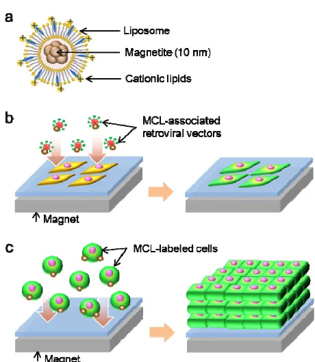
increasingly used in a number of biological and medical applications [5], [6]. Previously, we developed a magnetic tissue fabrication technique, in which magnetite cationic liposomes (MCLs; Fig. 1a) were used for magnetic labeling of cells, and the MCL-labeled cells were accumulated under a magnetic field to form multilayered sheet-like tissues [6]. We designated this technique as “magnetic force-based tissue engineering (Mag-TE)”. The advantage of this method is that cell-dense tissue constructs mimicking normal tissues can be created without the conventional scaffold-based procedures. On the other hand, magnetic nanoparticle-based gene transfer, magnetofection, has been applied to gene delivery and shown to enhance the transgene expression [7], [8]. In this method, interactions between cells and gene vector/magnetic nanoparticle complexes are promoted by applying a magnetic field, resulting in an accelerated gene transfer. Since MCLs have a positive surface charge, negatively-charged retroviral vectors can be captured by MCLs via electrostatic interactions. Thus, we previously reported that retroviral vector/MCL complexes could be used for gene transfer promoting retroviral transduction under a magnetic field [9].
In the present study, we examined the feasibility to fabricate VEGF gene-modified myoblast cell sheets using a combination of two magnetic biomanipulation techniques, magnetofection and Mag-TE. Additionally, we evaluated the angiogenic potential of the engineered tissues in a subcutaneous transplantation model.
II. MATERIALS AND METHODS
A. Cell Culture
Mouse myoblast C2C12 cells were cultured in low-glucose Dulbecco’s modified Eagle medium (DMEM; Sigma-Aldrich, St. Louis, MO, USA) supplemented with 10% fetal bovine serum (FBS), 0.1 mg/ml streptomycin sulfate and 100 U/ml penicillin G potassium (Wako Pure Chemical Industries, Osaka, Japan). Virus-producer 293FT cells were grown in high-glucose DMEM (Sigma-Aldrich) supplemented with 10% FBS, 0.1 mM MEM non-essential amino acids and 10 mM 2-[4-(2-hydroxyethyl)-1-piperazinyl] ethanesulfonic acid (HEPES; Dojindo laboratories, Kumamoto, Japan). Cells were cultured at 37°C in a 5% CO2
incubator.
B. MCL uptake by cells
The preparation of MCLs and the measurement of MCL uptake by cells were performed as described previously [10]. For the uptake measurements, C2C12 cells (3 × 10⁵/dish)
were seeded into a 100-mm culture dish (Greiner Bio-One,
Fabrication of Angiogenic Gene-modified Myoblast Cell
Sheets using Magnetic Tissue Engineering Techniques
Frickenhausen, Germany). After 24-h incubation, the medium was replaced with fresh medium containing MCLs (100 pg magnetite/cell). Subsequently, the cells were collected periodically. The magnetite concentrations and cell numbers were measured using the potassium thiocyanate method [11] and the trypan blue dye-exclusion method, respectively.
C. Vector construction
Total RNAs were extracted from C2C12 cells using a kit (QuickPrep Total RNA Extraction Kit; GE Healthcare, Buckinghamshire, UK) and reverse-transcribed with ReverTra Ace reverse transcriptase (Toyobo, Osaka, Japan). A DNA fragment of mouse VEGF isoform 164 (mVEGF164)
was amplified by PCR using the following primers: 5’ CGGGATCCACCATGAACTTTCTGCTGTCTTGGGT 3’
(forward) and 5’ CGGGATCCGAATTCACCGCCTCGGCT 3’ (reverse),
which append BamHI sites. A retroviral vector plasmid for VEGF expression (pQMSCV/CMV-VEGF-IRES-EGFP) was constructed by ligation of the mVEGF164 DNA fragment
into BamHI-digested pQMSCV/CMVHBD-3-IRES-EGFP plasmid [12]. In this vector, VEGF and green fluorescent protein (GFP) are bicistronically expressed under the control of a CMV promoter. The self-ligated pQMSCV/CMV-IRES-EGFP plasmid without the VEGF gene was used for the production of a control retroviral vector.
D. Retroviral magnetofection using MCLs
Retroviral vector particles were produced by transient transfection for 293FT cells as described previously [13]. For retroviral infection, C2C12 cells (3 × 10³ cells/well) were
seeded into wells of a 96-well plate (Greiner Bio-One), one day prior to infection. A schematic diagram of magnetofection is illustrated in Fig. 1b. The viral solution (150 μl) was mixed with MCLs (600 ng magnetite). The magnetite concentrations at infection corresponded to 100 pg/cell. After incubating for 30 min on ice, the solutions containing the retroviral vector/MCL complexes were added to the cell-cultured wells, and then a 96-magnet plate (Oz Biosciences, Marseille, France) was placed under the well plate to attract the complexes to cells. In some experimental conditions, polybrene was added to the viral solution at a concentration of 8 μg/ml.
For viral titration, C2C12 cells were infected using a 10-fold serial dilution of the viral solution, and GFP-expressing C2C12 cells were counted under a fluorescent microscope 48 h post-infection.
E. Fabrication of VEGF-gene engineered cell sheets A schematic diagram of cell sheet fabrication is shown in Fig. 1c. MCLs were added to the cells (100 pg/cell), and the cells were cultured for 4 h. Subsequently, the cells were harvested, and 500 μl of medium containing 1.2 × 10⁶ cells
was seeded inside a silicone rubber tube (inner diameter, 1 cm), which was placed at the center of a 35-mm culture dish (hydrophobic lumox dish, Greiner Bio-One). Immediately thereafter, a cylindrical neodymium magnet (diameter, 30 mm; height, 15 mm) was placed under the dish to accumulate MCL-labeled cells on the culture surface and to form a multilayered cell sheet.
Fig. 1 (a) Illustration of MCLs. Magnetite nanoparticles (10 nm) were encapsulated into cationic liposomes. (b) Magnetofection technique. Retroviral vector/MCL complexes were attracted toward the cultured cells using a magnet. (c) Mag-TE technique. MCL-labeled cells were accumulated to form a multilayered cell sheet using a magnet.
F. Detection of VEGF expression
Cell sheets and culture media were collected 24 h after the construction of cell sheets to determine VEGF expression.
Total RNAs were extracted from the cell sheets for semi-quantitative RT-PCR, and reverse-transcribed to prepare cDNAs, as described above. Using the cDNA samples, specific sequences were amplified by PCR using the following primers: mVEGF164 (forward, 5’
ACAGAACAAAGCCAGAAAATCACTG 3’; reverse, 5’ GTTTAACTCAAGCTGCCTCGCC 3’) and mGAPDH (forward, 5’ CTACCCCCAATGTGTCCGTC 3’; reverse, 5’ GCTGTTGAAGTCGCAGGAGAC 3’). PCR was initiated using G-Taq DNA polymerase (Cosmo Genetech, Seoul, Korea). The PCR products were electrophoresed on a 3% agarose gel and stained with ethidium bromide. The band intensity was quantified using ImageJ software (National Institute of Mental Health, Bethesda, MD, USA). A house keeping gene GAPDH was used as an internal loading control.
[image:2.595.312.542.68.332.2]To quantify VEGF secretion in the medium, the samples were analyzed using a kit based on an enzyme-linked immunosorbent assay (mouse VEGF ELISA kit; Ray Biotech, Norcross, GA, USA), according to the manufacturer’s instruction.
G. Transplantation
Female, 4- to 5-week-old KSN/Slc (Japan SLC, Shizuoka, Japan) nude mice were anesthetized by intraperitoneal injection of pentobarbital, and a small incision was created in the dorsal skin. Cell sheets were rinsed twice with PBS and harvested on thin films as a physical support. Subsequently, the cell sheet and film were inserted subcutaneously into the dorsal skin through the incision, and the film was removed, leaving the cell sheet on the underlying tissue. The incisions were then closed using silk sutures. At 14 days post-implantation, mice were sacrificed, and the grafts were resected. Animal experiments in this study were approved by the Ethics Committee for Animal Experiments of the Faculty of Engineering, Kyushu University (A19-114-1).
For histological examination, the grafts were fixed in 4% formaldehyde solution and embedded in paraffin. Thin slices (4 μm) were placed on silanized slides for immunofluorescent staining of CD31. The sections were blocked with 1% bovine serum albumin and were stained using anti-mouse CD31 antibody (Santa Cruz Biotechnology) and Alexa Fluor 488-conjugated anti-goat antibody (Invitrogen, Carlsbad, CA, USA). To compare the tissue vascularization, eight non-overlapping fields in each section of the graft were photographed under a fluorescent microscope at a high-power magnification. The pixel numbers of areas of CD31-positive vessels were calculated using image analysis software (Adobe Photoshop 6.0; Adobe Systems, San Jose, CA, USA). The total number of image pixels was 1.83 × 10⁶.
H. Statistical evaluation
All data are expressed as means ± SD. Statistical comparisons were evaluated using one-way analysis of variance (ANOVA); a P value of less than 0.05 was considered statistically significant and indicated by asterisks.
III. RESULTS
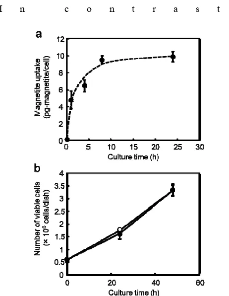
We first investigated the MCL uptake by C2C12 cells to elucidate the applicability of magnetofection and Mag-TE techniques. The magnetite amount taken up by C2C12 cells after MCL addition (100 pg-magnetite/cell) is shown in Fig. 2a. The uptake amount dramatically increased by 8 h after the addition, and virtually remained unchanged at the 24-h time point (uptake of 4.7, 6.4, 9.4 and 9.8 pg magnetite/cell at 1, 4, 8 and 24 h, respectively). At this concentration, MCLs did not inhibit the growth of C2C12 cells (Fig. 2b).
For magnetofection, MCLs were added to the viral solution, and retroviral vectors were captured by MCLs. The complexes were attracted toward the cultured C2C12 cells using a magnet. Table 1 shows the transduction efficiency compared to several conditions, including a conventional method using polybrene, when the efficiency without cationic agents is expressed as 1.0. There were small increases among the cases with polybrene and/or MCLs in the absence of a magnet, indicating that the cationic reagents
had minimal effects in the retroviral infection for C2C12 cells.
I n c o n t r a s t ,
Fig. 2 (a) Magnetite amounts taken up by C2C12 cells after the addition of MCLs (100 pg/cell). (b) The growth of C2C12 cells after the addition of MCLs. Open circles, no MCLs; Closed circles, with MCLs (100 pg/cell). Data are expressed as mean ± SD (n=3).
magnetofection enhanced the efficiency by 8.4-fold. This was a 6.7-fold improvement compared with the polybrene-mediated infection. Similar to the report by Scherer et al. [8], the addition of polybrene during the magnetofection halved the efficiency, which may be due to the competitive binding between polybrene and MCLs to the retroviral particles. These results indicate that magnetofection using MCLs has the potential for high-efficient gene transduction.
[image:3.595.306.537.60.358.2]Fig. 3 (a) Bright-field photographs of VEGF gene-engineered C2C12 cell sheets. The inset shows a bright-field micrograph of an H&E-stained section of the cell sheet. The scale bar in an inset, 50 μm. (b) Semi-quantitative RT-PCR analysis of VEGF expression. (c) Relative mRNA expression quantified from (b) using ImageJ software. (d) Measurements of VEGF secretions from the cell sheets by ELISA. Data are expressed as mean ± SD (n=3). *P < 0.05 vs. C2C12/GFP sheet group.
To detect exogenous VEGF gene expression, cell sheets were analyzed using semi-quantitative RT-PCR (Fig. 3b and c). A higher level of exogenous VEGF expression was detected for the C2C12/VEGF cell sheets. In the western blot analysis, a single band with a molecular weight of approximately 25 kDa was detected for the culture medium of C2C12/VEGF cell sheets, while it was an undetectable level for C2C12/GFP cell sheets. The VEGF amounts measured by ELISA were 2.98 ± 0.04 and 240 ± 29 ng/day for C2C12/GFP and C2C12/VEGF cell sheets, respectively (Fig. 3d). When human umbilical vein endothelial cells (HUVECs) were cultured in the conditioned medium collected from the culture of cell sheets, the cells promoted proliferation in the conditioned medium from the C2C12/VEGF cell sheets (data not shown). This observation indicates that the VEGF secreted from C2C12/VEGF sheets was biologically active and suggests that the VEGF gene-engineered cell sheets were successfully fabricated by combining the magnetofection and Mag-TE techniques.
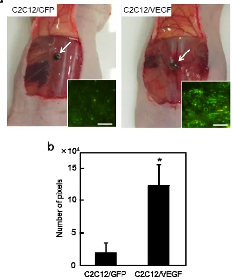
Finally, the subcutaneous transplantation of VEGF gene-engineered cell sheets into nude mice was performed for the evaluation of angiogenic potential in vivo (Fig. 4a and b). Fig. 5a shows bright-field photographs of the grafts at 14 days post-transplantation. Vascularization was observed for the sheet-grafts under a microscope. Capillary vessels with a remarkably higher density were observed from the CD31-stained sections within the C2C12/VEGF sheet-tissue (Fig. 5a, inset). The vessel area within the grafts of C2C12/VEGF sheets was significantly larger than that within C2C12/GFP sheet-grafts (Fig. 5b). The cross-sectional area of C2C12/VEGF sheet-grafts was 2.1-fold larger than that of
C2C12/GFP sheet-grafts, while both grafts maintained
Fig. 4 (a) The engineered cell sheets were transplanted subcutaneously into nude mice. (b) The grafts could be observed after the implantation. The white arrows indicate cell sheets.
Fig. 5 (a) Bright-field photographs of the grafts at 14 days post-transplantation. The white arrows indicate cell sheet-grafts. The insets show the representative images of sections of immunofluorescent staining of CD31 (green). The scale bar in the insets is 50 μm. (b) The vessel areas within the grafts. The pixel numbers of the areas occupied by CD31-positive vessels were analyzed. Data are expressed as mean ± SD (n=3). *P < 0.05 vs. C2C12/GFP sheet group.
cell-dense tissues (data not shown). These results indicate that VEGF gene-engineered cell sheets were provided sufficient tissue vascularization and promoted growth of grafts, and that the approach is useful for fabrication of gene-engineered tissue constructs.
IV. DISCUSSION
[image:4.595.59.287.69.315.2] [image:4.595.318.544.231.504.2]limiting the tissue engineering applications. In this study, we investigated the fabrication of VEGF gene-engineered myoblast cell sheets combining magnetofection and Mag-TE techniques using MCLs, and demonstrated the high angiogenic potential of these engineered cell sheets.
To achieve successful treatments by a tissue engineering approach accompanied with gene therapy, high-efficient gene delivery is essential. In this study, we utilized the magnetofection technique, in which retroviral vector/MCL complexes are attracted toward the cultured cells by applying a magnetic field. This technique increased the transduction efficiency by 8.4-fold compared with the infection without a cationic reagent, while the polybrene-mediated infection increased by only 1.3-fold. Considering that the MCL-mediated infection without a magnet increased slightly, it was assumed that the high level of transduction efficiency by magnetofection was due to acceleration of retroviral vector attachment to cells under a magnetic field. On the other hand, we previously showed that the spatial pattern of gene expression was created based on the magnetofection technique using a micro-patterned magnet [9], indicating that the method can be applicable for gene targeting. Thus, the magnetofection technique has wide-range applicability for biological and medical fields where gene therapy approaches are required.
For tissue fabrication, we applied the Mag-TE technique, in which MCL-labeled cells were accumulated to form a sheet-like tissue under a magnetic field. The benefit of this method is that cell-dense tissue constructs mimicking normal tissues can be created easily compared with conventional scaffold-based procedures. However, owing to this property, it may cause difficulty in mass transport. In this study, we evaluated the angiogenic potential of C2C12/VEGF cell sheets fabricated by combining magnetofection and Mag-TE techniques. At 14 days post-transplantation, high vascularization in the C2C12/VEGF sheet-grafts were observed compared with the C2C12/GFP sheet-grafts by both macroscopic observation and histological analysis. Moreover, the cross-sectional area of the C2C12/VEGF sheet-grafts was 2.1-fold larger than that of C2C12/GFP sheet-grafts, demonstrating a large variation in graft sizes. This finding indicates that the increased vascularization in the C2C12/VEGF sheet-graft improved the mass transport and formed a bulky tissue by cell proliferation. These results also suggest that VEGF gene-engineered cell sheets not only enhance angiogenic response but also stimulate growth of the graft.
As an alternative approach for tissue vascularization, several groups have proposed the fabrication of pre-vascularized networks within tissue constructs by co-culturing endothelial cells before transplantation [15], [16]. Such studies have demonstrated early vascularization in grafts with pre-vascularized networks by inoculating the host vasculature compared with grafts without networks. The pre-vascularized cell sheets have also been fabricated by Mag-TE [17]. Thus, we may need to examine whether a combination of the methods used in this study with a pre-vascularization method would be more effective for the induction of angiogenesis.
V. CONCLUSION
We demonstrated here a combinatorial approach of magnetofection and Mag-TE techniques. Magnetofection enhanced the gene transduction efficiency, and the Mag-TE technique enabled fabrication of cell-dense tissue constructs without the use of a scaffold. The VEGF gene-engineered cell sheets showed a high angiogenic potential when transplanted. We believe that this procedure may be useful for tissue replacement therapy, particularly where angiogenesis is necessary.
REFERENCES
[1] J. Rouwkema, N.C. Rivron, and C.A. van Blitterswijk, “Vascularization in tissue engineering,” Trends Biotechnol., vol. 26, Aug. 2008, pp. 434-441.
[2] A.M. Byrne, D.J. Bouchier-Hayes, and J.H. Harmey, “Angiogenic and cell survival functions of vascular endothelial growth factor (VEGF),” J. Cell. Mol. Med., vol. 9, Oct.-Dec. 2005, pp. 777-794.
[3] P. De Coppi, D. Delo, L. Farrugia, K. Udompanyanan, J.J. Yoo, and M. Nomi, et al., “Angiogenic gene-modified muscle cells for enhancement of tissue formation,” Tissue Eng., vol. 11, Jul.-Aug. 2005, pp. 1034-1044.
[4] Y. Lu, J. Shansky, M. Del Tatto, P. Ferland, X. Wang, and H. Vandenburgh, “Recombinant vascular endothelial growth factor secreted from tissue-engineered bioartificial muscles promotes localized angiogenesis,” Circulation, vol. 104, Jul. 2001, pp. 594-599. [5] J. Dobson, “Remote control of cellular behaviour with magnetic
nanoparticles,” Nat. Nanotechnol., vol. 3, Mar. 2008, pp. 139-143. [6] A. Ito, M. Shinkai, H. Honda, and T. Kobayashi, “Medical application
of functionalized magnetic nanoparticles,” J. Biosci. Bioeng., vol. 100, Jul. 2005, pp. 1-11.
[7] J. Dobson, “Gene therapy progress and prospects: magnetic nanoparticle-based gene delivery,” Gene Ther., vol. 13, Feb. 2006, pp. 283-287.
[8] F. Scherer, M. Anton, U. Schillinger, J. Henke, C. Bergemann, and A. Krüger, et al., “Magnetofection: enhancing and targeting gene delivery by magnetic force in vitro and in vivo,” Gene Ther., vol. 9, Jan. 2002, pp. 102-109.
[9] A. Ito, T. Takahashi, Y. Kameyama, Y. Kawabe, and M. Kamihira, “Magnetic concentration of a retroviral vector using magnetite cationic liposomes,” Tissue Eng. Part C Methods, vol. 15, Mar. 2009, pp. 57-64. [10] M. Shinkai, M. Yanase, H. Honda, T. Wakabayashi, J. Yoshida, and T. Kobayashi, “Intracellular hyperthermia for cancer using magnetite cationic liposomes: in vitro study,” Jpn. J. Cancer Res., vol. 87, Nov. 1996, pp. 1179-1183.
[11] C.S. Owen, and N.L. Sykes, “Magnetic labeling and cell sorting,” J. Immunol. Methods, vol. 73, Oct. 1984, pp. 41-48.
[12] A. Ito, T. Takahashi, Y. Kawabe, and M. Kamihira, “Human beta defensin-3 engineered keratinocyte sheets constructed by a magnetic force-based tissue engineering technique,” J. Biosci. Bioeng., vol. 108, Sep. 2009, pp. 244-247.
[13] A. Hotta, Y. Saito, K. Kyogoku, Y. Kawabe, K. Nishijima, and M. Kamihira, et al., “Characterization of transient expression system for retroviral vector production,” J. Biosci. Bioeng., vol. 101, Apr. 2006, pp. 361-368.
[14] M.W. Laschke, Y. Harder, M. Amon, I. Martin, J. Farhadi, and A. Ring et al., “Angiogenesis in tissue engineering: breathing life into constructed tissue substitutes,” Tissue Eng., vol. 12, Aug. 2006, pp. 2093-2104.
[15] P.L. Tremblay, V. Hudon, F. Berthod, L. Germain, and F.A. Auger, “Inosculation of tissue-engineered capillaries with the host's vasculature in a reconstructed skin transplanted on mice,” Am. J. Transplant., vol. 5, May. 2005, pp. 1002-1010.
[16] R.K. Jain, P. Au, J. Tam, D.G. Duda, and D. Fukumura, “Engineering vascularized tissue,” Nat. Biotechnol., vol. 23, Jul. 2005, pp. 821-823. [17] K. Ino, A. Ito, H. Kumazawa, H. Kagami, M. Ueda, and H. Honda,