Original Article
Raman spectroscopy of luminal subtype and basal
subtype muscle invasive bladder cancer
Di Jin1*, Xuetao Wang2*, Bing Fu2, Taihao Li3, Na Chen2, Zhenyi Chen2, Haige Chen1, Shupeng Liu2
1Department of Urology, Ren Ji Hospital, School of Medicine, Shanghai Jiao Tong University, No.1630 Dongfang
Road, Shanghai 200127, China; 2Key Laboratory of Specialty Fiber Optics and Optical Access Networks, Institute
of Biomedical Engineering, Joint International Research Laboratory of Specialty Fiber Optics and Advanced Communication, Shanghai Institute for Advanced Communication and Data Science, Shanghai University, 333 Nanchen Road, Shanghai 200444, China; 3College of Medical Instruments, Shanghai University of Medicine &
Health Sciences, Shanghai 201318, China. *Equal contributors.
Received December 17, 2018; Accepted January 7, 2019; Epub May 15, 2019; Published May 30, 2019
Abstract: Bladder cancer is the killer of human health, and its prevalence ranks second among all cancers. In order to improve the survival rate of bladder cancer, in addition to early detection, study on drugs, improvement of surgi-cal protocols, and good prognosis of high-grade bladder cancer are crucial. Surface Enhanced Raman spectroscopy (SERS) is not only a method of diagnosing bladder cancer, but also it can provide mechanisms of cancer and medi-cine for treatment. This paper explores the Raman spectra of luminal and basal subtype of muscle invasive bladder cancer. A total of 250 SERS spectra from muscle invasive bladder cancer (MIBC) were acquired from 24 luminal subtype subjects and 26 basal subtype subjects who were given a pathological diagnosis. The experimental results demonstrate that two categories of muscle invasive bladder cancer can be distinguished by Raman spectrum. Principal component analysis combined with linear discriminate analysis (PCA-LDA) was used to classify luminal and basal subtype, with an accuracy of 94% and the accuracy of cross validation was 86%. Surface Enhanced Raman spectrum combined with PCA-LDA algorithms is a relatively accurate method to separate different levels of cancer and it can be used to research the mechanism of cancer and medicine for treatment. It lays the foundation for clinical application.
Keywords: Bladder cancer, Raman spectroscopy, PCA-LDA
Introduction
Cancer is the human health killer and more than seven million people dying from cancer each year all over the world [1]. The prevalence rate of bladder cancer is second [2]. It’s impor-tant for diagnosing and treating bladder cancer
in early stage. The first recommended treat -ment plan for bladder cancer treat-ment in Eu- rope and America is chemotherapy. Neoad- juvant chemotherapy (NAC) based on platinum, gemcitabine, and paclitaxel can greatly improve the prognosis of muscle invasive bladder can-cer (MIBC) patients, reduce tumor recurrence and metastasis, and prolong median survival
time [3, 4]. But not all patients benefit from
chemotherapy. For those patients who are not sensitive to chemotherapy, this increases the
side effects and the cost of medical treatment, and even delays the surgical treatment time [5]. Currently, the main methods for diagnosing bl- adder cancer include ultrasound imaging [6], magnetic resonance imaging (MRI) [7], comput-ed tomography (CT) [8], endoscopy [9-11], and cytology [12] etc. These technologies are com-plex and require doctors to have extensive clini-cal experience and knowledge. The Raman spectrum is a scattering spectrum formed by the interaction of incident light, so the molecu-lar information is concluded in a Raman
spec-trum as molecular fingerprints with the advan
-tages of no damage, high sensitivity, and effi
By the detection and analy-sis of genomics and tran-scriptology of tumor tissue, some studies suggest that there are different molecu-lar subtypes in MIBC. At pr- esent, the mainstream cl-
assification includes
basal-like subtype, luminal
sub-type, and other classifica -tion subtypes [17, 18]. The study shows that the lumi-nal subtype is closer to the papillary growth of non-mu- scle invasive bladder can-cer. It is a potential popula-tion for bladder preserva-tion. The prognosis of Basal subtype is worse in general and should be operated Many studies on bladder cancer have been
done by using Raman spectroscopy in various ways. At present, most studies research
classi-fication bladder cancer patients and normal
people. The Raman spectra of bladder cancer tissue and normal bladder tissue may have more obvious difference in the intensity of characteristic peaks, which means that the
specific chemical component changes in the
process of cancer [13]. Many types of sample are related to bladder cancer, such as tissue, urine, and serum. Therefore, different methods of Raman spectroscopy are combined with dif-ferent type of sample. For example, bladder cancer is diagnosed from urine by Raman molecular imaging [14], and bladder cancer cells are detected effectively by Raman spec-troscopy with atomic force microscopy [15]. In order to apply Raman spectroscopy to clinic, different levels of tumor can be accurately
clas-sified with appropriate algorithms. At the
mo-ment, most of studies utilize principal compo-nent analysis combined with linear discriminate analysis (PCA-LDA), to support vector machi- ne combined with linear discriminate analysis (SVM-LDA) and genetic algorithms combined with linear discriminate analysis (GAs-LDA). For example, some studies explore that the SERS spectra of serum which acquired from 55 blad-der cancer patients and 36 normal people are
classified by genetic algorithms (GAs) combined
with linear discriminate analysis (LDA), and the
sensitivity and specificity are 90.9% and 100%
[16].
and supplemented with neoadjuvant chemo-therapy actively [19]. For patients with different molecular subtypes, individualized treatment, including surgery, NAC, and adjuvant chemo-therapy, or other targeted therapy and immuno-therapy, is an important breakthrough in the accurate treatment of bladder cancer. This study used SERS detected muscle invasive bladder cancer tissues and combined with PCA-LDA algorithms to discriminate between luminal subtype and basal-like subtype of MIBC on the resulting Raman spectra. Based on dis-tinguishing luminal subtype from basal-like subtype of muscle invasive bladder cancer, the difference between luminal and basal-like sub-type at the molecular level could be researched through SERS. The information of luminal and basal-like subtype in the molecular level could be provided with researching medicine for treatment and improving surgery program and adjuvant therapy.
Material and method
Preparation of the sample
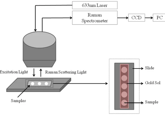
[image:2.612.92.382.70.274.2]sue samples were used in this experiment. There we- re 24 groups with luminal subtype and 26 groups wi- th basal-like subtype. The bladder cancer tissue sam-ples were cut into slices for
20 μm by freezing micro -tome and the slices were soaked in gold sol.
SERS measurement
A Raman microscope with a 50×objective and 633-nm excitation was utilized to obtain the spectrum with a 10 second integration time over the spectral range of 400-1800 cm-1. Each
sam-ple was measured at five
times to reduce noise and the side effect due to any instability in the spectrom-eter. The experimental se- tup is shown in Figure 1. Results and discussion
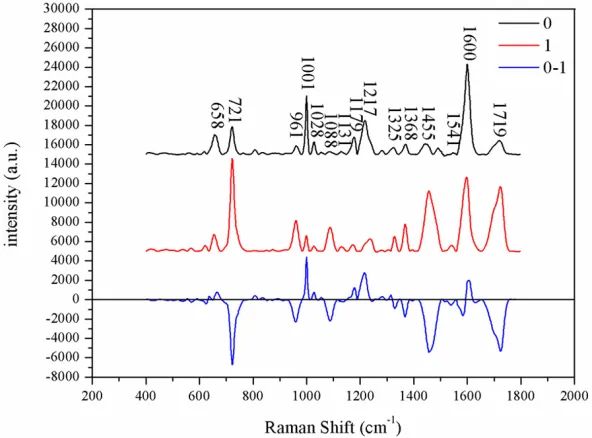
The Raman spectra were obtained from 24 patients of luminal subtype (0) and 26 patients of basal-like subtype (1). Two sets of sp-
ectra are removed the fluo
[image:3.612.91.387.71.290.2]-rescence background and averaged respectively and the results are shown in Figure 2. The black and red curves represent the Ra- man spectrum of luminal subtype and basal-like sub-type respectively, and the blue curve represents the difference between black and red curves.
To distinguish luminal type from basal-like sub-type further, the two sets of
data were classified by
PCA-LDA algorithm and the accuracy was 94% (Figure luminal subtype (0), basal-like subtype (1). A
[image:3.612.93.385.332.651.2]total of 50 muscle invasive bladder cancer tis- 3). As displayed in Table 2, the accuracy of cross validation was 86%. It can be seen that Figure 3. The classification result between luminal and basal-like subtype.
the luminal subtype can be differentiated from basal subtype by Raman spectr- um preliminarily.
According to the model of PCA-LDA, the ROC curve was drawn to evaluate the ability of classifying two ca- tegories of samples by PCA-LDA (Figure 4). The integral of the area under the curve was 0.973. Raman spectra of invasive bladder cancer tissue can be distributed by PCA-LDA well.
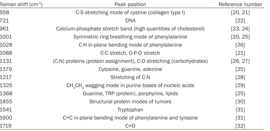
[image:4.612.94.531.83.296.2]As shown in Figure 2, the distribution of Raman char-acteristic peaks of the two sets of spectra is very simi-lar. The characteristic pe- aks on the Raman spectra of the invasive bladder can-cer were located at 658, 721, 961, 1001, 1028, 1088, 1131, 1179, 1217, 1325, 1368, 1455, 1541, 1600 and 1719 cm-1, as detailed in Table 1. The Ra- man peaks belong to bio-logical molecules, such as proteins, lipid, DNA.
Table 1. The Raman characteristic peak of invasive bladder cancer
Raman shift (cm-1) Peak position Reference number
658 C-S stretching mode of cystine (collagen type I) [20, 21]
721 DNA [22]
961 Calcium-phosphate stretch band (high quantities of cholesterol) [23, 24] 1001 Symmetric ring breathing mode of phenylalanine [20, 25] 1028 C-H in-plane bending mode of phenylalanine [26] 1088 C-C stretch, O-P-O- stretch [21]
1131 (C-N) proteins (protein assignment), C-O stretching (carbohydrates) [26, 27] 1179 Cytosine, guanine, adenine [25]
1217 Stretching of C-N [28]
1325 CH3CH2 wagging mode in purine bases of nucleic acids [29] 1368 Guanine, TRP (protein), porphyrins, lipids [25] 1455 Structural protein modes of tumors [30]
1541 Tryptophan [31]
1600 C=C in-plane bending mode of phenylalanine and tyrosine [31]
[image:4.612.90.386.322.681.2]1719 C=O [32]
Table 2. Classification between luminal and basal subtype
Classification
Expected Classification
Total Luminal
Subtype Basal-like Subtype Original label Luminal Subtype 24 0 24
Basal-like Subtype 3 23 26 Cross validation Luminal Subtype 21 3 24 Basal-like Subtype 4 22 26
Although the peaks of the Raman spectrum were similar between luminal and basal sub-type, the relative intensities of the peaks and peaks change. According to spectral range of 650-750 cm-1, 950-1100 cm-1 and 1400-1750 cm-1, it can separate two sets of data. 658/721 cm-1 (the ratio of 658 to 721 cm-1), 961/1001 cm-1, 1088/1217 cm-1, 1368/1455 cm-1 and 1600/1719 cm-1 all change. Subtle changes occurred in some biological molecules. In the luminal subtype group, the intensity of 658, 1001, 1028, 1179, 1217 and 1600 cm-1 was higher than the other group. In the basal sub-type group, the intensity of 721, 961, 1088, 1325, 1368, 1455 and 1719 cm-1 was higher than the group of basal subtype. These differ-ences can divide samples into two types poten-tially. The characteristic peak in 1455 cm-1 rep-resents the structural protein modes of tumors, which indicates the concentration of the pro-tein participating in reproduction, division of cancer cell in the group of basal subtype is higher than the other group. The peaks in 721, 1088, 1325 and 1368 cm-1 represent DNA, cytomembrane, which means that the rate of the reproduction, division of cancer cell in basal subtype is faster than luminal subtype.
Conclusion
In this study, Raman spectroscopy-molecular typing-chemosensitivity predictive system was established by using Surface-Enhanced Raman spectroscopy to explore the difference of differ-ent molecular typing and chemosensitivity of bladder tumor. The results show that higher level of tumor, luminal subtype, and basal sub-type of muscle invasive bladder cancer can be distinguished by Raman spectrum preliminarily. The prediction system is an important refer-ence indicator if muscle invasive bladder can-cer need for neoadjuvant chemotherapy preop-erative. It provides important evidence to the individual patient’s precise treatment. More- over, by using PCA-LDA algorithm to classify, the accuracy was 94% and the accuracy of
cross validation was 86%. It indicates benefit to
study the mechanism of cancers and drugs screening for treatment of cancers at present. In addition, it will be used in clinic basing on more precise algorithms and plenty of data- bases.
Acknowledgements
This work was funded by Natural Science Fo- undation of China (NSFC) (61575120, 6142-
2507, 61475095, 61520106014), and supp- orted by the Natural Sciences Fund of Shang- hai Municipal Science and Technology Com- mission (16ZR1420300). This work was sup-ported by Incubating Program for Clinical Re- search and Innovation of Ren Ji Hospital, Sc- hool of Medicine, Shanghai Jiao tong University (PYXJS16-011), and thanks for the support of the Key Laboratory of Specialty Fiber Optics and Optical Access Networks (SKLSFO2017-02).
Address correspondence to: Shupeng Liu, Key La- boratory of Specialty Fiber Optics and Optical Acce- ss Networks, Institute of Biomedical Engineering, Joint International Research Laboratory of Specialty Fiber Optics and Advanced Communication, Sha- nghai Institute for Advanced Communication and Data Science, Shanghai University, 333 Nanchen Road, Shanghai 200444, China. E-mail: liusp@shu. edu.cn; Haige Chen, Department of Urology, Ren Ji Hospital, School of Medicine, Shanghai Jiao Tong University, No.1630 Dongfang Road, Shanghai 20- 0127, China. E-mail: [email protected]
References
[1] Siegel R, Ma J, Zou Z, Jemal A. Cancer statis-tics, 2014. CA Cancer J Clin 2014; 64: 9-29. [2] Kallaway C, Almond LM, Barr H, Wood J, Hut-
chings J, Kendall C, Stone N. Advances in the clinical application of Raman spectroscopy for cancer diagnostics. Photodiagnosis Photodyn Ther 2013; 10: 207-19.
[3] Grossman HB, Natale RB, Tangen CM, Speights VO, Vogelzang NJ, Trump DL, deVere White RW, Sarosdy MF, Wood DP Jr, Raghavan D, Crawford ED. Neoadjuvant chemotherapy plus cystecto-my compared with cystectocystecto-my alone for locally advanced bladder cancer. N Engl J Med 2003; 349: 859-66.
[4] Reardon ZD, Patel SG, Zaid HB, Stimson CJ, Resnick MJ, Keegan KA, Barocas DA, Chang SS, Cookson MS. Trends in the use of periop-erative chemotherapy for localized and locally advanced muscle-invasive bladder cancer: a sign of changing tides. Eur Urol 2015; 67: 165-70.
[5] Vale CL. Neoadjuvant chemotherapy in inva-sive bladder cancer: update of a systematic review and meta-analysis of individual patient data. Eur Urol 2005; 48: 202-206.
[6] Oktem GC, Kocaaslan R, Karadag MA, Bag- cioglu M, Demir A, Cecen K, Unluer E. The role of transcavitary ultrasonography in diagnosis
and staging of nonmuscle-ınvasive bladder
[7] Wollin DA, Deng FM, Huang WC, Babb JS, Rosenkrantz AB. Conventional and diffusion-weighted MRI features in diagnosis of meta-static lymphadenopathy in bladder cancer. Can J Urol 2014; 21: 7454-9.
[8] Di Paolo PL, Vargas HA, Karlo CA, Lakhman Y, Zheng J, Moskowitz CS, Al-Ahmadie HA, Sala E, Bochner BH, Hricak H. Intradiverticular blad-der cancer: CT imaging features and their as-sociation with clinical outcomes. Clin Imaging 2015; 39: 94-8.
[9] Lopez A, Liao JC. Emerging endoscopic imag-ing technologies for bladder cancer detection. Curr Urol Rep 2014; 15: 1-8.
[10] Lerner SP, Goh A. Novel endoscopic diagnosis for bladder cancer. Cancer 2015; 121: 169-178.
[11] Witjes JA, Gomella LG, Stenzl A, Chang SS, Zaak D, Grossman HB. Safety of hexaminolev-ulinate for blue light cystoscopy in bladder can-cer. A combined analysis of the trials used for registration and postmarketing data. Urology 2014; 84: 122-6.
[12] Onal B, Han U, Yilmaz S, et al. The use of uri-nary nuclear matrix protein 22 (NMP22) as a diagnostic adjunct to urine cytology for moni-toring of recurrent bladder cancer-institutional experience and review. Diagnostic Cytopath- ology 2014; 43: 307-314.
[13] Jin D, Chen H, Cao M, Yang G, Xue W, Huang Y. SERS measurement of the bladder cancer cells with the nanoparticles. Pak J Pharm Sci 2015; 28 Suppl: 1853-6.
[14] Shapiro A, Gofrit ON, Pizov G, Cohen JK, Maier J. Raman molecular imaging: a novel spectro-scopic technique for diagnosis of bladder can-cer in urine specimens. Eur Urol 2011; 59: 106.
[15] Canetta E, Riches A, Borger E, Herrington S, Dholakia K, Adya AK. Discrimination of bladder cancer cells from normal urothelial cells with
high specificity and sensitivity: combined ap -plication of atomic force microscopy and mod-ulated Raman spectroscopy. Acta Biomater 2014; 10: 2043.
[16] Li S, Li L, Zeng Q, Zhang Y, Guo Z, Liu Z, Jin M, Su C, Lin L, Xu J, Liu S. Characterization and noninvasive diagnosis of bladder cancer with serum surface enhanced Raman spectroscopy and genetic algorithms. Sci Rep 2015; 5: 9582.
[17] Cancer Genome Atlas Research Netwrok. Co- mprehensive molecular characterization of urothelial bladder carcinoma. Nature 2014; 507: 315-22.
[18] Kurtova AV, Xiao J, Mo Q, Pazhanisamy S, Krasnow R, Lerner SP, Chen F, Roh TT, Lay E, Ho PL, Chan KS. Blocking PGE2-induced tu-mour repopulation abrogates bladder cancer chemoresistance. Nature 2015; 517: 209-13.
[19] Rebouissou S, Bernard-Pierrot I, de Reyniès A, Lepage ML, Krucker C, Chapeaublanc E, Hérault A, Kamoun A, Caillault A, Letouzé E, Elarouci N, Neuzillet Y, Denoux Y, Molinié V, Vordos D, Laplanche A, Maillé P, Soyeux P, Ofualuka K, Reyal F, Biton A, Sibony M, Paoletti X, Southgate J, Benhamou S, Lebret T, Allory Y, Radvanyi F. EGFR as a potential therapeutic target for a subset of muscle-invasive bladder cancers presenting a basal-like phenotype. Sci Transl Med 2014; 6: 244ra91.
[20] Stone N, Kendell C, Shepherd N, Crow P, Barr H. Near-infrared Raman spectroscopy for the
classification of epithelial pre-cancers and
cancers. Journal of Raman Spectroscopy 2002; 33: 564-573.
[21] Cheng WT, Liu MT, Liu HN, Lin SY. Micro-Raman spectroscopy used to identify and grade hu-man skin pilomatrixoma. Microsc Res Tech 2005; 68: 75-9.
[22] Binoy J, Abraham JP, Joe IH, Jayakumar VS, Petit GR, Nielsen OF. NIR-FT Raman and FT-IR spectral studies and ab initio calculations of the anti-cancer drug combretastatin-A4. Jo- urnal of Raman Spectroscopy 2004; 35: 939-946.
[23] Dukor RK. Vibrational spectroscopy in the de-tection of cancer. Biomedical Applications 2002; 5: 3335-3359.
[24] Chiang HP, Song R, Mou B, Li KP, Chiang P, Wang D, Tse WS, Ho LT. Fourier transform Raman spectroscopy of carcinogenic polycyclic aromatic hydrocarbons in biological systems: Binding to heme proteins. Journal of Raman Spectroscopy 1999; 30: 551-555.
[25] Stone N, Kendall C, Smith J, Crow P, Barr H.
Raman spectroscopy for identification of epi -thelial cancers. Faraday Discuss 2004; 126: 141-157.
[26] Notingher I, Green C, Dyer C, Perkins E, Hopkins N, Lindsay C, Hench LL. Discrimination between ricin and sulphur mustard toxicity in vitro using Raman spectroscopy. J R Soc In- terface 2004; 1: 79-90.
[27] Huang Z, McWilliams A, Lui H, McLean DI, Lam S, Zeng H. Near-infrared Raman spectroscopy for optical diagnosis of lung cancer. Int J Cancer 2003; 107: 1047-1052.
[28] Naumann D. Infrared and NIR Raman spec-troscopy in medical microbiology. Proc SPIE 1998; 3257: 245-257.
[29] Viehoever AR, Anderson D, Jansen D, Maha- devan-Jansen A. Organotypic raft cultures as an effective in vitro tool for understanding Raman spectral analysis of tissues. Photochem Photobiol 2003; 78: 517-524.
precan-cers. Applied Spectroscopy 2001; 55: 955-959.
[31] Stone N, Kendall C, Smith J, Crow P, Barr H.
Raman spectroscopy for identification of epi -thelial cancers. Faraday Discuss 2004; 126: 141.