Original Article
Roles of miR-433-5p in gastric carcinoma cell viability,
invasion, and apoptosis via targeting CLCN4 genes
Wei Wang1, Jing Pan2, Hai-Bing Jiang3
1Oncology Hematology Department, The Second Hospital University of South China, Hengyang, China; 2
Depart-ment of Pathogenic Biology and Immunology, Yongzhou Vocational Technical College, Yongzhou, China; 3
Depart-ment of Gastroenterology, The Second Hospital University of South China, Hengyang, China
Received March 29, 2019; Accepted June 10, 2019; Epub August 15, 2019; Published August 30, 2019
Abstract: Objective: The current study was designed to explore the mechanisms in which miR-433-5p affects vi-ability, invasion, and apoptosis of gastric cancer cells via targeting CLCN4. Methods: Gastric cancer tissues and adjacent tissues were collected. Bioinformatics tools and dual-luciferase reporter assays were utilized to confirm target genes of miR-433-5p. H&E staining was adopted to detect the histopathologic structure. CLCN4 expres-sion was determined by immunohistochemistry. Gastric cancer cell lines with the highest protein expresexpres-sion of CLCN4 were selected for the experiments. Cells were transfected with miR-433-5p mimics, miR-433-5p inhibitors, or CLCN4 shRNAs. A negative Control (NC) group was also set up. Cell viability and invasion abilities were evalu-ated using CCK-8 and Transwell assays. Q-PCR and Western blotting were used to quantify mRNA and protein expression levels of vimentin and MMP-2. Annexin V-FITC/PI staining was used to detect apoptosis levels of gastric cancer cells. Results: Results obtained from bioinformatics analysis and dual-luciferase reporter assays revealed that CLCN4 was the target gene of miR-433-5p. Immunohistochemical staining results indicated that, compared with para-cancerous tissues, expression of CLCN4 was significantly upregulated in cancer tissues. Cell experiments indicated that, compared with the NC group, cell viability, invasion, and metastasis were downregulated in the miR-433-5p mimics group and CLCN4 shRNA group. MMP-2 and vimentin mRNA and protein expression levels were also decreased, while cell apoptosis was induced in those two groups (all P < 0.05). Compared with the NC group, cell viability, invasion, and metastasis levels in the miR-433-5p inhibitor group were significantly upregulated. Moreover, mRNA and protein expression of MMP-2 and vimentin levels were increased. Cell apoptosis was inhibited (all P < 0.05). Conclusion: Findings obtained in the current study suggest that miR-433-5p functions as a tumor suppressor through targeting CLCN4 genes, resulting in the inhibition of cell viability, invasion, and metastasis.
Keywords: miR-433-5p, CLCN4, gastric cancer, cell viability, invasion and metastasis
Introduction
Gastric cancer, derived from gastric mucosal epithelium, is one of the most prevalent can-cers worldwide [1]. Its characteristic low cure rate has greatly troubled doctors [2]. Despite diagnosis and treatment strategies improving continuously, 5-year survival rates for advanced gastric cancer patients remain very low [3, 4]. Studies concerning the potential molecular mechanisms of this disease are necessary to provide effective therapeutic strategies for gas-tric cancer patients.
MicroRNAs, as master regulators of several essential cell biological processes, such as cell transcription, apoptosis, cell cycle regulation,
metabolism, and proliferation, have been re- ported to participate in the process of tumori-genesis [5-8]. A previous study indicated that expression of miR-433 was downregulated in gastric cancer, according to a microRNA micro-array. At the same time, low expression of miR-433 was shown to be relevant to adverse out-comes of gastric cancer [9]. Another study
confirmed that miR-433 expression was
(CLCN4) had been confirmed as a novel driver
of colon cancer migration, invasion, and metas-tases [11]. However, the roles in gastric cancer have not been fully presented.
The current study found that there was a bind-ing site between miR-433-5p and CLCN4. Given the above evidence, this study aimed to evaluate the mechanisms of miR-433-5p in tar-geting CLCN4 in gastric cancer.
Materials and methods
Ethical statement
All experiments were performed in accordance with the Declaration of Helsinki. The study was approved by the Ethics Committee of the Second Hospital University of South China (HengYang, Hunan, China). Informed consent was obtained from all patients and families.
Bioinformatics prediction
TargetScan7.2 (http://www.targetscan.org/ve-
rt_72/), miRWalk (
http://mirwalk.umm.uni-hei-delberg.de/), RNA22 (https://cm.jefferson.
edu/rna22/), and miRDB (http://www.mirdb.
org/) were used to predict downstream genes that might have binding sites with miR-433-5p. Subsequently the intersection was chosen using Venny2.1.0 (http://bioinfogp.cnb.csic.es/ tools/venny/index.html).
Luciferase reporter gene assay
The 3’UTR wild type (WT) and 3’UTR-mutant (MUT) fragments of the target genes were designed and synthesized by Beijing Genomics Institute (Beijing, China). The fragments were cloned into an upstream of pmirGLO (3577193, Promega, USA). The plasmid was extracted after sequencing. HEK-293T cells (CBP60441, Cobioer, China) were cultured into 24 well plates, then co-transfected with WT or MUT CLCN4 3’UTR fragments and combined with miR-433-5p mimics and negative controls (NC), according to the instructions of Lipofectamine 3000 (L3000001, Thermo Fisher, USA). Relative luciferase activity was detected using the Dual-Luciferase Reporter assay (E2920, Promega, USA) and chemiluminescence appa-ratus (DXI800, Beckman Coulter, USA), after culturing for 48 hours at 37°C. Relative lucifer-ase activity was calculated with the following
formula: Luciferase activity of firefly/luciferase
activity of renilla.
Tissue samples and cell culturing
Gastric cancer tissues and adjacent tissues (above 4 cm from cancer focuses) of 27 patients, diagnosed at the Second Hospital University of South China, between September 2016 and October 2017, were enrolled in this study. The study consisted of 14 males and 13 females. The median age of patients was 48 years (25-76 years old). In terms of the gastric
carcinoma classification, 9 cases were signet
ring cell cancer, 7 cases were adeno-squamous carcinoma, 7 cases were squamous cell noma, and 4 cases were undifferentiated carci-noma. A total of 24 cases were at stage I-II, while 3 cases were at III-IV, in accordance with tumor node metastasis (TNM) standards [12]. All collected tissues were cut into two pieces,
with one piece fixed in 10% formaldehyde solu -tion for pathological observa-tion. Another piece was preserved in liquid nitrogen. MGC-803 gas-tric cancer cell lines were purchased from American Type Culture Collection (CC-Y1353, ATCC, USA). SGC-7901 gastric cancer cell lines were obtained from Procell (CP-H048, Procell, China). MKN-45 gastric cancer cell lines and GES-1 gastric epithelium cells were received from the Third Xiangya Hospital of Central South University (Hunan, China). Gastric cancer cell lines were cultured in RPMI 1640 medium (61870-036, ThermoFisher, USA) containing
10% fetal bovine serum. GES-1 cells were cul -tured with gastric mucosal epithelial cells
spe-cific culture medium (CM-H048, Procell, China). Construction of gene silencing vector
CLCN4 gene sequencing was obtained from NCBI (Accession: NM_008561.3). CLCN4 sh- RNA sequences (5’-CACCGCAAGAGCAAGGA- GTCCATATCGAAATATGGACTCCTTGCTCTTG- C-3’) and negative control sequences (5’-CC- GGCCTAAGGTTAAGTCGCTCCTCGAGGAGCG- ACTTAACCTTAGGTG-3’) were designed using BLOCK-iT™ RNAi Designer. Subsequently, the two sequences were synthesized by Shenzhen BGI Health Technology Co., Ltd. (Shenzhen, China). Empty pcDNA3.1 (+) plasmid (#70219, Addgene, USA) was digested with EcoR I and BamH I restriction endonuclease. Next, the lin-ear plasmid was extracted and constructed by ligation of CLCN4 fragments with pcDNA3.1. Subsequently, the pcDNA3.1- CLCN4 plasmid
was further transfected and amplified in DH5α.
(B518225-0050, Sangon Biotech, China) was used to extract the recombinant vector after
identification by agarose gel electrophoresis. The purified recombinant plasmid was pre -served at -80°C.
Cell grouping and transfection
Gastric cancer cells were assigned into the fol-lowing groups: 1) NC group (transfected with negative control sequence); 2) miR-433-5p mimic group (transfected with miR-433-5p mimic); 3) miR-433-5p inhibitor group (trans-fected with miR-433-5p inhibitor); 4) CLCN4 shRNA group (transfected with CLCN4 shRNA); and 5) miR-433-5p inhibitor + CLCN4 shRNA group.
Transfection methods: Gastric carcinoma cells at the logarithmic phase were seeded into 24-well plates. Lipofectamine 3000 (L300- 0001, Thermo Fisher, USA) was used to
trans-fect the cells until cell confluence reached 80%,
in accord with manufacturer instructions.
Briefly, 100 pmoL of miR-433-5p mimic was
dissolved with 250 µL serum-free medium
Opti-MEM, with a final concentration of 50 nM (solu -tion A). The samples were then incubated at room temperature for 5 minutes. Next, 5 µL of Lipofectamine 3000 was dissolved with 250 µL serum-free medium Opti-MEM (solution B) and incubated at room temperature for 5 minutes. The above two solutions were mixed together and incubated at room temperature for 20 min-utes. The cells were then incubated at 37°C
containing 5% CO2. The RPMI1640 culture me- dium was changed after 6-8 hours. The cells continued to be cultured for 24-48 hours. Similar experimental steps were applied to NC sequences, miR-433-5p inhibitors, and CLCN4 shRNA.
H&E staining
Paraffin sections of gastric cancer and adja -cent tissue samples were produced and dried
Subsequently, the slices were differentiated
with 1% hydrochloric alcohol for 30 seconds
and rinsed with running water. The slices were then immersed in eosin staining solution for 2-3 minutes, then rinsed with running water. This was followed by dehydration using
gradi-ent ethanol (90%, 80%, 70%) for 4-6 minutes.
Finally, the slices were treated with xylene I and xylene II for 3-5 minutes and 5-10 minutes, respectively, and sealed with neutral gum.
Immunohistochemistry
Paraffin section of gastric cancer and adjacent
tissue samples were obtained and dried in an oven at 60°C for 1 hour. They were dewaxed using xylene I and xylene II for 5-10 minutes. The slices were hydrated using gradient
etha-nol (100%, 90%, 80%, 70%, 60%, 50%, 30%)
for 5 minutes. Subsequently, the slices were washed with H2O2 and distilled water for 1 min-ute. This was followed by immersion in 95°C 0.01 M citrate solution (pH 6.0). Afterward, the slices were washed with PBS and blocked with
5% bull serum albumin (BSA) for 20 minutes.
The rabbit anti-polyclonal antibody CLCN4 (1:1000, ab75008, ABCAM, UK) was added into the slices. The slices were then washed with PBS after incubating at 37°C for 20 minutes. Next, horseradish peroxidase (HRP) labeled goat anti-rabbit antibody IgG (1 µg/mL, ab- 99697, ABCAM, UK) was added into the slices and incubated at 37°C for 20 minutes. The sec-tions were washed with PBS for 3 minutes, then incubated with SABC at 37°C for 30 minutes. The slices were washed with PBS again 3 times for 5 minutes each time. Subsequently, the slic-es were developed with DAB, counterstained with hematoxylin, washed with running water, dehydrated with gradient ethanol, and sealed with neutral gum.
Staining intensities (SI) and positive percent-ages (PP) of the tissues were evaluated. SI was divided into 4 grades: Colorless, light yellow, pale brown, and tan, marked as 0, 1, 2, and 3, Table 1. Primer sequences
Gene Forward primer(5’-3’) Reverse primer(5’-3’) CLCN4 TCTGCCTTCTGGTATAGCCAT CAGCAGCTCCGACCATTTCT Vimentin CTCTCAAAGATGCCCAGGAG GCACGATCCAACTCTTCCTC MMP-2 TTTTCTCGAATCCATGATGG CTGGTGCAGCTCTCATATTT GAPDH TGCACCACCAACTGCTTAGC TCTTCTGGGTGGCAGTGATG
in an oven at 60°C for 1 hour. They were dewaxed using xylene I and xylene II for 5-10 minutes. The slices were hydrated using gradient ethanol
(100%, 90%, 80%, 70%) for 2 minutes.
respectively. PP was divided into 5 grades:
When positive cells were less than 5%, the slic -es were marked as 0. Slic-es were regarded as
1 when positive cells were 5%-25%. When posi
-tive cells were 25%-50%, the slices were
marked as 2. Slices were regarded as 3 when
positive cells were 51%-75%. When positive cells were above 75%, the slices were marked
as 4. The comprehensive score was the prod-uct of SI and PP: 0 was regarded as negative; 1 was marked as weakly positive (+); 2 was marked as moderately positive (++); Slices were marked as strongly positive when the comprehensive score was more than or equal to 3 [13].
otech, China). The reaction system was as
fol-lows: 10 μL 2 × TransStart Tip Green qPCR SuperMix, 1 μg RNA template, 0.4 μL of forward and reverse primer, 0.4 μL Transcript II One-Step RT/RI Enzyme Mix, 0.4 μL Passive Reference Dye (50 ×). Reaction conditions
were: Pre-denaturation at 50°C for 5 minutes, 40-45 cycles of denaturation at 94°C for 30 seconds, 94°C for 5 seconds, annealing at 50-60°C for 5 seconds, and extension at 72°C for 10 seconds. Primers were designed and synthesized by Shenzhen BGI Health Technology Co., Ltd. (Shenzhen, China). GAPDH was used as internal reference (Table 1). Specificity and reliability of PCR results were verified by disso -Figure 1. Bioinformatics tools combined with dual luciferase reporter system to predict the target gene of miR-433-5p. Note: (A) Four online prediction tools indicated that CASK, HIPK2, HSD3B7, CLCN4, DDHD1, TTF2, and CISD1 were the target genes of miR-433-5p; (B) Results obtained from dual-luciferase reporter assay system confirmed that miR-433-5p could downregulate the wild type fluorescence activity of CASK, HSD3B7, CLCN4, and TTF2. #
[image:4.612.91.521.70.229.2]com-pared with the NC group, P < 0.05.
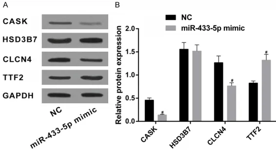
Figure 2. Western blotting was utilized to detect protein expression levels of target genes in SGC-7901. Note: (A) Protein bands of CASK, HSD3B7, CLCN4, and TTF2; (B) Quantification of protein expression of CASK, HSD3B7, CLCN4, and TTF2. #compared with the NC group, P < 0.05.
Reverse transcription quan-titative polymerase chain reaction (RT-qPCR)
[image:4.612.89.372.314.468.2]lution curve: ΔCt = CT (target gene) - CT (GAPDH), ΔΔCt = ΔCt (experimental group) - ΔCt (control
group). Expression of mRNA of related genes was calculated by 2-ΔΔCt. The experiment was
repeated 3 times.
Western blotting
RIPA lysate (R0010, Beijing Solarbio Science & Technology Co., Ltd., China) was added to the
PMSF before the experiment. The final concen -tration of PMSF was 1 mM. Gastric cancer cells were washed with PBS 2 times, then added
with RIPA lysate (2-5 × 10⁷ cells were added with 1 mL RIPA lysate). Gastric cancer cells were then incubated on ice and the lysates were centrifuged at 12,000 × g for 30 minutes. Next, protein concentrations were determined via the BCA kit (P0006, Beyotime Institute of Biotechnology, China). The proteins were trans-ferred to PVDF membranes after separating by sodium dodecyl sulfate polyacrylamide gel electrophoresis (SDS-PAGE). The membranes were then blocked with tris buffered saline with
Cell counting kit 8 (CCK8) assay
Cells were made into the cell suspension when
the cell density reached 80%. It was then added
into 96-well plates with each well 4 × 10³ cells
(200 μl per well). Six duplicated wells were set
and cultured in an incubator. Subsequently, each well was added with 5 g/L CCK-8 solution at 24, 48, and 72 hours post-incubation. It was then incubated for another 4 hours. Each well was added with 100 µL Dimethyl sulfoxide (DMSO). A microplate reader (SpectraMax iD5, Molecular Devices, USA) was used to detect optical density (OD) values at 490 nm. Cell growth curves were made in accordance with OD values. The experiment was repeated 3 times.
Transwell assay
[image:5.612.90.375.73.207.2]Matrigel (354230, Becton, Dickinson, and Company, USA) was diluted with serum-free MEM medium at a ratio of 1:10. The Transwell upper chamber was added with 100 μL diluted Figure 3. H&E staining results of gastric cancer and adjacent tissues (400
×).
Figure 4. Immunohistochemistry was used to detect CLCN4 protein expres-sion in cancer tissues and adjacent tissues (400 ×).
Tween 20 (TBST) containing
5% bovine serum albumin
for 1 hour. After abando- ning the blocking liquid, the membranes were incubated with primary antibody CL- CN4 (1:500, ab75008, AB- CAM, UK), vimentin (1:1000, ab52947, ABCAM, UK), MMP-2 (1 µg/mL, ab837150, ABCAM, UK), and GAPDH (1:1000, ab8245, ABCAM, UK). It was placed at 4°C overnight. The membranes were washed wi- th TBST for 3 × 10 minutes and incubated with goat anti-mouse IgG (1:5000, ab97040, ABCAM, UK) for 4-6 hours. Next, the membranes were washed with TBST for 3 × 5 minutes. ECL (PE0010, Beijing Solarbio Science & Technology Co., Ltd., China) was used to detect the chemilumines-cence signal. Target protein expression levels were
quanti-fied by determining the ratio of
[image:5.612.90.375.257.400.2]Matrigel and placed at room temperature for 2 hours. It was rinsed with 200 μL serum-free 1640 culture medium and resuspended using a serum-free DMEM medium. After the cells
were calculated and diluted to 3 × 10⁵ cells/
mL, 100 μL diluted cells were added to the
upper Transwell chamber. The lower chamber
was added with 600 μL DMEM containing 10%
serum. After incubating for 24 hours, crystal violet staining was performed. The Transwell chambers were collected and an optical micro-scope (CX21, Olympus, Japan) was utilized to
detect results. Invasion rate = cells passing through the Matrigel/total number of cells × 100%.
Annexin V-FITC/PI staining
Gastric cancer cells were washed with PBS after culturing for 48 hours. They were then
centrifuged at 2,000 × g for 5 minutes. Cell density was adjusted to 4 × 10⁵ cells/mL after removing the supernatant. The cells were
resuspended by adding with 500 μL binding buffer. This was followed by adding 5 μL Annexin-V-FITC and 5 μL PI dye liquid before flow cytometry analysis. It was mixed gently and
incubated in an ice bath in the dark for 10-12 minutes. Flow cytometry (CoulterCytoFLEX, Beckman Coulter, USA) was used to determine FITC and PI levels.
Statistical analysis
SPSS 21.0 software (IBM. Corp., Armonk, USA) was used for statistical data analysis. Quantitative data are presented as mean ± standard deviation (SD). Each experiment was conducted in triplicate. The Kolmogorov-Smirnov method was utilized to determine the normality of data distribution in each group. Student’s t-tests were used to compare group variables. One-way analysis of variance (ANOVA) was used for multiple groups analysis. LSD tests were used to compare inter-group vari-ables. Grade data were compared using
cancer through targeting CLCN4
Targetscan7.2, miRWalk, RNA22, and miRDB were used to explore potential target genes that might have binding sites with miR-433-5p. Moreover, 1,047, 14,645, 5,916, and 48 genes
were identified, respectively. Venn images
(Figure 1A) indicated that CASK, HIPK2, HSD3B7, CLCN4, DDHD1, TTF2, and CISD1 were the intersection. Subsequently,
dual-lucif-erase reporter assays were used to confirm tar -geting regulatory relationships between miR-433-5p and the above genes. Results indicated that miR-433-5p have binding sites with CASK, HSD3B7, CLCN4, and TTF2 (Figure 1B). Next, SGC-7901 was transfected with miR-433-5p mimics. Negative controls were also set up. Western blotting was adopted to determine protein expression levels of CASK, HSD3B7, CLCN4, and TTF2. Results indicated that (Figure 2A, 2B), compared with the NC group, protein expression of CASK and CLCN4 was
sig-nificantly downregulated. TTF2 protein expres -sion was upregulated in the miR-433-5p mimic group (all P < 0.05). There were no significant
differences in HSD3B7 protein expression lev-els between NC and miR-433-5p mimic groups (P > 0.05).
[image:6.612.86.395.97.162.2]MicroRNAs always function as negative regula-tors in post transcriptional gene silencing (PTGS) through base pairing with target mRNAs. This results in mRNA cleavage and translation-al inhibition. Thus, it can be speculated that other ways may participate in the regulating effects of miR-433-5p on HSD3B7 and TTF2. Therefore, CASK and CLCN4 were selected as candidate genes in this study. The protein encoded by CASK was calcium/calmodulin dependent serine protein kinase, a member of the membrane associated guanylate kinase family. A previous study proved that CASK was highly expressed in esophageal cancer and related to progression and worse prognosis in colorectal cancer [14, 15]. In addition, CASK Table 2. CLCN4 expression in gastric cancer tissues and adjacent
tis-sues
CLCN4 expression
P Negative
(-) Weakly positive (+) positive (++)Medium positive (+++)Strong
Adjacent tissue 15 8 3 1 0.001
Gastric tissue 5 6 13 3
Wilcoxon rank-sum tests.
P < 0.05 indicates
statis-tical significance. Results
had been reported to participate in the growth and metastasis of gastric cancer via regulation by miR-203 [16]. Thus, the regulatory mecha-nisms of CASK in gastric cancer are relatively clear. CIC-4 chloride channel (CLCN4) gene is a member of voltage gated chloride chan- nels (CICs) family. Previous studies have
con-firmed that CLCN4 could induce the invasion
and metastasis of colon cancer using gene trapping technology. There existed gender dif-ferences in copy number variations of CLCN4 in
colon cancer [11]. However, the specific molec -ular mechanisms of CLCN4 in gastric cancer remain largely unknown. The present study focused on the function and regulation of miR-433-5p in gastric cancer through targeting CLCN4.
H&E staining results
According to results obtained from H&E stain-ing (Figure 3), gastric cancer tissues showed
sue invaded the gastric fundus glandular mucosa.
CLCN4 expression was upregulated in gastric cancer
Immunohistochemical staining was performed to examine expression of CLCN4 in tumor tis-sues and para-cancerous tistis-sues. Results showed that (Figure 4, Table 2) CLCN4 was mainly expressed in signet cells, as well as the cytoplast and nucleolus. Compared to adjacent normal tissues, elevated CLCN4 expression was found in cancer tissues.
Highest levels of CLCN4 found in SGC-7901 cells
Western blotting was utilized to detect CLCN4 expression. Results indicated that (Figure 5), compared with GES-1, levels of CLCN4 in other
cell lines were significantly upregulated. The
highest level of CLCN4 expression was found in SGC-7901 cell lines. Thus, SGC-7901 was selected for the experiments.
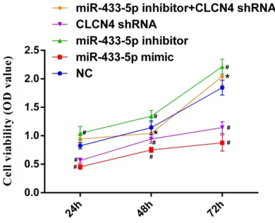
miR-433-5p inhibited cell viability of SGC-7901 through targeting CLCN4
To evaluate the impact of miR-433-5p in SGC-7901 cell line, cells were transfected with miR-433-5p inhibitors, miR-miR-433-5p mimics, or CLCN4 shRNA. CCK-8 assays were performed to determine the cell viability in each group. Results indicated that (Figure 6), compared
with the NC group, cell viability was significantly
[image:7.612.91.374.73.179.2]decreased after the cells were transfected with miR-433-5p mimics or CLCN4 shRNA, at each time point (all P < 0.05). However, cell viability in the miR-433-5p inhibitor group was obvious-ly increased, compared with the NC group. Figure 5. CLCN4 protein expression in different gastric cancer cells, Note: (A)
The protein bands of CLCN4 in different gastric cancer cell lines; (B) Quan-tification of CLCN4 protein in different gastric cancer cell lines. &P < 0.05.
Figure 6. Cell viability of SGC-7901 in each group. Note: compared with NC group, #P < 0.05; compared
with miR-433-5p inhibitor group, *P < 0.05.
[image:7.612.90.285.242.400.2]tis-Figure 7. Invasion ability of SGC-7901 in each group. Note: (A) Transmembrane cells stained by crystal violet in each group; (B) Quantification of invasive rate in each group. Compared with NC group, #P < 0.05; compared with
[image:8.612.97.521.72.252.2]miR-433-5p inhibitor group, *P < 0.05.
Figure 8. mRNA and protein expression of vimentin and MMP-2 in each group of SGC-7901 cell. Note: (A) mRNA expression of vimentin and MMP-2 in each group; (B) Protein bands of vimentin and MMP-2 in each group; (C) Quan-tification of vimentin and MMP-2 protein expression in each group. Compared with NC group, #P < 0.05; compared
with miR-433-5p inhibitor group, *P < 0.05.
Effects of miR-433-5p inhibitors on SGC-7901 cells can be reversed by silencing of CLCN4.
miR-433-5p inhibited invasion and metastasis of SGC-7901 by targeting CLCN4
Transwell assays were adopted to determine invasion abilities in each group. Results revealed that (Figure 7), compared with the NC group, invasion abilities of the miR-433-5p mimic group and CLCN4 shRNA group were
sig-nificantly decreased (all P < 0.05). However, invasion abilities in the miR-433-5p inhibitor group were obviously enhanced (P < 0.05). Moreover, CLCN4 silencing could reverse tumor invasion induced by miR-433-5p inhibitors. Western blotting and Q-PCR were performed to quantify the mRNA and protein expression
lev-els of vimentin and MMP-2. Results indicated that (Figure 8), compared with the NC group, vimentin and MMP-2 mRNA and protein
expres-sion levels were significantly downregulated in
the miR-433-5p mimic group and CLCN4 shRNA group (all P < 0.05). However, levels in the miR-433-5p inhibitor group were obviously upregulated (P < 0.05). CLCN4 silencing could partly reverse the induction of vimentin and MMP-2 induced by miR-433-5p inhibitors.
miR-433-5p could promote SGC-7901 apopto-sis through targeting CLCN4
Annexin V-FITC/PI staining was utilized to detect apoptosis levels of SGC-7901 in each group. Results obtained from this experiment indicat-ed that (Figure 9), compared with the NC group,
[image:8.612.93.520.311.431.2]in the miR-433-5p mimic group and CLCN4 shRNA group (all P < 0.05). However, cell apop-tosis was inhibited when cells were transfected with miR-433-5p inhibitors. CLCN4 silencing could partly reverse SGC-7901 apoptosis induced by miR-433-5p inhibitors.
Discussion
Prognosis of patients with advanced gastric cancer remains extremely low despite great advancements made in targeting preparations and cytotoxic drugs [17-19]. Thus, it is impor-tant to reveal the underlying pathological mech-anisms of gastric carcinoma, providing novel diagnostic biomarkers and new strategies for gastric cancer therapy. Moreover, miR-433 has
been reported to play a significant role in the
progression of gastric carcinoma [10]. Guo LH
et al. confirmed that miR-433 exerted a sup -pressor role in gastric cancer [20]. Thus, the current study focused on miR-433-5p and its potential mechanisms in gastric cancer. A series of experiments were performed. Results obtained from this study indicate that miR-433-5p could inhibit viability, as well as invasion and metastasis, of gastric cancer cells. It may also induce cell apoptosis through downregulation of CLCN4.
Initially, bioinformatics tools and dual-lucifer-ase reporter assays were used to predict the
target genes of miR-433-5p. CLCN4 was even-tually selected. A study designed by Ishiguro et al. found that CLCN4 could induce invasion and metastasis of colon cancer [11]. Thus, this study focused on roles in gastric cancer. Evidence obtained from immunohistochemical tests demonstrated that CLCN4 expression was upregulated in gastric cancer tissues, com-pared with para-cancerous tissues. SGC-7901 cell lines were used for subsequent experi-ments and transfected with miR-433-5p mim-ics, miR-433-5p inhibitors, or CLCN4 shRNA. CCK8 assay and Transwell assay results proved that miR-433-5p overexpression or CLCN4 silencing could inhibit the viability and invasion abilities of gastric cancer cells. Metastasis and recurrence are the most important factors that affect the prognosis of cancer patients. Degradation of the basement membrane is a key point for tumor metastasis and invasion [21-23]. Matrix metalloproteinases (MMPs), a calcium-dependent endopeptidase, has been proven to have the ability to degrade the extra-cellular matrix, promoting metastasis and inva-sion of tumors [24-27]. MMP2 is an important member of the MMPs family. In this study, it was found that mRNA and protein expression
levels of MMP-2 were significantly decreased
[image:9.612.92.521.75.279.2]a kind of cytoskeletal protein. Accumulating evi-dence has demonstrated that vimentin is expressed in a variety of tumor cells [28-31]. Detection of vimentin expression has been an important measure in diagnosing or screening
of cancer [32, 33]. Results also confirmed that
miR-335-5p could downregulate expression of vimentin through inhibition of CLCN4. Annexin V-FITC/PI staining results showed that, com-pared with the NC group, cell apoptosis was increased in the miR-433-5p mimic group and CLCN4 shRNA group, while decreased in the miR-433-5p inhibitor group. Present results suggest that miR-433-5p could promote cell apoptosis by inhibiting CLCN4.
Collectively, key findings obtained in this study
indicate that miR-433-5p can act as a tumor suppressor in gastric cancer. It can inhibit gas-tric cancer cell viability, invasion, and metasta-sis. At the same time, it may induce gastric can-cer cell apoptosis through downregulation of
CLCN4. Current findings demonstrate the vital
roles of miR-433-5p and CLCN4 in gastric can-cer, suggesting that miR-433-5p and CLCN4 could serve as a pivotal therapeutic targets in gastric cancer management. More in vivo and in vitro studies are necessary to further vali-date the clinical utility.
Disclosure of conflict of interest
None.
Address correspondence to: Hai-Bing Jiang, De- partment of Gastroenterology, The Second Hospital University of South China, No.35 Jiefang Avenue, Huaxin Development Zone, Hengyang 421001, China. Tel: 0734-8899680; E-mail: haibingji-ang680@163.com
References
[1] Hu B, Wang B, Zhao B, Guo Q, Li ZH, Zhang XH, Liu GY, Liu Y, Tang Y, Luo F, Du Y, Chen YX, Ma LY and Liu HM. Thiosemicarbazone-based se-lective proliferation inactivators inhibit gastric cancer cell growth, invasion, and migration. Medchemcomm 2017; 8: 2173-2180.
[2] Coburn N, Cosby R, Klein L, Knight G, Maltha-ner R, Mamazza J, Mercer CD and Ringash J. Staging and surgical approaches in gastric cancer: a systematic review. Cancer Treat Rev 2018; 63: 104-115.
[3] Wang W, Li Z, Wang J, Du M, Li B, Zhang L, Li Q, Xu J, Wang L, Li F, Zhang D, Xu H, Yang L, Gong
W, Qiang F, Zhang Z and Xu Z. A functional poly-morphism in TFF1 promoter is associated with the risk and prognosis of gastric cancer. Int J Cancer 2018; 142: 1805-1816.
[4] Dugue PA, Bassett JK, Joo JE, Jung CH, Ming Wong E, Moreno-Betancur M, Schmidt D, Makalic E, Li S, Severi G, Hodge AM, Buchanan DD, English DR, Hopper JL, Southey MC, Giles GG and Milne RL. DNA methylation-based bio-logical aging and cancer risk and survival: Pooled analysis of seven prospective studies. Int J Cancer 2018; 142: 1611-1619.
[5] Zhang X, Li Z, Xuan Z, Xu P, Wang W, Chen Z, Wang S, Sun G, Xu J and Xu Z. Novel role of miR-133a-3p in repressing gastric cancer growth and metastasis via blocking autophagy-mediated glutaminolysis. J Exp Clin Cancer Res 2018; 37: 320.
[6] Daneshpour M, Karimi B and Omidfar K. Si-multaneous detection of gastric cancer-in-volved miR-106a and let-7a through a dual-signal-marked electrochemical nanobiosensor. Biosens Bioelectron 2018; 109: 197-205. [7] Zhang H, Mao F, Shen T, Luo Q, Ding Z, Qian L
and Huang J. Plasma 145, 20a, miR-21 and miR-223 as novel biomarkers for screening early-stage non-small cell lung can-cer. Oncol Lett 2017; 13: 669-676.
[8] Chen YL, Xu QP, Guo F and Guan WH. MicroR-NA-302d downregulates TGFBR2 expression and promotes hepatocellular carcinoma gr- owth and invasion. Exp Ther Med 2017; 13: 681-687.
[9] Ueda T, Volinia S, Okumura H, Shimizu M, Tac-cioli C, Rossi S, Alder H, Liu CG, Oue N, Yasui W, Yoshida K, Sasaki H, Nomura S, Seto Y, Kamin-ishi M, Calin GA and Croce CM. Relation be-tween microRNA expression and progression and prognosis of gastric cancer: a microRNA expression analysis. Lancet Oncol 2010; 11: 136-146.
[10] Luo H, Zhang H, Zhang Z, Zhang X, Ning B, Guo J, Nie N, Liu B and Wu X. Down-regulated miR-9 and miR-433 in human gastric carcinoma. J Exp Clin Cancer Res 2009; 28: 82.
[11] Ishiguro T, Avila H, Lin SY, Nakamura T, Yama-moto M and Boyd DD. Gene trapping identifies chloride channel 4 as a novel inducer of colon cancer cell migration, invasion and metasta-ses. Br J Cancer 2010; 102: 774-782.
[12] Shparyk IaV. [New classification of malignant tumors TNM (5th edition)]. Klin Khir 1998; 36-38.
[13] Konno R, Yamakawa H, Utsunomiya H, Ito K, Sato S and Yajima A. Expression of survivin and Bcl-2 in the normal human endometrium. Mol Hum Reprod 2000; 6: 529-534.
CASK correlates with progression and poor prognosis of colorectal cancer. Tumour Biol 2014; 35: 9185-9194.
[15] Wang Q, Lu J, Yang C, Wang X, Cheng L, Hu G, Sun Y, Zhang X, Wu M and Liu Z. CASK and its target gene Reelin were co-upregulated in human esophageal carcinoma. Cancer Lett 2002; 179: 71-77.
[16] Lozovatsky L, Abayasekara N, Piawah S and Walther Z. CASK deletion in intestinal epithelia causes mislocalization of LIN7C and the DLG1/Scrib polarity complex without affecting cell polarity. Mol Biol Cell 2009; 20: 4489-4499.
[17] Zhong J, Zhao W, Ren F, Qi S, Wang X, Lv T, Su Z, Yin H, Ren J and Huan Y. Lymph node metas-tasis in patients with gastric cancer: a multi-modality, morphologic and functional imaging study. Am J Transl Res 2016; 8: 5601-5609. [18] Lee MH, Cho Y, Kim DH, Woo HJ, Yang JY, Kwon
HJ, Yeon MJ, Park M, Kim SH, Moon C, Tharma-lingam N, Kim TU and Kim JB. Menadione in-duces G2/M arrest in gastric cancer cells by down-regulation of CDC25C and proteasome mediated degradation of CDK1 and cyclin B1. Am J Transl Res 2016; 8: 5246-5255.
[19] Ito H, Sato J, Tsujino Y, Yamaguchi N, Kimura S, Gohda K, Murakami K, Onimaru M, Ohmori T, Ishikawa F and Inoue H. Long-term prognostic impact of circulating tumour cells in gastric cancer patients. World J Gastroenterol 2016; 22: 10232-10241.
[20] Guo LH, Li H, Wang F, Yu J and He JS. The tu-mor suppressor roles of miR-433 and miR-127 in gastric cancer. Int J Mol Sci 2013; 14: 14171-14184.
[21] Zhang L, Kim S, Ding W, Tong Y, Zhang X, Pan M and Chen S. Arsenic sulfide inhibits cell mi -gration and invasion of gastric cancer in vitro and in vivo. Drug Des Devel Ther 2015; 9: 5579-5590.
[22] Xu X, Chen L, Xu B, Xie Q, Sun M, Deng X, Wu C and Jiang J. Increased MT2-MMP expression in gastric cancer patients is associated with poor prognosis. Int J Clin Exp Pathol 2015; 8: 1985-1990.
[23] Kosaka Y, Mimori K, Fukagawa T, Ishikawa K, Etoh T, Katai H, Sano T, Watanabe M, Sasako M and Mori M. Clinical significance of molecu -lar detection of matrix metalloproteinase-1 in bone marrow and peripheral blood in patients with gastric cancer. Ann Surg Oncol 2012; 19 Suppl 3: S430-437.
[24] Torres-Martinez AC, Gallardo-Vera JF, Lara-Hol-guin AN, Montano LF and Rendon-Huerta EP. Claudin-6 enhances cell invasiveness through claudin-1 in AGS human adenocarcinoma gas-tric cancer cells. Exp Cell Res 2017; 350: 226-235.
[25] Wang Z, Li G, Wu H, Sun K, Chen J, Feng Y, Chen C, Cai S, Xu J and He Y. CXCL1 from tu-mor-associated lymphatic endothelial cells drives gastric cancer cell into lymphatic system via activating integrin beta1/FAK/AKT signal-ing. Cancer Lett 2017; 385: 28-38.
[26] Wei Y, Zhao L, He W, Yang J, Geng C, Chen Y, Liu T, Chen H and Li Y. Benzo [a] pyrene pro-motes gastric cancer cell proliferation and me-tastasis likely through the Aryl hydrocarbon re-ceptor and ERK-dependent induction of MMP9 and c-myc. Int J Oncol 2016; 49: 2055-2063. [27] Xu J, E C, Yao Y, Ren S, Wang G and Jin H.
Ma-trix metalloproteinase expression and molecu-lar interaction network analysis in gastric can-cer. Oncol Lett 2016; 12: 2403-2408. [28] Kumar C, Rasool RU, Iqra Z, Nalli Y, Dutt P,
Sat-ti NK, Sharma N, Gandhi SG, Goswami A and Ali A. Alkyne-azide cycloaddition analogues of dehydrozingerone as potential anti-prostate cancer inhibitors via the PI3K/Akt/NF-kB path-way. Medchemcomm 2017; 8: 2115-2124. [29] Xu ZF, Sun XK, Chen G, Han C, Wang F and
Zhang YD. Oroxyloside inhibits human glioma progression by suppressing proliferation, me-tastasis and inducing apoptosis related path-ways. Biomed Pharmacother 2018; 97: 1564-1574.
[30] Zheng P, Li H, Xu P, Wang X, Shi Z, Han Q and Li Z. High lncRNA HULC expression is associated with poor prognosis and promotes tumor pro-gression by regulating epithelial-mesenchymal transition in prostate cancer. Arch Med Sci 2018; 14: 679-686.
[31] Shi W, Wang X, Ruan L, Fu J, Liu F and Qu J. MiR-200a promotes epithelial-mesenchymal transition of endometrial cancer cells by nega-tively regulating FOXA2 expression. Pharmazie 2017; 72: 694-699.
[32] Li MY, Liu JQ, Chen DP, Li ZY, Qi B, Yin WJ and He L. p68 prompts the epithelial-mesenchymal transition in cervical cancer cells by transcrip-tionally activating the TGF-beta1 signaling pathway. Oncol Lett 2018; 15: 2111-2116. [33] Wu F, Zhu J, Mao Y, Li X, Hu B and Zhang D.