https://www.scirp.org/journal/wjcs ISSN Online: 2164-3210
ISSN Print: 2164-3202
DOI: 10.4236/wjcs.2019.99013 Sep. 16, 2019 108 World Journal of Cardiovascular Surgery
Initial Experience with Open Heart Surgery in
Sub-Saharan Africa: Challenges in Mali with
Minimum Standards for Practice
Seydou Togo
1*, Moussa Abdoulaye Ouattara
1, Abdoul Aziz Maïga
1, Moussa Bazongo
1,
Issa Boubacar Maïga
1, Cheik Ahmed Sékou Touré
2, Ibrahim Coulibaly
1, Sounkalo Diop
1,
Allaye Ombotimbe
1, Sitta Illiassou
1, Souleymane Coulibaly
1, Mamadou Solo Koita
1,
Koumba Nelly Dora Ignanga
1, Sanibé Dramane Koné
1, Moussa Oscar Kamano
1,
Fatoumata Konaté
1, Adama Issa Koné
1, Amadou Sidibé
2, Ahmadou Dramé
2, Nouhoum Oueloguem
3,
Bourama Kané
4, Boubacar Dramé
5, Sékou Koumaré
6, Zimogo Zié Sanogo
6, Sadio Yéna
11Department of Thoracic and Cardiovascular Surgery, Mali Hospital, Bamako, Mali 2Department of Anesthesiology and Critical Care, Mali Hospital, Bamako, Mali 3Department of Cardiologie, Mali Hospital, Bamako, Mali
4Department of Pediatrics, Mali Hospital, Bamako, Mali
5Department of Biomedical Laboratory, Mali Hospital, Bamako, Mali 6Department of Surgery “A”, Point G Hospital, Bamako, Mali
Abstract
Introduction: There has been limited experience with Open Heart Surgeries (OHS) in Sub-Saharan Africa. In west Africa especially in Mali, most fled-gling centers are unable to overcome the myriad of challenges encountered in establishing OHS though there is a high prevalence of surgically correctable heart diseases. The aim of this paper is to review our initial experience of our first cases in developing OHS program and discuss the challenges and pros-pects that need to be overcome to further develop it. Methods: A total of 6 patients who underwent OHS during the first “cardiac mission” in July 2016 were included in this retrospective study. The medical records of the patients were examined and data on age, sex, diagnosis, EuroSCORE, type of surgery, cardiopulmonary bypass details, complications and length of hospital stay were extracted. Results: Six patients with a male to female ratio of 1, ages ranging between 12 and 35 years (mean of 22.5 ± 12 years) were studied. The mean of EuroSCORE was 6 ± 41. Pericardial patch closure of isolated atrial septal defect was performed in one patient. One patient had mitral valve re-pair for rheumatic mitral regurgitation consisting of chordal shortening with How to cite this paper: Togo, S., Ouattara,
M.A., Maïga, A.A., Bazongo, M., Maïga, I.B., Touré, C.A.S., Coulibaly, I., Diop, S., Ombo-timbe, A., Illiassou, S., Coulibaly, S., Koita, M.S., Ignanga, K.N.D., Koné, S.D., Kamano, M.O., Konaté, F., Koné, A.I., Sidibé, A., Dramé, A.I., Oueloguem, N., Kané, B., Dramé, B., Koumaré, S., Sanogo, Z.Z. and Yéna, S. (2019) Initial Experience with Open Heart Surgery in Sub-Saharan Africa: Challenges in Mali with Minimum Standards for Practice. World Journal of Cardiovascular Surgery, 9, 108-118.
https://doi.org/10.4236/wjcs.2019.99013
DOI: 10.4236/wjcs.2019.99013 109 World Journal of Cardiovascular Surgery a tricuspid valvuloplasty. Three patients had mitral valve replacement with tricuspid valvuloplasty. Four patients had mitral valve replacement. Sixty-day mortality was 0%. Conclusion: Safe conduct of open heart surgery in Mali Hospital setting is feasible. Grant financial aid is required for rapid growth of Open-Heart Surgery in this part of Sub-Saharan Africa.
Keywords
Open-Heart, Surgery, Mali, Africa
1. Introduction
Open Heart Surgery (OHS) is defined as “surgical repair of the heart during which the blood circulation is often maintained mechanically requiring Cardi-opulmonary Bypass (CPB)” [1]. There has been limited experience with OHS in West Africa with only a few established cardiac centers [2] [3]. In Mali most fled-gling centers are unable to overcome the myriad of challenges encountered in establishing OHS though there is a high prevalence of surgically correctable heart diseases. OHS is relatively expensive as income is low in Mali. It has been demonstrated that in sub-Saharan Africa, gross domestic product (GDP) per ca-pita remains low comparing to OHS cost, which remains beyond the reach of our population’s financial capacities [4]. Confronted with this deficit, funding from our States is insignificant or does not exist at all in some of them [5]. Car-diothoracic practice in Mali faces multiple challenges that need to be overcome to enable sustainable practice. A seed fund was provided by the Malian govern-ment which was used as the start of specialized cardiac training abroad. A small fraction of patients are sponsored or are able to fund their own surgery abroad but the goal for any country has to be to establish its own programs that can be developed and sustained. Despite some early attempts to develop OHS in Mali [6], this has not been sustained. An OHS program is in the first time being per-formed at the Mali University Teaching Hospital. It is encouraging to see the surge in OHS activity but we need to transit from cardiac missions. Successful OHS requires 24-hour laboratory support, an active blood bank and cardiac ca-theterization support. Access to these various support facilities were very limited in sub-Saharan Africa. The aim of this article is to share the result of our first cases in developing OHS program and discuss the challenges and prospects that need to be overcome to further develop and sustain it.
2. Patients and Methods
Surgical treatment for heart diseases did not exist in Mali located in sub-Saharan Africa and early death was inevitable. For several years the Mali government has sponsored few needy patients to undergo OHS abroad. Since 2009 a makeshift cardiac centre was under construction in Mali without a serious sustain program Copyright © 2019 by author(s) and
Scientific Research Publishing Inc. This work is licensed under the Creative Commons Attribution International License (CC BY 4.0).
ur-DOI: 10.4236/wjcs.2019.99013 111 World Journal of Cardiovascular Surgery gent care for any potentially serious symptoms. Recovery at home after leaving the hospital usually takes between 4 to 6 weeks. The healthcare team guide reha-bilitation and advice on medications and restrictions on physical activity. Then patients has regular checkup one’s a month for about 1 year. In echographic fol-low-up comparison is made to show the relation between preoperative left ven-tricular diameter and the possibility of postoperative venven-tricular recuperation after mitral valve replacement. Patients consent was obtained before this study and permission was obtained from the Ethics Committee of the Mali University Teaching Hospital for use of the patient data from the database. The results are presented below, as well as the challenges encountered in achieving these results. Data are expressed as absolute values, percentages, or mean ± SD where appro-priate. Fisher exact test was used for statistical analysis and P < 0.05 was statisti-cally significant.
3. Results
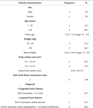
A total of 6 OHS cases were performed during 7 days in our hospital. The pa-tients had a mean age of 22.5 ± 12 years (range: 12 - 33 years) with a male to fe-male ratio of 1. The patients in all had a mean weight of 25.8 ± 10.9 and a body surface area of 0.56 ± 0.5 m2. Three patients had mild clinical cardiac
decom-pensation (50.0%) in New York Heart Association (NYHA) class 2 while they were no NYHA class 3 and NYHA class 4 in the patients selected. The various anatomical lesions in echocardiography were dilatation of the mitral ring, dilata-tion of the heart chambers, thrombi in the atria, atrial septal defect, and valve calcification, leakage and stenosis. Sociodemographic and clinical characteristics were found in Table 1. Pericardial patch closure of isolated atrial septal defect was done in 1 patient. One patient with rheumatic mitral regurgitation aged 12 years had mitral valve repair for rheumatic mitral regurgitation consisting of chordal shortening with tricuspid valvuloplasty using the De Vega method. Four patients had mitral valve replacement with tricuspid valvuloplasty by the De Vega method. Surgical removal of the thrombi and closure of the left atrial ap-pendage were performed after mitral valve replacement in 2 patients. Table 2 shows age, euroscore the various pathologies encountered and types of OHS procedures performed.
DOI: 10.4236/wjcs.2019.99013 112 World Journal of Cardiovascular Surgery
Table 1. Sociodemographic and clinical characteristics.
Patients characteristics Frequency %
Sex
Male 3 50
Female 3 50
Age (years)
5 - 20 2 33.3
>20 4 66.7
Mean age: 22.5 ± 12 (range: 12 - 33)
Weight (Kg)
10 - 30 2 33.3
>30 4 66.7
Mean weight: 25.8 ± 10.9 (range: 15 - 62) Body surface area (m2)
0.5 - 1.0 m2 4 66.7
1.0 - 1.5 m2 2 33.3
Mean body surface area: 0.56 ± 0.5 m2 New York Heart Association class
2 6 100
Diagnosis - Congenital heart disease:
ASD (secundum = 2.5 mm) 1 16.7
- Acquired heart disease:
Severe rheumatic mitral stenosis 1 16.7
Severe rheumatic mitral regurgitation + tricuspid insufficiency 4 66.6 ASD: Atrial Septal Defect.
Table 2. Shows age, EuroSCORE, the various pathologies encountered and types of OHS procedures performed.
Patient
number Age EuroSCORE Diagnosis Pathology Surgical procedure
1 24 3 Atrial septal defect Secundum (25 mm) Autologous pericardium closure
2 12 6 MV disease MV regurgitation + TV regurgitation Mitral valve repair + tricuspid valvuloplasty by “De Vega” Method
3 23 4 Mitral valve disease MV stenosis + TV regurgitation Mechanical Mitral valvereplacement + Tricuspid Valvuloplasty by “De Vega” Method
4 15 10 Mitral valve disease MV stenosis + TV regurgitation + thrombi in left atrium Mechanical Mitral valve replacement + Tricuspid Valvuloplasty by “De Vega” Method + thrombi removal and left atrial appendage closure 5 29 5 Mitral valve disease MV stenosis Mechanical Mitral valve replacement
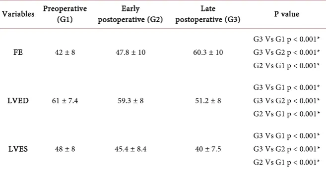
[image:5.595.55.530.519.709.2]DOI: 10.4236/wjcs.2019.99013 113 World Journal of Cardiovascular Surgery infection in 1 patients (7.8%). Table 3 is a summary of the procedures which in-cludes the distribution of cardiopulmonary bypass time, cross clamp time and postoperative complications. The post-operative hospital stay was 9.1 ± 2.4 days and the overall mortality in the series was null. The patients were symptom-free at the 6-month follow-up clinic visit and trans-thoracic echocardiography done then did not show any deterioration in the degree of mitral valve or tricuspid valve regurgitation. All of the patients (100%) were in NYHA class I. Long term (4 years) follow-up is being done on these patients. The comparative echographic result before and after surgery are presented in Table 4.
4. Discusions
[image:6.595.207.538.299.453.2]Since 1950s, one of the major challenges was to accomplish correction of intra- cardiac lesions within a bloodless heart using a heart-lung machine. Throughout
Table 3. Operative details, complications and mortality.
Patient
number Age Operations (min) CPB X Clamp (min) Complications Mortality
1 24 ASD 92 73 Superficial wound infection 0
2 12 MV repair + TV 146 96 - 0
3 23 MVR + TV + LAC 149 101 Fever 0
4 15 MVR + TV 132 95 Pericardial effusion 0
5 29 MVR 136 98 Atrial fibrillation (tachycardia) 0
6 35 MVR + TV + LAC 151 105 Ventricular fibrillation 0 ASD: Atrial Septal Defect. MV: Mitral Valve. TV: Tricuspid Valvuloplasty. CBP: Cardio-Pulmonary Bypass time. MVR: Mitral Valve Replacement. X Clamp: Aortic cross Clamp Time. LAC: Left Atrial Closure.
Table 4. Comparison of Ejection fraction, left ventricular end-systolic diameter, left
ven-tricular end-diastolic diameter in patients with mitral valve replacement.
Variables Preoperative (G1) postoperative (G2) Early postoperative (G3) Late P value
FE 42 ± 8 47.8 ± 10 60.3 ± 10
G3 Vs G1 p < 0.001* G3 Vs G2 p < 0.001* G2 Vs G1 p < 0.001*
LVED 61 ± 7.4 59.3 ± 8 51.2 ± 8
G3 Vs G1 p < 0.001* G3 Vs G2 p < 0.001* G2 Vs G1 p < 0.001*
LVES 48 ± 8 45.4 ± 8.4 40 ± 7.5
G3 Vs G1 p < 0.001* G3 Vs G2 p < 0.001* G2 Vs G1 p < 0.001*
[image:6.595.209.538.521.696.2]DOI: 10.4236/wjcs.2019.99013 117 World Journal of Cardiovascular Surgery and the empowerment of the Malian teams for the relay. This autonomy will, in the near future, give the Malian population access to cardiac surgery at a lower cost. Regarding to this operating result in our hospital, further cardiac missions were starting organized in others hospitals to performed OHS in Mali.
5. Conclusion
By encouraging international humanitarian services for OHS in developing coun-tries without any capacity to take care of patients with acquired or congenital heart disease, OHS can be done with success in Mali. Grant financial aid to the care of the poorest patients by public, governmental or private initiatives is required for rapid growth of Open-Heart Surgery in sub-Saharan Africa.
Conflicts of Interest
The authors declare no conflicts of interest regarding the publication of this pa-per.
References
[1] Open-Heart Surgery (2012) Collins English Dictionary—Complete & Unabridged. 10th Edition. Dictionary.com.
http://dictionary.reference.com/browse/open-heartsurgery
[2] Anyanwu, C.H., Ihenacho, H.N., Okoroma, E.O., et al. (1982) Initial Experience with Open Heart Surgery in Nigeria. Tropical Cardiology, 8, 123-127.
[3] Adebonojo, S.A., Grillo, I.A. and Osinowo, O. (1981) Initial Experience with Open Heart Surgery at University College Hospital, Ibadan, Nigeria. Tropical Cardiology,
7, 49–54.
[4] Adebonojo, S.A. (2012) How Viable Are the Cardiac Programmes in West Africa Today? In: Adebonojo, S.A., Ed., Development of Open Heart Surgery in West Africa: A Historical Perspective, Acecool Medical Publishers: Nigeria, Eruwa, 43-46. [5] Diakité, A., Sidibé, N. and Diarra, M.B. (2009) Epidemiological and Clinical Aspects
of Congenital Heart Disease. Mali Médical, 24, 67-68.
[6] Coulibaly, B., Diarra, M. and Dicko, M. (2014) Angers-Bamako Cooperation in the Context of Cardiovascular Surgery: Situation and Perspectives: Is Open Heart Sur-gery Feasible in Mali? Journal de la SFCTCV, 18, 55-58.
[7] Yangni-Angate, K.H. (2016) Open heart surgery in Sub-Saharan Africa: Challenges and Promise. Cardiovascular Diagnosis and Therapy, 6, S1-S4.
https://doi.org/10.21037/cdt.2016.10.05
[8] Jean-François, M. (2007) Man Seeking Humanity. Presses Rebirth, Paris.
[9] Mvondo, C.M., Pugliese, M., Giamberti, A., Chelo, D., Kuate, L.M., Boombhi, J. and Dailor, E.M. (2016) Surgery for Rheumatic Mitral Valve Disease in Sub-Saharan African Countries: Why Valve Repair Is Still the Best Surgical Option. Pan African Medical Journal, 24, 307.https://doi.org/10.11604/pamj.2016.24.307.7504
[10] Masuda, M., Kado, H., Tatewaki, H., Shiokawa, Y. and Yasui, H. (2004) Late Results after Mitral Valve Replacement with Bileaflet Mechanical Prostheses in Children: Evaluation of Prosthesis-Patient Mismatch. The Annals of Thoracic Surgery, 77, 913-917.https://doi.org/10.1016/j.athoracsur.2003.09.066
DOI: 10.4236/wjcs.2019.99013 118 World Journal of Cardiovascular Surgery
of Care and Cost Containment. Anaesthesiology, 88, 1429-1433.
https://doi.org/10.1097/00000542-199806000-00002
[12] Neirotti, R.A., Jones, D., Hackbarth, R. and Paxson Fosse, G. (2002) Early Extuba-tion in Congenital Heart Surgery. Heart, Lung and Circulation, 11, 157-161.
https://doi.org/10.1046/j.1444-2892.2002.00144.x
[13] Falase, B., Sanusi, M., Majekodunmi, A., et al. (2013) Open Heart Surgery in Nige-ria: A Work in Progress. Journal of Cardiothoracic Surgery, 8, 6.
https://doi.org/10.1186/1749-8090-8-6
[14] Chelo, D., Nguefack, F., Ndombo, P.O., et al. (2016) Challenges of Surgical Man-agement of Childhood Cardiac Diseases in Sub-Saharan Africa, Experience of a Pe-diatric Cardiology Unit in Yaounde, Cameroon. Pediatric Research, 1, 103. [15] Howson, C.P., Reddy, K.S., Ryan, T.J., et al. (1998) Control of Cardiovascular
Dis-eases in Developing Countries Research, Development, and Institutional Streng-thening for Control of Cardiovascular Diseases in Developing Countries. National Academy Press, Washington DC. https://www.nap.edu/read/6218/chapter/1 [16] Pezzella, T.A. (2005) Global Expansion of Cardiothoracic Surgery. The African
Challenge. African Annals of Thoracic and Cardiovascular Surgery, 1, 9-11. [17] Vahanian, A., Alfieri, O., Andreotti, F., et al. (2012) Guidelines on the Management
of Valvular Heart Disease (Version 2012). European Heart Journal, 33, 2451-2496.
https://doi.org/10.1093/eurheartj/ehs109
[18] Camm, J.A., Kirchhof, P., Lip, G.Y.H., et al. (2010) Guidelines for the Management of Atrial Fibrillation. ESC Guidelines. European Heart Journal, 31, 2369-2429. [19] Diez, C., Koch, D., Kuss, O., et al. (2007) Risk Factors for Mediastinitis after Cardiac
Surgery—A Retrospective Analysis of 1700 Patients. Journal of Cardiothoracic Sur-gery, 2, 23.https://doi.org/10.1186/1749-8090-2-23
[20] Nigeria Country Data Profile. World Bank. http://web.worldbank.org
[21] Adebonojo, S.A., Grillo, I.A., Osinowo, O., Adebo, O.A., Akinyemi, O., Famewo, C.E., Idowu, A.L. and Osanyintuyi, S.O. (1981) Initial Experience with Open Heart Surgery at University College Hospital, Ibadan, Nigeria. Tropical Cardiology, 7, 49-54. [22] Une, D., Mesana, L., Chan, V., Maklin, M., Chan, R., Masters, R.G., et al. (2015)
Clinical Impact of Changes in Left Ventricular Function after Aortic Valve Re-placement. Circulation, 132, 741-747.
https://doi.org/10.1161/CIRCULATIONAHA.115.015371
[23] Morris, J.J., Schaff, H.V., Mullany, C.J., Rastogi, A., McGregor, C.G., Daly, R.C., et al. (1993) Determinants of Survival and Recovery of Left Ventricular Function after Valve Replacement. The Annals of Thoracic Surgery, 56, 22-30.