On the effect of functional electrical stimulation upon
spasticity and gait in the individual with incomplete spinal
cord injury.
SCOTT, Elaine M.
Available from Sheffield Hallam University Research Archive (SHURA) at:
http://shura.shu.ac.uk/20805/
This document is the author deposited version. You are advised to consult the
publisher's version if you wish to cite from it.
Published version
SCOTT, Elaine M. (2003). On the effect of functional electrical stimulation upon
spasticity and gait in the individual with incomplete spinal cord injury. Masters,
Sheffield Hallam University (United Kingdom)..
Copyright and re-use policy
ProQuest Number: 10702909
All rights reserved
INFORMATION TO ALL USERS
The quality of this reproduction is dependent upon the quality of the copy submitted.
In the unlikely event that the author did not send a com plete manuscript and there are missing pages, these will be noted. Also, if material had to be removed,
a note will indicate the deletion.
uest
ProQuest 10702909
Published by ProQuest LLC(2017). Copyright of the Dissertation is held by the Author.
All rights reserved.
This work is protected against unauthorized copying under Title 17, United States C ode Microform Edition © ProQuest LLC.
ProQuest LLC.
789 East Eisenhower Parkway P.O. Box 1346
On the effect of Functional Electrical Stimulation upon
Spasticity and Gait in the Individual with Incomplete Spinal
Cord Injury
Elaine May Scott
A thesis submitted in partial fulfilment of the requirements of
Sheffield Hallam University
for the degree of Master of Philosophy
Abstract
Functional electrical stimulation (FES) has been used for many years as a method of improving walking ability in individuals with neurological damage. In spite of this, its use in mainstream physiotherapy practice continues to be limited. One of the possible reasons for this may be the persistent belief that FES somehow increases spasticity in this subject group.
This study had two main aims: to investigate the effects of FES upon spasticity, and upon the walking abilities in the individual with incomplete spinal cord injury (ISCI). Review of the literature relating to FES, spasticity and gait resulted in the following conclusions. FES has not been shown to increase spasticity; in fact it is far more likely to decrease it via the activation of spinal inhibitory neuronal mechanisms. FES has been found to have an overall beneficial affect on gait parameters. Although it is perceived as a substantially disabling impairment, spasticity is a hugely complex phenomenon that has proven difficult to measure. Conclusions as to the effects of spasticity upon gait need to be made with care. Due to this final point consideration was also given to the theoretical links between spasticity and gait.
As the measurement of spasticity was shown to be substantially problematic, a review of the psychometric properties of the measures chosen to answer the research questions was undertaken.
Given the stated aims of the project, two research questions were asked:
1. What changes in spasticity does an individual who receives FES as a treatment experience?
2. What changes in gait does an individual who receives FES as a treatment experience?
The chosen methodology was that of a single subject experimental design. Ten subjects with incomplete spinal cord injury were recruited to the study; eight completed the programme. FES systems were applied cutaneously to improve the walking abilities of all subjects. The Modified Ashworth Scale (MAS) and isokinetic dynamometric analysis of lower limb resistance to movement were used as measures of spasticity. The Rancho Los Amigos Observational Gait Analysis System (OGA) was chosen to analyse walking ability. TELER Gait Indicators were developed, also to analyse gait, due to the perceived issues with the Rancho Los Amigos system.
The results of this study show that spasticity, when measured by the MAS, did not increase in 7 out of 8 subjects. When considered as a group, the subjects demonstrated substantial improvement in their walking abilities. When considered individually the degree of improvement varied substantially.
Acknowledgments
Many thanks to Dr Sue Mawson and Professor Anne Parry at Sheffield Hallam University for their excellent advice, support and patience.
Thanks also to Dr Ben Heller, initially for his advice and technical knowledge o f FES systems, more recently for his proof reading skills.
This study could not have been undertaken without the participation and support o f the subjects who participated in this study and the staff o f the Physiotherapy Department at the Northern General Hospital - in particular those o f the Princess Royal Spinal Injuries Unit.
Thanks to Wendy Dickens, senior physiotherapist, for her invaluable advice on aspects o f gait analysis, and in the development o f the TELER Gait Indicators.
Table of Contents
Abstract...i
Acknowledgements...H Table o f Contents...iii
List o f Figures...viii
List o f Tables...ix
Chapter 1. Introduction ...1
1.1 Background to this study...1
1.2 Aims and objectives...4
1.3 Summary...5
Chapter 2. Literature Review...6
2.1 Spinal Cord Injury...6
2. l.a. An Overview...6
2.1 .b Aetiology and Incidence...7
2.1 .c Incomplete injuries...9
2. l.d Health and Life Expectancy...11
2. l.e Spasticity in the Spinal Cord Injured Subject...12
2. I f Section summary...14
2.2 Functional Electrical Stimulation...14
2.2. a Demand for FES treatment in Spinal Cord Injury...15
2.2. b Physiological effects o f FES...16
2.2.c Section summary...19
2.3 Spasticity...19
2.3. a Definition...19
2.3. b Spasticity as part o f the Upper Motor Neurone Syndrome...21
2.3.c Pathophysiology o f spasticity...22
2.3.c.i Changes in neural inhibitory mechanisms following neurological injury ...24
2.3. c. ii Plasticity o f the spinal cord...27
2.3. c. iii Summary o f pathophysiological changes...28
2.3.d Measurement o f spasticity...29
2.4 Measures for this study ...24
2.5 Functional Electrical Stimulation and Spasticity ...36
2.6 Functional Electrical Stimulation and Gait...40
2.7 Spasticity and Gait...43
2.8 Chapter Summary...44
Chapter3. Measurement...46
3.1 Criteria o f measuring scales...46
3.2 The Modified Ashworth Scale...48
3.2.a Validity...49
3.2.b Reliability...51
3.2.c Responsiveness...51
3.2.d Feasibility...51
3.2. e Summary...52
3.3 Isokinetic Dynamometry...52
3.3. a Validity...52
3.3.b Reliability...55
3.3.C Responsiveness...56
3.3.d Feasibility...56
3.3. e Summary...56
3.4 Observational Gait Analysis...56
3.4. a Validity...57
3.4.b Reliability...59
3.4.c Responsiveness...60
3.4.d Feasibility...60
3.4.e Summary...61
3.5 TELER Gait Indicators...61
3.5. a Validity...63
3.5.b Reliability...65
3.5.c Responsiveness...65
3.5.d Feasibility...65
3.5.e Summary...66
Chapter 4 Methodology...67
4.1 Study Design...67
4.2 Study Validity...68
4.2.a Internal Validity...69
4.2.a.i Competing independent variables...70
4.2.b External Validity...72
4.3 Pilot Study...73
4.4. Subject Selection...74
4.5 Process...75
4.5. a An Overview... 75
4.5.b Test Protocols...76
4.5. b. i The Modified Ashworth Scale...77
4.4.b.ii Torque measurements using Isokinetic Dynamometry...77
4.5. b. iii Rancho Los Amigos Observational Gait Analysis...80
4.5.b.iv TELER Gait Indicators...81
4.5. c Intervention...81
4.6 Statistical analysis...85
4.7 Chapter Summary...87
Chapter 5 Results...88
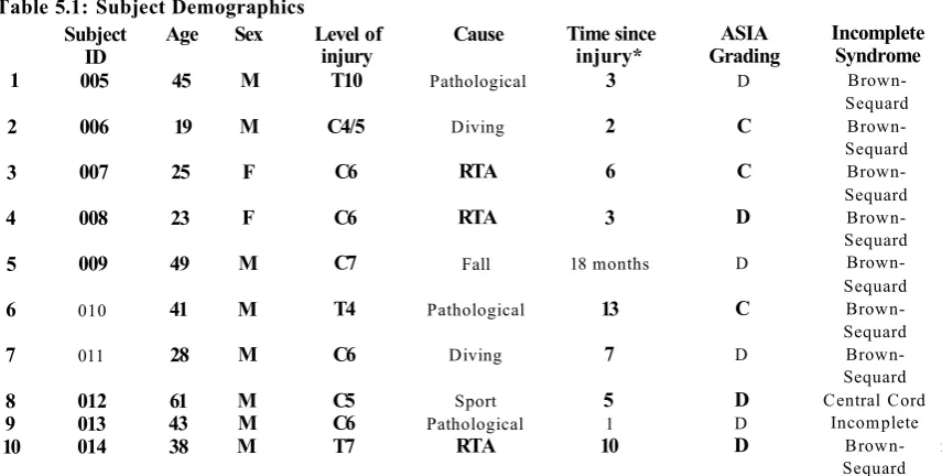
5.1 Subjects for this study...88
5.2 FES and Spasticity...89
5.2. a The Modified Ashworth Scale...89
5.2.a.i.MAS results for Subject 005...89
5.2.a.ii Summary o f MAS results for all subjects...91
5.2.b Isokinetic Dynamometry...92
5.3 FES and Gait...95
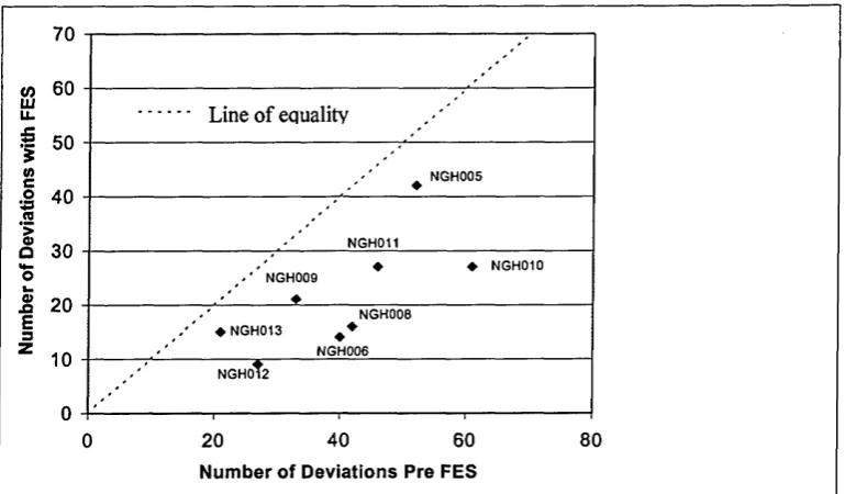
5.3. a Rancho Los Amigos Observational Gait Analysis...95
5.3.a.i. Results from Inter-observer testing o f OGA D ata...96
5.3.b TELER Gait Indicators...98
5.3.b.i Analysis o f TELER data: Subject 005... 98
i aDie or uonienis
5.3.b.iii TELER Data: group analysis...101
5.4 Chapter Summary...103
Chapter 6 Discussion...105
6.1 Thesis development...105
6.2 Individual results by subject...106
6.2.a.i Subject 005...106
6.2.a.ii Subject 006....107
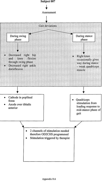
6.2.a. iii Subject 007...108
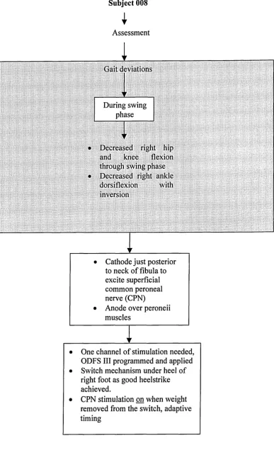
6.2.a.iv Subject 008...109
6.2.a.v Subject 009...110
6.2.a.vi Subject 010...110
6.2.a.vii Subject O il...I l l 6.2.a.viii Subject 012...112
6.2. a. ix Subject 013...113
6.2.a.x Subject 014...114
6.2. b The Modified Ashworth Scale - general observations...114
6.2.c TELER Indicators - general observations...115
6.2.d Isokinetic dynamometry...117
6.3 Results for subjects as a group...118
6.3. a Observational gait analysis...118
6.3.b TELER Gait Indicators...120
6.3.c Group versus individual analysis...120
6.4 Clinical significance...120
6.5 Valid and reliable measurement o f spasticity...121
6.6 Comparison o f results with published literature...122
6.7 Limitations to this study...126
6.8 Conclusions...127
Appendices
Appendix I - The TELER® Gait Indicators...Appendix 1.1-6 Appendix I I - Treatment aims and interventions by subject. Appendix II. 1-18
Appendix III-M odified Ashworth Scale data analysis...Appendix III. 1-16 Appendix IV - Isokinetic torque data graphs Appendix IV. 1-7
Appendix V — Summary o f torque range data. Appendix V. 1-2
Appendix VI—Rancho Los Amigos OGA data collection form s Appendix VI. 1-13
List of Figures
2.1 Causative Factors...8
2.2 Mechanisms o f Injury...8
2.3 Age at Injury...8
2.4 Gender...8
2.5 Incomplete syndromes...10
2.6 ASIA Grades on admission to PRSIU....9
2.7 The effects o f electrical stimulation...18
2.8. a The la inhibitory interneurone...25
2.8.b The Renshaw cell...25
3.1 The Kin-Com isokinetic dynamometer...53
3.2 Observational gait analysis data collection form...58
3.3 Format o f a Component Indicator...64
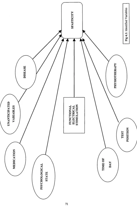
4.1 Identified variables...71
4.2 Data collection sheet MAS...78
4.3 Decision tree for FES strategy...83
4.4 Odstock 2-channel stimulator...84
4.5 CREST stimulator...84
5.1 Example o f torque data: Subject 005- graphed torque data for 3 consecutive test sessions pre-FES at 60°/s...93
5.2. a Torque data details: subject 005 607s...94
5.2.b Torque data details: subject 005 1207s...94
5.3 OGA data pre- and post-FES intervention for all subjects...95
5.4. a Observed gait data from Assessors 1 and 2: pre-FES....97
List o f Tables
1.1 Hierarchy o f strength o f evidence...5
2.1 The ASIA impairment scale...9
2.2 The features o f the upper motor neurone syndrome...22
2.3 Examples o f outcome measures used in spasticity management...30
2.4 Schematic illustration o f paradigmatic approach to quantitative measurement o f spasticity ...35
3.1 Modified Ashworth Scale for grading spasticity...49
3.2 The Ashworth Scale...49
4.1 Summary o f subject attendance and intervention...75
5.1 Subject demographics...88
5.2 MAS data by grade pre- and post intervention...89
5.3 Number and nature o f change episodes post-FES intervention...90
5.4 Subject 005: MAS grades above and below the pooled median...91
5.5 Summary o f descriptive and statistical analysis o f MAS data for all subjects...91
5.6 Summary o f visual analysis o f mean torque data by subject...94
5.7 Paired t-test: data analysis...96
5.8 Spearman's rank correlation coefficient: data analysis...97
5.9 Subject 005: TELER grades pre and post-intervention...99
5.10 Worksheet for TELER index for subject 005...100
5.11 Summary o f TELER data analysis for all subjects...101
5.12 TELER codes pre- and post-intervention for subjects as a group...101
5.13 Calculation o f Chi2 expected values for subjects as a group...102
5.14 Chi2 analysis o f TELER data for subject group...103
CHAPTER 1: INTRODUCTION
1.1 Background to this study
The purpose of this study was to investigate the use of functional electrical stimulation applied to subjects with incomplete spinal cord injury to improve their walking ability. The effects of this treatment upon spasticity and gait were considered.
Spinal cord injury (SCI) is seen as a result of trauma or pathological damage to the spinal cord. There are an approximated 600-900 new injuries in this country every year. The majority of injuries treated in Spinal Injury Units are due to trauma, the age group often young. SCI results in a very substantial degree of disability for the individual, having a devastating impact upon all areas of their lifestyle. Damage to the spinal cord may result in a complete or incomplete injury. Incomplete injuries (ISCI) have some degree of either motor or sensory sparing below the level of damage to the cord.
Functional electrical stimulation (FES) has been used as a treatment modality for subjects with neurological conditions, including spinal cord injury, since Liberson’s work in the early 1960’s. Kidd (1992) defined FES as:
“A form o f electrical stimulation that will cause a muscle to generate a force adequate to perform an artificial function expected o f it ”
Electrical stimulation can also be used for pain relief, muscle training and diagnostic nerve testing. For the purposes of this study FES was cutaneously applied to the lower limbs to enhance gait in the chosen subject population. Whilst most literature relating to the subject suggests that FES is likely to decrease spasticity, there are some articles that report increases in spasticity.
time attempting to decrease and control its effects. Any intervention that may be considered as increasing spasticity is likely to be discarded as a viable treatment option. With spasticity as one variable under consideration, its valid and reliable measurement was seen as the key to the overall validity of the study. Definitions of spasticity and tools for its measurement are varied and disparate. Spasticity is often used not as an exclusive term, but rather as an umbrella term for the features of the upper motor neurone syndrome (a complex collection of pathological sequelae to CNS damage). To avoid confusion and aid clarity in this thesis Lance’s (1980) definition of spasticity was used:
‘Spasticity is a motor disorder characterised by a velocity dependent increase in tonic stretch reflexes ( ‘muscle tone’) with exaggerated tendon jerks, resulting from hyperexcitability o f the stretch reflex, as one component o f the upper motor
neurone syndrome ’
The research study for this thesis ran in conjunction with a three-year European Union funded study titled ‘Clinical Rehabilitation using Electrical Stimulation via Telematics’ (CREST). This project ran from 1997-2000 and investigated both the use of FES for gait enhancement in spinal cord injury and the exchange of computerised clinical information between treatment centres. The Princess Royal Spinal Injuries Unit at the Northern General Hospital Trust in Sheffield was one of five European Centres, specialising in the treatment of spinal cord injuries, involved in the CREST study. The ten CREST study subjects chosen for Sheffield’s part in the project were used for this authors study. The chosen methodology of the CREST study was that of repeated single case studies. A large variety of parameters were measured for the CREST project - for example - muscle strength, spasticity, gait and disability status. The Modified Ashworth Scale and Rancho Los Amigos Observational Gait Analysis System specified in the CREST study were therefore two of the measures used in this authors study. Due to the perceived issues with these tests further measures of spasticity and gait were chosen. Isokinetic dynamometry was used to quantify spasticity during passive movement of the knee joint. TELER® Normal Gait Indicators were developed as an observational gait analysis tool.
the literature, the potential place for the findings of this study within that evidence-base and the implications for clinical practice. In 1993 the Department of Health’s Research and Development Strategy for the National Health Service stated that its main aim was ‘to see that research became an integral part of healthcare’. This was so that practitioners, managers and other staff found it natural to rely on the results of research in their day-to-day decision-making and long term strategic planning. It went on to comment that there remained an issue where ‘belief-based views’ rather than relevant knowledge from reliable sources still had a major effect upon the provision of healthcare. The clearly stated intent in this document was that research-based evidence should become an integral part of healthcare provision. There have been many debates in the literature since the publication of this research report regarding the benefits or otherwise of evidence-based practice. Detractors fear that it will negatively affect clinical autonomy, that treatment would become an oversimplified set of guidelines and that clinicians would be reduced to mere technicians. Other authors see this as the chance to bring research-based practice more formally into the working lives of therapists and to enhance patient care.
In her article from 1997, Newham took the view that research is of vital importance to direct treatment. However, little attention appears to be given to the experience of the clinician in the process of treatment. Other authors, whilst promoting the need for
evidence-based practice (EBP), emphasise the role of the clinician. Sackett et al(1996)
defined evidence-based medicine as ‘ the conscientious, explicit and judicious use o f current best evidence in making decisions about the care o f individual patients The authors went on to discuss the importance of the expert clinician in the interpretation of research data and the appropriate application or rejection of it dependent upon the individual patient’s situation.
The research strategy of the Chartered Society of Physiotherapy (Chartered Society of Physiotherapy, 1995) stated that research is an integral and essential aspect of physiotherapy. Many authors concur with this statement (Moore, 1997, Bury & Mead,
day-to-day clinical practice, Sumsion (1997) echoes Sackett et a ls’ (1996) emphasis on the importance of the patient in this process. Due to increased levels of awareness, patients are less and less the passive recipients of clinicians perceived ‘best practice’. Any intervention must be appropriate to their needs.
Evidence-based practice should ensure the use of research to optimise patient care appropriate to the individual. For research to be directed towards improving care, clinicians and academics must collaborate to benefit from each other’s specialist knowledge and to ensure dissemination of research findings. However, Bannigan & Bryar’s (2002) recent review of research utilization, the final stage of the evidence- based practice process, discovered that allied health professionals in clinical practice seldom use research findings. A number of possible reasons were given for this, conflicting reports and lack of clarity of implications for practice in the literature being two stated possibilities. There remains a gap between research and clinical practice. The pinnacle of research is often seen as the randomised controlled trial. The results from such research may be of limited use to the therapist working in a clinical setting and dealing on a daily basis with a host of enmeshed, possibly confounding issues. By its very nature the randomised controlled trial gives information on the ‘average’ in a very sanitised setting. Within a clinical neurorehabilitative setting treatment is not ‘by rote’ or ‘applied’, but rather an active process of informed negotiation that directs treatment towards the individual’s needs and particular problems.
1.2 Aims and Objectives
necessitated a change in direction as a natural development of the study. The reviewed objectives were therefore to:
1. Investigate the effects of FES upon spasticity in the individual with incomplete spinal cord injury
2. Investigate the effects of FES upon the walking abilities of the individual with incomplete spinal cord injury
Consideration was also given to the theoretical links between spasticity and walking ability.
As the purpose of this project was to evaluate the effects of FES in the individual, the chosen methodology was that of a single subject experimental design (SSED). Measurements were taken at initial and final baselines (AB design). Ten subjects were initially recruited, giving a multiple baseline design across subjects (Ottenbacher, 1986). Whilst the chosen methodology may not meet the upper echelons of the hierarchy of research evidence as recognised by the Chartered Society of Physiotherapy (table 1.1), it is hoped that the strength of the SSED in answering clinical questions for the individual will be demonstrated.
Table 1.1: Hierarchy of strength of evidence (Ref. Moore, 1995)
I Strong evidence from at least one systematic review o f multiple well-designed randomised control trials
II Strong evidence from at least one properly designed randomised control trial o f appropriate size
III Evidence from well-designed trials without randomisation, single group pre-post, cohort, time series or matched case-controlled studies
IV Evidence from well-designed non-experimental studies from more than one centre or research group
V Opinions o f respected authorities, based on clinical evidence, descriptive studies or reports of expert committees
1.3 Summary
CHAPTER 2: LITERATURE REVIEW
The intent of this study was to investigate the effects of functional electrical stimulation (FES) upon spasticity. In this instance FES was applied to improve the gait of individuals with incomplete spinal cord injury, so the effect of spasticity change upon gait was also to be considered. This chapter presents a review of the literature relating to spinal cord injury (SCI), FES, spasticity and gait, pulling together links between all four topics.
The aetiology and incidence of spinal cord injury are introduced, along with the resultant effects of the pathology upon the individual. The increasing population sustaining, and living with, incomplete spinal cord injury is also presented, as this was the subject group under study.
The clinical uses and physiological effects of FES as a treatment for neurological conditions are explored. The final sections of this chapter consider the literature relating FES to spasticity and to gait, and to the links between spasticity and gait.
The section relating to spasticity is substantial. Initial reading on the subject of spasticity showed that the subject is hugely complex and poorly understood. Given that this was one of the phenomena to be measured the author considered that a comprehensive understanding of the neuropathology of spasticity was necessary both to be able to chose appropriate outcome measures and to be able to consider the possible effect of FES upon this impairment.
2.1 Spinal Cord Injury
2. l.a A n Overview:
Spinal cord injury (SCI) occurs due to traumatic or pathological damage to the spinal
cord or cauda equina. It may result in a loss of motor, sensory or autonomic function, or
In the UK, patients with SCI are managed in one of the ten Spinal Injury Units scattered across the country. Sir Ludwig Guttman, a neurologist, set up the first Spinal Injuries Unit at Stoke Mandeville Hospital in Aylesbury in 1944 for the treatment of ex- servicemen. He prescribed systems for the management and prevention of the major complications of SCI and emphasised the importance of specialist nursing and therapy management of such injuries. He strongly advocated that these patients should be managed within specialist units. Other units in the UK, the United States and Australia soon opened. This approach to the management of SCI brought about huge positive change in the life and health expectancy of those sustaining spinal cord damage (Bedbrook, 1985).
2.1. b A etiology and Incidence:
The incidence of SCI in this country is 10-15 cases per million of the population
(Grundy et al, 1986). This means that there are approximately 600-900 new injuries per
annum: 80% of these injuries are due to trauma; 20% to pathology. The main cause of traumatic injury is road traffic accidents (up to 50%). Industrial accidents, falls at home, sport, assaults and self-harm are some of the other main causes. There are two peak age ranges: 16-30 year olds and the 60-plus age group. The male to female ratio is 4:1.
Cervical spine injuries result in tetra/quadriplegia - a paralysis that involves all four limbs. Neck injuries account for up to 50% of all Spinal Injury Unit admissions. Injuries to the thoracic, lumbar or sacral spine result in paraplegia - a paralysis that affects lower limbs and trunk to a greater or lesser degree. Level of injury is described in terms of the last intact neurological segment, for example, C5 complete (an injury complete below the 5th cervical level) and T9 incomplete (an injury with some degree of sparing below the 9th thoracic level).
Zejdlik (1992) reports spinal cord injury statistics for the USA similar to those in the UK. The main difference seen in the States is the percentage of injuries due to gunshot wounds and assault in some sectors of the population.
Princess Royal Spinal Injuries Unit
Admission Statistics 1995-2000
D Traum atic Pathological a P o st Surgical
Figure 2.1: Causative Factors
□ RTA Driver
a RTA M/C □ Domestic Fall □ Industrial Fall □ Other
Figure 2.2: Mechanism of Injury
25
20
10
5
0
Figure 2.3: Age at Injury
10 15 20 25 30 35 40 45 50 55 60 65 70 75 80 85 90
Age
Figure 2.4: Gender
(PRSIU), where this project was conducted. The statistics are similar to those for the
UK (Grundy et al, 1986).
2.1. c In co m p lete In ju ries:
An incomplete injury is one in which any combination o f motor, sensory or autonomic function remains intact below the level of the lesion. There are a number o f ways of defining incomplete injuries - either by describing the pattern o f paralysis following injury (Figure 2.5) or by categorising the neurology remaining to the patient such as the
American Spinal Injuries Association Impairment Scale (Maynard et al, 1997).
T ab le 2.1: A S IA Im p airm en t Scale (M aynard et a l 1997) Classification Neurological sparing
A - complete No motor or sensory function is preserved below the level of the lesion B - incomplete Sensory but not motor function is preserved below the neurological level and
includes the sacral segments S4-5
C - incomplete Motor function is preserved below the neurological level, more than half of key muscles below this have a muscle grade less than 3
D - incomplete Motor function is preserved below the neurological level, at least half of key muscles below the level have a muscle grade of 3 or more
E - normal Motor and sensory function is normal
For the purposes o f this study the American Spinal Injuries Association (ASIA) impairment scale definitions (Table 2.1) were used to give a broad definition o f the level o f neurology remaining to the subject. This is a widely recognised scale, which is in frequent use in clinical practice. This scale was used in the CREST project.
° A ■ B
□c
□ d
■ e
A nterior Cord Syndrom e - the anterior part of the cord is damaged due to a flexion injury, producing an anterior dislocation or a compression wedge fracture. Corticospinal and spinothalamic tracts are damaged due to direct trauma and spinal artery compression leads to ischaemia. Clinical picture is of loss of power and reduced pain and temperature sensation below the level of the lesion.
Central Cord Syndrome - typically seen in older patients with cervical spondylosis. Mechanism is a hyperextension injury that compresses the cord between osteophytic vertebral body and intervertebral disc and the ligamentum flavum. Grey matter at the level of the lesion and centrally situated cervical tracts are most damaged. Classic picture is of flaccid weakness of arms with relatively strong but spastic trunk and legs.
Posterior Cord Syndrome - most common in hyperextension injuries where the spinal arch has been damaged. This results in loss of proprioception due to posterior column damage. Patients may have good power and pain and temperature but profound ataxia.
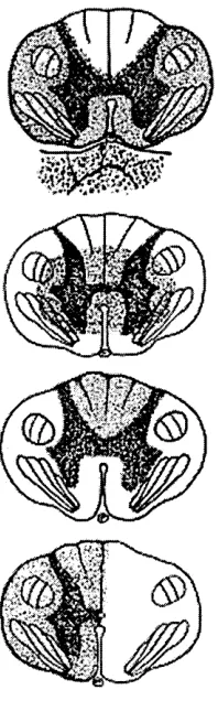
[image:23.612.96.195.105.423.2]Brown-Sequard Syndrome - classically due to stab injuries but also occurs where the lateral mass fractures of the vertebrae have occurred. Signs are those of a hemisection of the cord. Reduced or absent power and proprioception but good pain and temperature on the side of the injury as the spinothalamic tract decussates in the cord. The uninjured side has good power and proprioception but reduced or absent pain and temperature.
Edwards (1991) stated that a considerable percentage of patients with spinal cord injury had incomplete injuries- on average 50% of patients on a SIU at any one time may have some degree of sparing. Unpublished work from the Princess Royal Spinal Injuries Unit (Ash, unpublished) showed that approximately 70% of the admissions to the Unit during the period 1995-2000 were incomplete injuries (Figure 2.6). This work mirrors the findings of the author in a small study over an 18-month period in the early 1990’s where it was again found that over 70% of admissions to the Unit were incomplete.
Incomplete spinal cord injuries can also be divided into upper or lower motor neurone lesions. Upper motor neurone lesions occur where the spinal cord is damaged at or above the level of the first lumbar vertebrae. The spinal cord terminates at this level in the conus medullaris, then cauda equina, which compromises the nerve roots of the lumbar, sacral and coccygeal segments. Damage below T12 therefore results in a lower motor neurone lesion whose clinical presentation is effectively that of a peripheral injury - with a complete loss of spinal reflexes and often profound muscle wasting.
Subjects for this project were all classified as either ASIA C or D lesions. All had injuries above the level of T12.
2.1. d H ealth and Life Expectancy;
An Egyptian physician noted the first recorded case of spinal cord injury in 2500BC when he described the signs and symptoms of a young man with tetraplegia. His final words were ‘...an ailment not to be treated’. This very much remained the popular perception, as the prognosis following injury was so poor. Statistics from World War I showed that 90% of patients suffering a spinal cord injury died within one year. Approximately 1% were still alive 20 years post-injury. The main causes of death were either early complications such as respiratory distress, paralytic ileus or deep venous thrombosis, or ‘any time’ complications such as decubitus ulcers leading to septicaemia or poorly managed bladder and bowel leading to renal failure or bowel complications.
medical science have undoubtedly also added to life expectancy for this patient population.
Bedbrook (1985) emphasised the need for total care in specialist units. It is not just the acute management that is of importance but the provision of a comprehensive rehabilitation package and follow-up, which ensures early identification, and treatment of possible complications. The Princess Royal Spinal Injuries Unit, like many others, provides a ‘cradle to grave’ service. Following discharge from hospital, patients return for annual review. They can also self refer for assessment. Where complications develop - such as pressure sores, urological problems, fractures or long term respiratory problems - patients are managed on the Spinal Unit.
Where septicaemia following pressure sores and urological complications leading to renal failure were once the main causes of mortality, vascular disease and cancers are an increasing cause of death in individuals with spinal cord injury. Life expectancy is now within a few years of the expected average for the rest of the population.
2.1.e Spasticity in the Spinal Cord Injured Subject
Spasticity in spinal cord injury is a topic to which much time and space in literature is devoted. Studies show that approximately 78% of people with spinal cord injury develop some level of spasticity by discharge from hospital. 40% of these subjects report it as ‘problematic’- that is affecting activities of daily living, linked to pain, and not well controlled by medication. Reports of problematic spasticity correlate with level of injury (cervical and upper thoracic injuries) and degree of incompleteness (ASIA C and D categories). All the subjects for this study are categorised as ASIA C or D injuries. There is therefore a substantial possibility of these candidates already having a ‘problematic’ level of spasticity. This is of importance as it is these categories of subjects, who have some ability or potential to walk, who are subjects for this study.
A small number of studies have been published which consider the epidemology of
spasticity. Maynard et al (1990) published the results of two studies they had
subjects’ first annual follow-up. The scale used was an ordinal one, where no spasticity was graded as 0 and ‘problematic’ spasticity (that which had been treated with unsatisfactory results) was graded as 3. The assessing physician graded spasticity. The second study followed the same basis as above. 466 patients with traumatic spinal cord injury were assessed. Both sets of results were collated by level of injury and by Frankel grading for classification of degree of incompleteness. Study 1 found that 67% of subjects developed spasticity by discharge from hospital with 37% needing medication by that time. By follow up 78% presented with spasticity with 49% medicated. Study 2 found that 26% of subjects were medicated by discharge and 46% by follow up. The most frequently medicated subjects fell into Frankel categories C and D with 50% and 52% respectively being treated. The ASIA classification system is based upon that developed by Frankel, C and D grades from each system are very similar.
Skold (1999) gained similar results. This author studied 354 SCI subjects and found that 65% of subjects reported developing spasticity, 43% of who said their spasticity was problematic. Skold’s operational definition of spasticity was as above but included pathological radiation of reflexes. There were significant links between reports of problematic spasticity and incomplete injuries. As well as subjects’ self reports on spasticity levels, assessment was undertaken using the Modified Ashworth Scale (MAS) (Bohannon & Smith, 1987). Only 60% of the subjects reporting it were found to have elicitable spasticity on assessment.
Johnson et al (1998) again corroborated the above findings. They undertook initial assessments then telephone questionnaire follow-ups over a five-year period and found that approximately 30% of subjects reported spasticity as problematic.
Maynard et al (1990) identified some of the main issues with their studies: the
subjectivity of the measurement scale, the discrepancy between the two sets of subjects being medicated by discharge (37% and 26%). They believed that this could have been due to different criteria used for indications for treating spasticity. Skold (1999) suggested that the reason for the discrepancy between self-reported and clinician elicited spasticity could be that the MAS may only measure one aspect of spasticity.
understandings - operationalisation - of what spasticity is between groups of clinicians and subjects may also be expected to cause differences in results. However, in spite of these issues these three independent studies have produced remarkable similar findings as to the epidemiology of ‘spasticity’ within the spinal cord injured population. The accurate definition of spasticity and its valid measurement are key issues to this thesis.
2 .1 .f Section Sum m ary
SCI affects a very small but significant percentage of the population each year. Life expectancy is now almost normal, and there is a growing population of people living with SCI. The relative percentage of incomplete injuries - who may have the potential to walk - is increasing. Spasticity is significantly problematic following injury, in particular amongst those with incomplete injuries, adding to disability levels. The main group affected by SCI is the young. The percentage returning to work is low. As Trieschmann (1986) states SCI is a ‘low-incidence but high-cost disability that makes tremendous changes in a person’s lifestyle’. Treatments, such as FES, which may have a beneficial impact upon levels of handicap and disability need to be fully explored and, if appropriate, considered as possible treatment options.
2.2 Functional Electrical Stimulation
Early experiments by Swammerdam and Galvani in the 17th and 18th centuries respectively demonstrated that the electrical excitation of nerve caused muscle to contract. The knowledge needed to evolve neural prostheses of a size small enough to be practical in a clinical setting became available with the development of the transistor in the 1950s. Since that time there has been a substantial development in the technology needed to produce function via electrical stimulation. FES has found its way into everyday clinical practice in the form of pacemakers, bladder stimulators and phrenic nerve stimulators. Other applications have included its use in the facilitation of standing and gait.
FES has been used in the treatment of many neurological conditions. Burridge et al
(1997a) reported on their work with stroke patients. Bajd et al (1999) and Granat et al
head injury and multiple sclerosis (e.g. Dimitrijevic & Dimitrijevic, 1992). Any pathology that results in motor loss due to damage to the upper motor neurone is potentially responsive to FES.
Electrical stimulation has been used for many years in the treatment of SCI. The literature records a wide variety of possible uses in both upper and lower limb rehabilitation. In the 1980’s a group of bio-engineers based in Ljubljana, Slovenia, produced a large body of work relating to the use of electrical stimulation, particularly for SCI. The therapeutic effects of electrical stimulation and its use in the management of spasticity have also been investigated by authors such as Vodovnik (1981).
FES is now used in both research centres and specialist clinical centres. Its more general use in the clinical field however is still limited. The potential demand for FES as a treatment option and possible reasons for its limited use in clinical practice shall be considered in the following section.
2.2. a D em and fo r Functional E lectrical Stim ulation in Spinal C ord Injury
Patient demand for FES systems has been assessed in two studies. Jaeger et al (1990) undertook a retrospective study of medical records for 192 patients with spinal cord injury. They estimated a minimum of 10.4% and a maximum of 25% (dependent upon relaxation of some inclusion criteria) of their subjects as possible FES users. However, they were considering FES for complete injuries from T4-T12 spinal levels. These authors also estimated their subject group to make up 45% of the SCI population. The above study was therefore of limited use to this thesis where the aim was to enhance gait in incomplete subjects who already had some walking ability. The percentage of incomplete spinal cord injury (approximately 70%) apparent in recent studies may also make the above study less relevant in the present day. As part of the CREST project, Maxwell et al (1999) published the results of a questionnaire survey looking at the potential demand for FES devices. Whilst 19% of respondents stated that they had used functional electrical stimulation, only 2% had done so for walking. 16% of respondents stated that they could walk independently but less than half of these could do so for more than 50 metres. 13% had used some form of orthosis. Whilst this article was slightly confusing in its data presentation and should have included a section on level of
reasonable to conclude that only a tiny percentage of potential users were using FES systems for gait enhancement. The sample group were self selected members of the Spinal Injuries Association.
In spite of the apparent potential population who could benefit from FES and the increasing sophistication of commercially available systems this is not a treatment option that is often chosen in clinical practice. In this author’s opinion the following reasons may add to the general reluctance of physiotherapists to use such systems: • Lack of underpinning knowledge in and the potential uses of FES
• Few reliable, flexible systems available for purchase
• A generally held belief that FES increases spasticity - ‘in clinical practice there even exists a widespread attitude that one should not stimulate an already ‘overstimulated’ spastic muscle’ (Stefanovska et al, 1989)
Whatever the actual underlying issues, FES is infrequently used in the clinical workplace. Given the emphasis put upon controlling and decreasing spasticity in the clinical management of neurologically damaged subjects, the final point may well be enough to prevent physiotherapists exploring this as an option for treatment.
2.2.b Physiological effects o f FES:
The FES systems used for this project make muscle contract via stimulation of nervous tissue. Whilst it is possible to stimulate denervated muscle, this is not practical in the clinical setting at present because the amplitude of current or stimulus pulse width needs to be far greater than that which will stimulate muscle with an intact nerve supply. Electrical stimulation can be applied via surface or implanted electrodes. There are benefits and potential risks with both options. This study used surface applied electrodes. This is a non-invasive technique that is relatively simple to apply.
to the nerve temporal and/or spatial summation of action potentials occur and the muscle is caused to contract. Stimulation will travel both proximally and distally from the point of stimulation. Muscle contraction can therefore be directly affected by the motor efferent or indirectly by a reflex response to the stimulation.
Electrical stimulation initially recruits large diameter axons as they offer lower impedance to current (Baker, 1993). Smaller diameter axons are likely to be the last recruited into activity. This is the opposite pattern of recruitment to that seen in normal physiological muscle contraction. Electrical stimulation is not specific when applied to peripheral, mixed nerve. All types of nerve fibres will potentially be excited. As well as alpha motor neurones, group la and II afferents from intrafusal muscle fibres, lb afferents from golgi tendon organs, sensory, cutaneous and sympathetic nerve fibres are all likely to be stimulated. The effect of electrical stimulation is therefore complex.
Figure 2.7 summarises the potential effects of electrical stimulation (Vodovnik & Stefanovska, 1992). The fourth effect of electrical stimulation ‘change in cell membrane transport’ relates to trophic systems within nerve and muscle and is not addressed by this thesis. Restorative functional stimulation is also known as therapeutic stimulation.
The potential effects of FES can be considered in two timescales. In the short-term, FES will produce a contraction of muscle and potentially functional movement. This is often described as orthotic stimulation. With continued use over time, many other therapeutic physiological effects have been reported
FES can address the loss or negative aspects of upper motor neurone damage seen in SCI - that is fatigue, weakness and atrophy (Dimitrijevic & Dimitrijevic, 1992). SCI subjects demonstrate a decreased ability to produce smooth, efficient contraction of muscle and they fatigue quickly. Relatively smaller percentages of fatigue-resistant motor units are present in the muscles of SCI subjects than in the ‘normal’ population. Disturbances in motor unit recruitment and modulation of anterior horn cell firing rates have also been shown (Heckman, 1994). Reported benefits with the use of FES have included changes in muscle phenotype from fast-fatiguable to fatigue-resistant fibres (e.g. Pette & Vrbova, 1999,Gordon & Mao, 1994), increases in muscle bulk (e.g. Mohr
Ef fe re nt In flo w CD r "
— — 2
g £ t$ g i |
I g? i s
**- . 2 o ci ♦ ;
£o 5- i E2 ™ I ° §
# .i
3
O W . *
c >» ~ <D O TO O ‘J= as c w o ro ro
■o _ T3
. l l l §i- X2 T3 J S ^ rS3 C Cl!
E
(D
> — C
"m 2 °ro c 4=
a3 .2 J 5
c o 3 8 > § i
CD
O C
E O JS o ■5"CD Jt=
5= T3
' £
.2 ^
m
CD C O O 1 oI— '§ ) 'ro. 2 o 3 CD o n= z ■(0 TD >» O
■ S .S
(0 c CD c '■ e o CD 4—» CD Q . o O « = Q . T 3 CD O 3 2 toCO
+ 4
c CD o o o (0 CD 3 i_-*-<
C O O c S ifc <
o g -e
.£ E §.
® -i c g® 2 js s » -o CM CT> (J) CD co > o c .CD C/3 OS ‘c >o •Oo > c o E CO Id o 'u O _cd HI •4—O <0 o LU CD tv. CM 2 3 O) c
CD .2 •E l _CD
O 3
As well as the negative features that are related to a loss of motor activity, FES has been reported as having an effect on the ‘motor overactivities’ (see table 2.2) that follow
central nervous system damage. Evidence of both increases (e.g. Robinson et al, 1988)
and decreases (e.g. Burridge et al 1997b, Stefanovska et al, 1989) in spasticity have been reported. Daly et al (1996) described the use of electrical stimulation as a tool for neuroplastic change and motor ‘learning’ at cord level.
2.2.c Section Sum m ary
FES has been used as a therapeutic tool for many years in the treatment of a variety of neurological conditions. It has not, however, come into widespread use in clinical practice. This may be for a variety of reasons, one of which may be a lack of understanding of its complex neurophysiological effects. The above review would suggest that FES is capable of improving gait by both its immediate orthotic effect of producing movement by making muscle contract and in applying a ‘training effect’ - improving muscle bulk, phenotype and blood supply. It may also be that FES can address the effects of spasticity at a synaptic level. To understand the possible effects of FES upon spasticity an understanding of its underlying neurophysiology - what spasticity is, how it develops and how the central nervous system attempts to compensate for it - is necessary.
2.3 Spasticity
2.3.a Definition:
Spasticity is a much discussed phenomenon in the field of neuroscience and rehabilitation literature. It occurs following damage to the upper motor neurone within the brain or spinal cord. Sheean (1998) described spasticity as hypertonia that shows as an increased resistance to movement. In spite of its being a common sequela to neurological disease, and its being recognised by many practitioners in the field as a substantially disabling one, it is poorly defined in the literature. According to Young
(1994) this is ‘presumably because the neurobiology o f the motor system remains
The term spasticity is frequently used as a generic one to describe an increase in muscle tone or other tonal abnormalities in individuals with central nervous system (CNS) damage. This confusion does not assist in the definition of the impairment. Clarity of definition is of vital importance in the recognition and measurement of spasticity so that clinicians can direct treatment appropriately (Sheean, 1998). From a research viewpoint definition is important so that the validity of measurement tools can be ensured. If validity is considered in its broadest sense as ‘the extent to which an item actually measures what the researcher purports it to measure’ (Krebs, 1987) then the importance of definition becomes apparent. Accurate measurement depends, in part, upon a clear definition of the dimension in question. If this is clearly defined appropriate measurement tools can be chosen. Much of the literature relating to spasticity is unclear in its definition and therefore hazy in its use of measurement tools and outcomes.
To clarify this issue and for the purposes of this project Lance’s (1980) definition of spasticity will be used:
‘Spasticity is a motor disorder characterised by a velocity dependent increase in tonic stretch reflexes ( ‘muscle tone’) with exaggerated tendon jerks, resulting from hyperexcitability of the stretch reflex, as one component of the upper motor neurone syndrome ’
This definition, although under some debate, is still the most commonly referred to in the literature relating to spasticity.
Given the extremely complex nature of spasticity, the often apparently confused use of the term in both clinical practice and research literature, and its importance to this study as the impairment being investigated, a deeper knowledge and understanding of this impairment was necessary to ensure valid measurement and therefore study validity.
2.3.b Spasticity as p a rt o f the Upper M otor Neurone Syndrom e:
The upper motor neurone syndrome (UMNS) is a useful but non-specific concept which describes a number of phenomena seen when the upper motor neurone is damaged. Upper motor neurones (UMN) are motor neurones in any long, descending tract that control or influence movement and muscle tone. They have a variably direct influence upon the excitability of the lower motor neurone (LMN) or anterior horn cell. The LMN has an enormous dendritic tree with a large surface area covered with thousands of synaptic boutons. The main role of these neurones is to ‘translate the huge variety of input from afferent and descending fibres and from interneurones into an output that will precisely control the contraction of muscle fibres in the development of force or patterns of movement’ (Burke 1990). The influence of UMN’s is effected by either direct synapse onto the LMN or, more commonly, via a network of interneurones. They modulate important segmental motor reflex activity in the spinal cord. The majority of the positive phenomena (Table 2.2) of the UMNS are due to interruption of supraspinal control of these reflexes (Sheean, 1998).
From the breakdown shown in Table 2.2, it can be seen that spasticity is a positive feature of the UMNS. The other positive features, which result in motor overactivities, are frequently mislabelled ‘spasticity’. Whilst they tend to occur together, there are clear differences in the pathophysiology of many of these phenomena. It is beyond the scope of this project to describe these differences. Sheean, in ‘Spasticity Rehabilitation’ (1998), gives a good overview. The use of the terms ‘positive’ and ‘negative’ is unfortunately misleading. Both terms relate to abnormal pathological changes, positive to excessive activity, negative to a resultant lack of activity. A positive change in this context is not beneficial.
therefore appropriate, efficient functional movement. Effectively a ‘disuse’ atrophy of musculature is seen with attendant soft tissue change.
Table 2.2: Features of the upper motor neurone syndrome (Sheean 1998)
Ne g a t iv e
Acute Hypotonia (Shock)
Weakness due to inadequate muscle activation Loss o f dexterity
Loss o f cutaneous reflexes Fatiguability
Po s i t iv e
At rest in response to peripheral stimulation
Proprioceptive
Increased tendon reflexes with radiation Clonus
Spasticity
During movement (spastic dystonias)
Dyssynergic patterns o f co-contraction Associated reactions
Flexor withdrawal reflexes Positive support reaction Extensor thrust
‘Pushing’ reactions
The next section shall consider the pathophysiological changes that are believed to lead to spasticity.
2.3. c Pathophysiology o f spasticity
Motor systems have three levels of control: the spinal cord, the brainstem and the brain. The spinal cord is considered as the lowest in the hierarchy. These systems are organised serially and in parallel. Spinal segmental mechanisms have an important role to play in the production and control of movement. They are modulated by both proprioceptive and higher centre input through a complex system of interneurones. (Kandel, Schwartz & Jessell, 2000). In the case of CNS damage the normal balance of these mechanisms is lost, spasticity being one of the possible results from this damage.
There are many descending tracts within the brainstem and spinal cord. Those believed to be particularly involved with the development of spasticity and other features of the UMNS are the dorsal reticulospinal tract (inhibitory to spinal stretch reflexes) and the ventral reticulospinal and vestibulospinal tracts (mainly excitatory). These tracts have an effect on the lower motor neurone (LMN) or anterior horn cell via a complex system
Nociceptive
of interneurones. It is the balance between these descending tracts which controls the spinal stretch reflexes and flexor and extensor reflexes. Loss or partial loss results in an imbalance of this normal control mechanism.
Sheean (1998) states that spasticity is believed to result from an enhanced tonic stretch reflex (TSR). The TSR is a polysynaptic reflex which occurs in response to a stretch of a relatively long duration (when compared to the phasic or ‘tendon jerk’ reflex). In the normal neurological system, the excitability of this reflex is dependent upon the balance of higher centre input as described above. It is believed that should a movement need to be adjusted the stretch reflex can be enhanced or diminished, as the situation requires to fine-tune activity (Davidoff, 1992). In spasticity, the individual may be unable to control the reflex which then responds to stretch whether it is relevant or not (Carr & Shepherd, 1998). The tonic stretch reflex can therefore be considered as having an important role to play in functional activity.
There are three key elements likely to affect the sensitivity of TSR pathway:
1. The muscle spindle sensitivity
2. Intrinsic excitability of the alpha motorneuron
3. Interneuronal processing of the la afferent information within the spinal cord
Where it was previously believed that the muscle spindle caused the increase in muscle tone due to enhanced fusimotor drive, more recent work has shown that there is no increase in this. It has been shown that it is the processing of la afferent information that is enhanced (Sheean 1998).
2.3.c.i Changes in neural inhibitory m echanism s follow in g neurological injury
Much has been written about the spinal mechanisms controlling stretch reflex
excitability. Kandel, Schwartz & Jessell (2000) stated that inhibitory interneurones played important roles in the co-ordination of reflex activity and therefore normal control of movement. Loss of such inhibition is believed to lead to the development of spasticity and other positive, or overactivity, features (see table 2.2) of the UMNS. This section considers the effects that damage to the CNS has upon the function of specific interneurones.
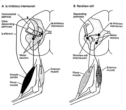
Although many complex and poorly understood mechanisms have been implicated (see section 2.3.a), current literature concentrates particularly upon the activity of the la inhibitory interneurone. This mechanism is involved with the control of reciprocal inhibition of muscle agonist-antagonist activity and therefore in the degree of co contraction around joints during movement.
Ia fibres from muscle spindles excite motor nerves from that muscle and also those with a similar action. At the same time Ia fibres inhibit antagonistic muscles via Ia inhibitory interneurones (Figure 2.8A). This is the basis for reciprocal inhibition. This system is of importance to the production of controlled voluntary movement. Inhibition can be increased or decreased to allow freer movement (increased reciprocal inhibition) or to increase stability (co-contraction or co-activation) at a joint. Ia inhibitory interneurones also receive both inhibitory and excitatory information from descending pathways.
Studies investigating the activity of the Ia inhibitory intemeurone have shown (Morita,
A la inhibitory interneuron B Renshawcetl
Corticospinal pathway Other descending pathways
ia afferent
-fa inhibitory interneuron
Motor neurons
L
Muscle
Flexor muscle
y Extensor muscle
[image:38.612.111.518.106.460.2]o
Figure 2.8: Spinal inhibitory interneurons A - la inhibitory interneuron
They found that, compared to their control group of normal subjects, the subjects with ISCI demonstrated impaired reciprocal inhibition and a related large degree of co activation of soleus during dorsiflexion. This effect was particularly active during fast, alternating ankle movement, such as those seen during walking.
Faist et al (1994) also investigated the effect of changes in Ia presynaptic inhibition upon spasticity development. They found that the subject group with SCI demonstrated a decreased level of presynaptic inhibition. They did not however find a correlation between this and their clinical measure of spasticity - the Ashworth Scale (Ashworth 1964). This led them to deduce that the changes in presynaptic inhibition did not have a substantial effect upon spasticity. This conclusion begs the question as to whether or not the Ashworth Scale can be considered as more of an analogue to true spasticity than electrophysiological testing of interneuronal activity. The discrepancy between research laboratory and clinical measures of spasticity is one which many authors highlight. This is an important issue and will be returned to in the section upon measurement of spasticity later in this chapter.
This section has summarised the role of the Ia inhibitory interneurone in reciprocal inhibition and therefore co-contraction of muscle groups during active movement in normal subjects and those with spasticity. The role of the Renshaw cell and presynaptic inhibition upon Ia intemeuronal functioning has also been considered.
2.3.c.ii Plasticity o f the spinal cord
Immediately following spinal cord injury a period of spinal shock is seen. Hiersemenzel
et al (2000) investigated the development of upper motor neurone syndrome features with patients in the acute stages of spinal cord injury. They undertook a series of clinical and electrophysiological tests. Three phases were described:
Spinal Shock: this phase lasted up to three weeks. It was identified clinically by loss of tendon reflexes and hypotonia.
Transition Phase: this lasted for 3-8 weeks in the group studied. Clinically identified by increasing excitability of tendon tap reflexes, increasing muscle tone and frequency of spasms.
Spastic State: clinically shows as exaggerated tendon taps, increased muscle tone and involuntary muscle contractions.
The authors stated that there was a smooth transition from one state to the next. Chapman & Wiesendanger (1982) saw this period as the time in which plastic adaptation of the nervous system took place.
Where higher centres have long been accepted as being capable of plastic adaptation to injury the spinal cord has not been considered as such (Wolpaw & Tennissen, 2001). However research into the recovery of spinal cord function suggest the presence of four mechanisms active in this process:
• Sprouting • Synaptogenesis
• Restoration of function to uninjured and uncrossed fibres
• Remyelination of demyelinated injured fibres, as well as restoration of the ability to conduct impulses through injured fibres in the absence of myelin (Dimitrijevic, 1988)
activity-dependent and injury-induced plasticity. There was evidence that spinal circuitry undergoes significant and widespread alterations after spinal injury. These changes include an increase in responsiveness and amplitude of both mono- and polysynaptic stretch reflexes. However, not all injury-induced physiological changes are detrimental. These authors believed that it was possible for plastic change to be guided by activity, even in the damaged spinal cord, and that to modify spinal circuitry for a specific task movement performed during rehabilitation should be executed as normally as possible.
Wolpaw and Tennissen’s (2001) article reviewing the plasticity of the spinal cord reiterated the above points. The authors also stated that the spinal cord is capable of activity-dependent plasticity. The guidance of positive plastic change following spinal cord injury is important in maximising function.
There is therefore a growing body of evidence to support the plastic capabilities of the spinal cord. The spinal cord is capable of both neural and synaptic plasticity. Higher centre and peripheral inputs can direct this plasticity. Physiotherapists aim to maximise positive change via mainly peripheral inputs directed towards producing goal-orientated movement in as normal a manner as possible. It may be that the use of electrical stimulation in a functional setting such as used in this study, gait enhancement, may bring about positive plastic change within the spinal cord.
2.3. c. iii Sum m ary o f pathophysiological changes
2.3. d M easurem ent o f spasticity
The complexity of the underlying neurophysiological mechanisms, the gaps in our
knowledge of some of these systems, and the lack of correlation between laboratory and
clinical measures (e.g. Priebe et al 1996, Hiersemenzal et al 2000) make the
measurement of spasticity problematic. However the quantification of this substantially disabling phenomenon is seen as important by those working in research and clinical fields so that efficacy of treatment can be ensured.
Haas (1994) undertook a postal questionnaire in an attempt to survey current clinical practice of healthcare professionals invo