Acta Cryst.(2002). E58, o331±o333 DOI: 10.1107/S1600536802003392 Bond and Davies C9H10
o331
organic papers
Acta Crystallographica Section E Structure Reports Online
ISSN 1600-5368
a
-Methylstyrene
Andrew D. Bond* and John E. Davies
Department of Chemistry, University of Cambridge, Lensfield Road, Cambridge CB2 1EW, England
Correspondence e-mail: [email protected]
Key indicators
Single-crystal X-ray study T= 180 K
Mean(C±C) = 0.005 AÊ Rfactor = 0.085 wRfactor = 0.250
Data-to-parameter ratio = 10.8
For details of how these key indicators were automatically derived from the article, see http://journals.iucr.org/e.
#2002 International Union of Crystallography Printed in Great Britain ± all rights reserved
The crystal structure of -methylstyrene, C9H10, has been determined at 180 (2) K followingin situcrystal growth from the liquid. In space group P21/n, the structure consists of herring-bone-packed layers within which intermolecular CÐ H interactions are evident.
Comment
As part of a study devoted to improving techniques for determining the crystal structures of substances that are liquid at room temperature, we have recently reported the structure of styrene, C8H8(Bond & Davies, 2001; Yasudaet al., 2001). We report here the crystal structure of the-methyl deriva-tive, C9H10, determined at 180 (2) K followingin situ crystal growth from the liquid.
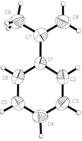
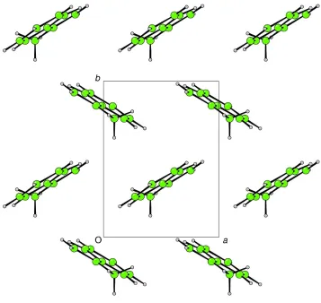
±Methylstyrene, (I), crystallizes in the monoclinic space groupP21/nwith one whole molecule in the asymmetric unit (Fig. 1). The propenyl substituent lies approximately coplanar with the phenyl ring [the angle between the least-squares planes through C1±C6 and C7±C9 is 1.8 (3)]. Molecules of (I) form herring-bone-packed layers parallel to (001) (Fig. 2). These layers may be considered to stack in an ABAB
arrangement (Fig. 3). CÐH interactions are evident between molecules within layers [H5 cent(C7±C8)i= 3.06 AÊ, C5ÐH5 cent(C7±C8)i= 153; H6 cent(C1±C6)i= 3.06 AÊ, C6ÐH6 cent(C1±C6)i = 137; H8B cent(C1±C6)ii = 3.03 AÊ, C8ÐH8B cent(C1±C6)ii= 138; symmetry codes: (i)
ÿ1/2ÿx, 1/2+y, 1/2ÿz; (ii) 1/2ÿx,ÿ1/2+y, 1/2ÿz; cent denotes the centroid of the indicated ring]. A similar arrangement is observed in styrene itself, but in that case, the-hydrogen also acts as a CÐH donor; this interaction is clearly prohibited in (I).
Experimental
The sample (99%) was obtained from the Lancaster company and used without further puri®cation. The crystal was grown in a 0.3 mm glass capillary tube atca247.4 K (a temperature only slightly less than the melting point of the solid in the capillary) using a technique described earlier (Davies & Bond, 2001). Once grown, the crystal was
organic papers
o332
Bond and Davies C9H10 Acta Cryst.(2002). E58, o331±o333cooled to 180 (2) K for data collection. The length of the cylindrical crystal was not estimated, but it exceeded the diameter of the colli-mator (0.35 mm).
Crystal data C9H10
Mr= 118.17
Monoclinic,P21=n
a= 5.795 (1) AÊ
b= 7.829 (1) AÊ
c= 15.820 (1) AÊ
= 93.23 (1)
V= 716.60 (16) AÊ3
Z= 4
Dx= 1.095 Mg mÿ3
MoKradiation Cell parameters from 2680
re¯ections
= 1.0±22.5
= 0.06 mmÿ1
T= 180 (2) K Cylinder, colourless 0.15 mm (radius) Data collection
Nonius KappaCCD diffractometer Thin-slice!and'scans Absorption correction: none 3208 measured re¯ections 895 independent re¯ections 676 re¯ections withI> 2(I)
Rint= 0.089
max= 22.3
h=ÿ5!6
k=ÿ7!8
l=ÿ15!16
Re®nement Re®nement onF2
R[F2> 2(F2)] = 0.085
wR(F2) = 0.250
S= 1.15 895 re¯ections 83 parameters
H-atom parameters constrained
w= 1/[2(F
o2) + (0.1448P)2
+ 0.2267P]
whereP= (Fo2+ 2Fc2)/3
(/)max< 0.001
max= 0.37 e AÊÿ3
min=ÿ0.27 e AÊÿ3
H atoms were placed geometrically and allowed to ride during subsequent re®nement withUiso(H) =xUeq(C) (x= 1.2 for alkene and
phenyl H, and 1.5 for methyl H). The methyl group was allowed to rotate about its local threefold axis. No signi®cant diffracted intensity was observed beyond 22.5in(equivalent to 0.93 AÊ resolution) and the data were truncated at this point; the precision of the result is reduced accordingly.
Data collection:COLLECT(Nonius, 1998); cell re®nement:HKL SCALEPACK(Otwinowski & Minor, 1997); data reduction: HKL DENZO and SCALEPACK (Otwinowski & Minor, 1997); program(s) used to solve structure:SIR-92 (Altomareet al., 1994); program(s) used to re®ne structure:SHELXL97 (Sheldrick, 1997); molecular graphics:XP(Sheldrick, 1993) andCAMERON(Watkinet al., 1996); software used to prepare material for publication: SHELXL97.
We thank the EPSRC for ®nancial assistance towards the purchase of the Nonius CCD diffractometer.
Figure 2
Projection on to (001) of a single layer of (I), showing the herring-bone packing arrangement (CAMERON; Watkinet al., 1996).
Figure 1
The molecular unit in (I), showing displacement ellipsoids at the 50% probability level for non-H atoms (XP; Sheldrick, 1993).
Figure 3
References
Altomare, A., Cascarano, G., Giacovazzo, C., Guagliardi, A., Burla, M. C., Polidori, G. & Camalli, M. (1994).J. Appl. Cryst.27, 435.
Bond, A. D. & Davies, J. E. (2001).Acta Cryst.E57, o1191±o1193. Davies, J. E. & Bond, A. D. (2001).Acta Cryst.E57, o947±o949. Nonius (1998).COLLECT. Nonius BV, Delft, The Netherlands.
Otwinowski, Z. & Minor, W. (1997). Methods in Enzymology, Vol. 276,
Macromolecular Crystallography, Part A, edited by C. W. Carter and R. M. Sweet, pp. 307±326. London: Academic Press.
Sheldrick, G. M. (1993).XP. University of GoÈttingen, Germany. Sheldrick, G. M. (1997).SHELXL97. University of GoÈttingen, Germany. Watkin, D. J., Prout, C. K. & Pearce, L. J. (1996).CAMERON. Chemical
Crystallography Laboratory, University of Oxford, England.
Yasuda, N. Uekusa, H. & Ohashi, Y. (2001).Acta Cryst.E57, o1189±o1190.
Acta Cryst.(2002). E58, o331±o333 Bond and Davies C9H10
o333
supporting information
sup-1
Acta Cryst. (2002). E58, o331–o333supporting information
Acta Cryst. (2002). E58, o331–o333 [doi:10.1107/S1600536802003392]
α
-Methylstyrene
Andrew D. Bond and John E. Davies
S1. Comment
As part of a study devoted to improving techniques for determining the crystal structures of substances that are liquid at
room temperature, we have recently reported the structure of styrene, C8H8 (Bond & Davies, 2001; Yasuda et al., 2001).
We report here the crystal structure of the α-methyl derivative, C9H10, determined at 180 (2) K following in situ crystal
growth from the liquid.
α-Methylstyrene, (I), crystallizes in the monoclinic space group P21/n with one whole molecule in the asymmetric unit
(Fig. 1). The propenyl substituent lies approximately coplanar with the phenyl ring [the angle between the least-squares
planes through C1–C6 and C7–C9 is 1.8 (3)°]. Molecules of (I) form herring-bone packed layers parallel to (001) (Fig.
2). These layers may be considered to stack in an ABAB arrangement (Fig. 3). C—H···π interactions are evident between
molecules within layers [H5···cent(C7–C8)i = 3.06 Å, C5—H5···cent(C7–C8)i = 153°; H6···cent(C1–C6)i = 3.06 Å, C6—
H6···cent(C1–C6)i = 137°; H8B···cent(C1–C6)ii = 3.03 Å, C8—H8B···cent(C1–C6)ii = 138°; symmetry codes: (i) -0.5 - x,
0.5 + y, 0.5 - z; (ii) 0.5 - x, -0.5 + y, 0.5 - z; cent denotes the centroid of the indicated ring]. A similar arrangement is
observed in styrene itself, but in that case, the α-hydrogen also acts as a C—H···π donor; this interaction is clearly
prohibited in (I).
S2. Experimental
The sample (99%) was obtained from the Lancaster company and used without further purification. The crystal was
grown in a 0.3 mm glass capillary tube at ca 247.4 K (a temperature only slightly less than the melting point of the solid
in the capillary) using a technique described earlier (Davies & Bond, 2001). Once grown, the crystal was cooled to
180 (2) K for data collection. The length of the cylindrical crystal was not estimated, but it exceeded the diameter of the
collimator (0.35 mm).
S3. Refinement
H atoms were placed geometrically and allowed to ride during subsequent refinement with Uiso(H) = xUeq(C) (x = 1.2 for
alkene and phenyl H, and 1.5 for methyl H). The methyl group was allowed to rotate about its local threefold axis. No
significant diffracted intensity was observed beyond 22.5° in θ (equivalent to 0.93 Å resolution) and the data were
supporting information
[image:5.610.175.441.69.523.2]sup-2
Acta Cryst. (2002). E58, o331–o333Figure 1
The molecular unit in (I), showing displacement ellipsoids at the 50% probability level for non-H atoms (XP; Sheldrick,
supporting information
[image:6.610.126.483.68.406.2]sup-3
Acta Cryst. (2002). E58, o331–o333Figure 2
Projection on to (001) of a single layer of (I), showing the herring-bone packing arrangement (CAMERON; Watkin et al.,
1996).
Figure 3
[image:6.610.126.484.454.637.2]supporting information
sup-4
Acta Cryst. (2002). E58, o331–o333isopropenylbenzene
Crystal data
C9H10
Mr = 118.17 Monoclinic, P21/n
a = 5.795 (1) Å
b = 7.829 (1) Å
c = 15.820 (1) Å
β = 93.23 (1)°
V = 716.60 (16) Å3
Z = 4
F(000) = 256
Dx = 1.095 Mg m−3
Melting point: 250 K
Mo Kα radiation, λ = 0.7107 Å Cell parameters from 2680 reflections
θ = 1.0–22.5°
µ = 0.06 mm−1
T = 180 K
Cylinder, colourless 0.15 mm (radius)
Data collection
Nonius KappaCCD diffractometer
Radiation source: fine-focus sealed tube Graphite monochromator
Thin–slice ω and φ scans 3208 measured reflections 895 independent reflections
676 reflections with I > 2σ(I)
Rint = 0.089
θmax = 22.3°, θmin = 3.7°
h = −5→6
k = −7→8
l = −15→16
Refinement
Refinement on F2
Least-squares matrix: full
R[F2 > 2σ(F2)] = 0.085
wR(F2) = 0.250
S = 1.15 895 reflections 83 parameters 0 restraints
Primary atom site location: structure-invariant direct methods
Secondary atom site location: difference Fourier map
Hydrogen site location: inferred from neighbouring sites
H-atom parameters constrained
w = 1/[σ2(F
o2) + (0.1448P)2 + 0.2267P]
where P = (Fo2 + 2Fc2)/3
(Δ/σ)max < 0.001
Δρmax = 0.37 e Å−3
Δρmin = −0.27 e Å−3
Special details
Experimental. Crystal grown in situ at 247.4 K in 0.3 mm diameter glass capillary tube.
Geometry. All e.s.d.'s (except the e.s.d. in the dihedral angle between two l.s. planes) are estimated using the full covariance matrix. The cell e.s.d.'s are taken into account individually in the estimation of e.s.d.'s in distances, angles and torsion angles; correlations between e.s.d.'s in cell parameters are only used when they are defined by crystal symmetry. An approximate (isotropic) treatment of cell e.s.d.'s is used for estimating e.s.d.'s involving l.s. planes.
Least-squares planes (x,y,z in crystal coordinates) and deviations from them (* indicates atom used to define plane) 3.0239 (0.0069) x + 6.6056 (0.0061) y + 1.5219 (0.0192) z = 1.4245 (0.0060)
* 0.0002 (0.0019) C1 * -0.0008 (0.0020) C2 * -0.0005 (0.0022) C3 * 0.0024 (0.0022) C4 * -0.0030 (0.0021) C5 * 0.0018 (0.0020) C6
Rms deviation of fitted atoms = 0.0018
3.0744 (0.0127) x + 6.5936 (0.0096) y + 1.0447 (0.0850) z = 1.3655 (0.0095) Angle to previous plane (with approximate e.s.d.) = 1.78 (0.27)
* 0.0000 (0.0000) C7 * 0.0000 (0.0000) C8 * 0.0000 (0.0000) C9 Rms deviation of fitted atoms = 0.0000
Refinement. Refinement of F2 against ALL reflections. The weighted R-factor wR and goodness of fit S are based on F2,
conventional R-factors R are based on F, with F set to zero for negative F2. The threshold expression of F2 > σ(F2) is used
only for calculating R-factors(gt) etc. and is not relevant to the choice of reflections for refinement. R-factors based on F2
supporting information
sup-5
Acta Cryst. (2002). E58, o331–o333Fractional atomic coordinates and isotropic or equivalent isotropic displacement parameters (Å2)
x y z Uiso*/Ueq
C1 0.0133 (5) 0.1575 (3) 0.22612 (18) 0.0411 (10)
C2 0.1594 (5) 0.0765 (4) 0.2865 (2) 0.0529 (11)
H2 0.2931 0.0192 0.2690 0.064*
C3 0.1140 (6) 0.0778 (4) 0.3716 (2) 0.0579 (11)
H3 0.2161 0.0216 0.4116 0.070*
C4 −0.0787 (6) 0.1603 (4) 0.3982 (2) 0.0593 (11)
H4 −0.1095 0.1618 0.4565 0.071*
C5 −0.2261 (6) 0.2404 (4) 0.3398 (2) 0.0563 (11)
H5 −0.3602 0.2967 0.3576 0.068*
C6 −0.1790 (6) 0.2391 (4) 0.2549 (2) 0.0535 (11)
H6 −0.2817 0.2960 0.2153 0.064*
C7 0.0638 (6) 0.1560 (4) 0.1350 (2) 0.0518 (11)
C8 0.2540 (6) 0.0716 (5) 0.1078 (2) 0.0685 (12)
H8A 0.2814 0.0681 0.0492 0.082*
H8B 0.3581 0.0167 0.1477 0.082*
C9 −0.0938 (7) 0.2390 (5) 0.0749 (2) 0.0731 (13)
H9A −0.0436 0.2199 0.0175 0.110*
H9B −0.0961 0.3618 0.0867 0.110*
H9C −0.2492 0.1918 0.0795 0.110*
Atomic displacement parameters (Å2)
U11 U22 U33 U12 U13 U23
C1 0.0422 (19) 0.0306 (16) 0.050 (2) −0.0042 (12) −0.0015 (14) −0.0005 (12)
C2 0.048 (2) 0.0472 (18) 0.063 (2) 0.0071 (14) −0.0020 (16) −0.0025 (14)
C3 0.066 (2) 0.055 (2) 0.051 (2) 0.0043 (16) −0.0110 (17) 0.0051 (15)
C4 0.075 (3) 0.053 (2) 0.050 (2) −0.0062 (17) 0.0070 (17) −0.0032 (15)
C5 0.050 (2) 0.057 (2) 0.062 (2) 0.0041 (15) 0.0075 (17) −0.0044 (15)
C6 0.051 (2) 0.0481 (19) 0.060 (2) 0.0013 (14) −0.0086 (17) 0.0020 (14)
C7 0.060 (2) 0.0428 (18) 0.052 (2) −0.0090 (14) −0.0020 (16) −0.0013 (13)
C8 0.077 (3) 0.070 (2) 0.061 (2) 0.0117 (19) 0.0174 (19) 0.0002 (17)
C9 0.073 (3) 0.093 (3) 0.053 (3) 0.001 (2) −0.0033 (19) 0.0014 (18)
Geometric parameters (Å, º)
C1—C6 1.384 (4) C5—H5 0.950
C1—C2 1.393 (4) C6—H6 0.950
C1—C7 1.487 (4) C7—C8 1.375 (5)
C2—C3 1.386 (5) C7—C9 1.436 (5)
C2—H2 0.950 C8—H8A 0.950
C3—C4 1.376 (5) C8—H8B 0.950
C3—H3 0.950 C9—H9A 0.980
C4—C5 1.374 (5) C9—H9B 0.980
C4—H4 0.950 C9—H9C 0.980
supporting information
sup-6
Acta Cryst. (2002). E58, o331–o333C6—C1—C2 117.0 (3) C1—C6—C5 121.9 (3)
C6—C1—C7 121.9 (3) C1—C6—H6 119.0
C2—C1—C7 121.1 (3) C5—C6—H6 119.0
C3—C2—C1 121.4 (3) C8—C7—C9 120.1 (3)
C3—C2—H2 119.3 C8—C7—C1 120.8 (3)
C1—C2—H2 119.3 C9—C7—C1 119.1 (3)
C4—C3—C2 120.1 (3) C7—C8—H8A 120.0
C4—C3—H3 119.9 C7—C8—H8B 120.0
C2—C3—H3 119.9 H8A—C8—H8B 120.0
C5—C4—C3 119.5 (3) C7—C9—H9A 109.5
C5—C4—H4 120.2 C7—C9—H9B 109.5
C3—C4—H4 120.2 H9A—C9—H9B 109.5
C4—C5—C6 120.0 (3) C7—C9—H9C 109.5
C4—C5—H5 120.0 H9A—C9—H9C 109.5
C6—C5—H5 120.0 H9B—C9—H9C 109.5
C6—C1—C2—C3 0.0 (4) C7—C1—C6—C5 179.9 (3)
C7—C1—C2—C3 179.8 (3) C4—C5—C6—C1 0.6 (5)
C1—C2—C3—C4 −0.1 (5) C6—C1—C7—C8 −178.9 (3)
C2—C3—C4—C5 0.4 (5) C2—C1—C7—C8 1.3 (4)
C3—C4—C5—C6 −0.6 (5) C6—C1—C7—C9 −1.2 (4)