FORMULATION OF
IN-SITU
GELLING OPHTHALMIC DROPS OF
MOXIFLOXACIN
Ashwini Mahadeo Chincholkar*, Tanaji Dilip Nandgude, Sushilkumar Sharatchandra Poddar
Department of Pharmaceutics, Dr.D. Y. Patil Institute of Pharmaceutical Science and
Research, Pimpri, Pune, Maharashtra, India-411018.
ABSTRACT
The aim of the present work was to obtain an ophthalmic delivery
system with improved mechanical and mucoadhesive properties that
could provide prolonged retention time for the treatment of ocular
diseases. The most frequently used dosage forms i.e. ophthalmic
solutions and suspensions are compromised in their effectiveness by
several limitations, leading poor ocular bioavailability. In-situ gels are
instilled as drops into the eye and undergoes a sol to gel transition in the
cul-de-sac, improved ocular bioavailability by increasing the duration of
contact with corneal tissue, thereby reducing the frequency of
administration. The purpose of the present work was to develop
pH-triggered an ophthalmic drug delivery system using combination of
gelling agents with different mechanisms for in-situ gelation of
Moxifloxacin. In-situ gels were prepared by simple dispersion method
using carbopol 940 along with HPMC E15 LV in factorial design and then evaluated for pH,
gelling capacity, drug content, Viscocity, and in-vitro dissolution studies and
Muccoadhession study. Among formulation batches F1- F9; optimized formulation F7
imparted sustained release property to the gel formed in-situ and effective other evaluation
parameters. The developed formulations were therapeutically efficacious, stable, non-irritant
and provided sustained release of the drug overcoming convential drawbacks leading to better
patient acceptance.
KEYWORDS: In-situ forming systems; ophthalmic In-situ gel; Moxifloxacin; carbopol 940, HPMC E15 LV.
Volume 5, Issue 11, 712-725. Research Article ISSN 2277– 7105
*Corresponding Author
Ashwini Mahadeo
Chincholkar
Department of
Pharmaceutics, Dr.D. Y. Patil Institute of
Pharmaceutical Science and Research, Pimpri, Pune, Maharashtra, India-411018. Article Received on
24 August 2016,
Revised on 13 Sept. 2016, Accepted on 03 Oct. 2016
INTRODUCTION
Topical administration of anti-infective drug is the treatment of choice for diseases of anterior
segments of the eye.When a drug solution is dropped into the eye, effective tear drainage and
blinking result in a 10-fold reduction of drug concentration in 4-20 minutes. The limited
permeability and rapid elimination results in low absorption and short duration of the
therapeutic regimen.[1].Occular therapy could be significantly improved if the pre-corneal residence time of drugs could be increased. Various ophthalmic vehicles such as inserts,
ointments, suspensions, and aqueous gels lengthen the residence time of instilled dose but
have some drawbacks such as blurred vision from ointments or low patient compliance from
inserts. This problem can be overcome by using in-situ gel forming ophthalmic drug delivery
systems prepared from polymers that exhibit reversible phase transitions and pseudoplastic
behaviour to minimize interference with blinking.[2] Depending on the method employed to cause sol to gel phase transition on the ocular surface, the following three types of systems
have been recognized: pH-triggered - The polymers used in this system are Pseudolatexes -
Carbomer (carbopol), Temperature-dependent-Poloxamers (Pluronic,Tetronics), Cellulose
derivatives (MCHPMC), Xyloglucan. Ion-activated induced - Alginates, Gelrite® (Gellan
gum). Such a system can be formulated as liquid as solution upon exposure to physiological
pHcondition of eye, shifts to gel phase which has a higher viscosity thus increasing the pre-
corneal residence and can improve patient compliance.[2].With the advent of new generation of flouroquinolone.[3] such as Moxifloxacin, the treatmentof gram positive bacterial infections has been achieved. This drug shows increased potency than all other topical antibiotics
making it able to eradicate methicillin-resistant Staphylococcus 61The Pharma Innovation
Journal species. Moxifloxacin penetrates at very high level into occular tissues including the
tear film, cornea, anterior chamber, and ciliary body due to its biphasic nature i.e.soluble in
both lipid and aqueous solutions.[4] Therefore,it can achieve very high concentration in the eye. Hence, it was thought of combining the benefits of the drug with pHsensitive /
mucoadhesive polymers such as carbopol 940 and HPMC E15 LV viscosity enhancing agent
to come out with a formulation, which might outperform the conventional eye drops of the
same drug. The formulation would be useful to treat external infections of the eye such as
acute and subacute conjunctivitis, bacterial keratitis, bacterialendophthalmitis, and
MATERIAL AND METHOD MATERIAL
Drug: Moxifloxacin HCl (gifted by SAVA pharmaceutical Pvt.Ltd.) Polymers: Carbopol 940,
HPMC E15 LV (gifted by Himedia Laboratories Excipients: Sodium acetate, Citric acid,
sodium chloride, calcium chloride, sodium bicarbonate (procured from LobachemiePvt. Ltd.,
Mumbai Preservative: Benzalkonium chloride (procured from LobachemiePvt. Ltd. Mumbai.
METHOD
Determination of Absorbance of Moxifloxacin by UV Spectrophotometer
A drug solution of 8 g/ml in simulated tear fluid (pH 7.4) was prepared, scanned and UV
spectrum was recorded in range of 200-400 nm.
Calibration Curve of Moxifloxacin in Simulated Tear Fluid (pH 7.4)
The stock solution was prepared by dissolving 10 mg of drugin 100 ml of STF to get 1 mg/ml
concentration solution. From the above solution, remove anddiluted suitably to acquire final
concentration from 1 to 10g/ml. All the solutions were scanned through UV
Spectrophotometer and absorbances were taken against blank of STF at max of 288 nm.
Compatibility Study[5],[6]
The formulation of a dosage form requires consideration of the physical, chemical and
biological characteristics of all drug substances and excipients to be used in the formulation
of the product. The drug and excipients must be compatible with one another to produce a
stable, efficacious, and safe product. The interaction study of prepared in situ gel
formulations was carried out using infrared spectroscopy following KBR dispersion method.
The spectrum of dried mixture of drug and potassium bromide was then runfollowed by drug
with excipients in the wavelength region between 4000 and 400 cm-1. The drug-polymers compatibility was confirmed by differential scanning calorimetric (DSC), which was carried
out by heating drug and the physicalmixture of drug with polymers separately from 25 °C to
275°C at the heating rate of 10 °C/min in a nitrogen environment. The instrument used was
METTLER differential scanning calorimeter with Stare SW 8.10 software.
Formulation of In-situ gel.[7]
Preparation of solution A: Accurately weighed quantity (0.25gm) of the Moxifloxacin was dissolved in 30 ml distilled water. The Mannitol and Benzalkonium Chloride were added to
Preparation of solution B: The Carbopol 940 and Methocel E15 LV were sprinkled over 50 ml of boiling water and was allowed to hydrate for 15 min to produce a clear solution.
Compounding of ophthalmic solution: The solution B was mixed slowly to solution A with continuous mechanical stirring to produce clear and transparent solution. The pH of
formulation was checked and adjusted with 0.1 N HCL and volume was made up with
distilled water to 100ml.
Aseptic filling to container: The formulation was aseptically transferred to previously sterilized glass vials and sealed with pre treated rubber closure
Optimization by 32 Factorial Design.[8]
A 32 full factorial design was constructed where the amounts of carbopol 940(X1) and HPMC E15 LV (X2) were selected as the factors. The levels of the two factors were selected
on the basis of the preliminary studies carried out before implementing the experimental
design. All other formulation and processing variables were kept invariant throughout the
study. Table 1 gives the Amount of variables in 32 factorial design batches, Table 2 gives the Selected Concentration Ranges of Independent Variables and table 3 gives Contents of
formulations.
Table 1: Amount of variables in 32 factorial design batches Sr. No. Variables Factors
1
Independent
X1 Carbopol 940
X2 Methocel E15LV
2
Dependent
Y1 Viscosity
Y2 Release
Table 2: Selected Concentration Ranges of Independent Variables
Table 3 : Contents of formulations Sr. No. Formulation
Code Moxifloxacin (% w/v) Carbopol 940 (% w/v) Methocel E15 LV (%w/v)
BKC (%w/v)
Mannitol (% w/v)
1 F1 0.25 0.4 1 0.01 5
2 F2 0.25 0.4 1.5 0.01 5
3 F3 0.25 0.4 2 0.01 5
Sr. No. Independent variables Concentration range (%)
1 Carbopol 940 0.4-0.6
4 F4 0.25 0.5 1 0.01 5
5 F5 0.25 0.5 1.5 0.01 5
6 F6 0.25 0.5 2 0.01 5
7 F7 0.25 0.6 1 0.01 5
8 F8 0.25 0.6 1.5 0.01 5
9 F9 0.25 0.6 2 0.01 5
Evaluation of Prepared In-Situ Gelling System Visual Appearance and Clarity.[9]
Clarity is one of the most important characteristic features of ophthalmic preparations. The
formulations were examined for visual appearance and clarity by visual observation against a
white and black background to check the presence of any particulate matter.
pH
The preparation to be instilled into eye should be non-irritating to the eye. To ensure that the
preparation has same pH as that of lacrimal fluid, the pH of the prepared in-situ gelling
system after addition of all the ingredients was measured using digital pH meter
In-Vitro Gelation Studies
The gelling capacity of the prepared system containing different concentrations of carbopol
940 and HPMC E15 LV was evaluated. It was performed by placing a drop of system in
vials containing 1 ml of simulated tear fluid, freshly prepared, and visually assessing the gel
formation and noting the time for gelation as well as time taken for the gel formed to
dissolve.The Composition of Simulated tear fluid (STF) was sodium chloride (0.67g), sodium
bicarbonate (0.2 g), calcium chloride dihydrate (0.008 g) and distilled water q.s.100.0
g.Physiological pH (7.4) was adjusted by adding the required amount of 0.1 N HCl.
Drug Content Uniformity.[10]
The vials containing the preparation were shaken for 2-3 min and 1 ml of preparation was
transferred to 100 ml volumetric flask and volume was made up with simulated tear fluid pH
7.4. Aliquot of sample was withdrawn and further diluted to 10 ml with same simulated tear
fluid pH 7.4. The concentration of Moxifloxacin was determined at 288 nm by using
UV-Visible spectrophotometer (Pharmspec, 1700, Shimadzu, Japan).
Rheological Studies.[11]
The viscosity of the gel was determined using programmable viscometer (Brookfield
was attached to the lower shaft of the viscometer. The motor was turned on and spindle was
rotated within the container containing 20 ml of performed gel. The helipath movement was
controlled to avoid touching of the spindle to any part of the sample holder especially the
bottom. A typical run involved changing the angular velocity from 0.5 to 100rpm at a
controlled speed which was changed after every 10 seconds (0.5 to 100rpm). The viscosity
values at each rpm were noted from the display window.
Mucoadhesive strength measurement by Brookfield texture analyser.[12,13]
Adhesive properties of in-situ gel formulations were carried out using a texture analyser with
a 10gm load cell. Texture analysis is a useful tool and has been used as a valid methodology
for mechanical characterization of pharmaceutical mucoadhesive dosage forms. Goat eye
cornea was used as the corneal surface. Goat eye cornea was collected immediately after
slaughter of the animals and was rapidly frozen (−200C) and stored in isotonic phosphate-buffered saline pH 6.8. Before testing, goat eye cornea was defrosted at room temperature.
The goat eye cornea was then placed on the base of the texture analyser with the corneal
membrane facing upward. Gels which were to be tested attached to the base of an aluminium
probe (using double sided adhesive tape) fixed to the mobile arm of the texture analyser as
shown in fig.7.4. The area of contact on the cornea was moistened with solution. The tablet
was lowered at a rate of 0.1mms−1 until contact with the corneal tissue was made. A contact force of 10gm was maintained for 10 secs. After which the probe was withdrawn from the
corneal membrane at a rate of 5 mms−1. The peak Force of Adhesion (N) and the Mucoadhesive Force (gms) was recorded.
Microbiological study.[14,15]
2.5 gm of dextrose agar was weighed and transferred in a 250 ml of conical flask containing
100 ml of purified water and was heated to dissolve it completely. Further, it was sterilized at
121°C and 15 lb pressure in autoclave for about 20 min. Then, it was cooled to room
temperature and the bacterial strain (staphylococcus aureaus) was dispersed in the medium (1
ml). The medium (0.1 ml) was poured in the petridish and allowed to cool until it solidifies at
room temperature. The cups were bored in petridish with the help of sterile steel bore of
6mm. The test formulations were added to these wells and plates were incubated for 48 h at
37°C in incubators. The zone of inhibition was observed and the radius of the zone of
In-Vitro Release Studies.[16,17]
The in-vitro release of formulation was studied through dialysis membrane using a modified
USP XXIII dissolution testing apparatus. The dissolution medium used was PBS 7.4 freshly
prepared. Dialysis membrane, previously soaked overnight in the dissolution medium, was
tied to one end of a specifically designed glass cylinder (open at both ends and of 1.75 cm
diameter). 1 ml volume of the formulation was accurately added into this assembly. The
cylinder was attached to the metallic driveshaft and suspended in 50 ml of dissolution
medium so that the membrane just touched the receptor medium surface. The shaft was
rotated at 50 rpm. Aliquots, each of 1 ml volume, were withdrawn at one hour intervals and
replaced by an equal volume of the receptor medium. The aliquots were diluted with receptor
medium and analyzed by UV spectrophotometry at respected maximum wavelength.
RESULT AND DISCUSSION
Estimation of Moxifloxacin by UV Spectrophotometer
The drug solution was scanned for UV absorption between 200-400 nm. The spectrum was
recorded, which showed the absorbance maxima (max) at 288 nm.
Construction of Calibration Curve of Moxifloxacin
Calibration curve of the drug in simulated tear fluid (pH 7.4) was plotted by recording the
absorbance of solutions of different concentrations (1-10 μg/ml). The Beers and Lamberts
range was found to be in the range of 1-10 μg/ml and the coefficient of correlation was 0.998
and slope 0.089 as shown in fig.1.
[image:7.595.151.445.531.717.2]The moxifloxacin were subjected to FT-IR studies for the purpose of characterization. The
infrared spectra of the drug were recorded by potassium bromide dispersion technique using
FTIR with diffuse reflectance attachment (FTIR-8400S). Drug was mixed with potassium
[image:8.595.147.449.190.376.2]bromide and spectra were obtained in range of 400-4000 cm-1. The baseline correction was carried out using dried potassium bromide.
Fig. 2: IR spectrum of Moxifloxacin
Evaluation of Prepared In Situ Gelling System Visual appearance, Clarity, and pH
The clarity of all formulations was found to be satisfactory.The formulations were light
yellow in colour. Terminal sterilization with autoclaving had no effect on the
physicochemical properties of the formulations. PH of the formulations did not vary
considerably.
Drug Content Uniformity
The drug content was found to be in the acceptable range forall the formulations. Percent
drug content for all nineformulations was in the range of 98.53-100.0.5% indicating uniform
distribution of the drug inTable 4.
Table 4: Drug content and gelling capacity of formulations
Sr.No. Formulation Code %Drug content Gelling ability
1 F1 99.17±0.91 +
2 F2 99.17±1.90 +
3 F3 98.79±1.90 ++
4 F4 100.05±1.48 ++
6 F6 98.53±2.30 ++
7 F7 99.50±1.32 +++
8 F8 101.5±0.21 +++
9 F9 98.67±1.23 +++
In- vitro Gelation Studies
Table 4 indicatets F1, F2 and F3 exhibited very weak gelation. F4 and F5 showed more
suitable gelling capacity, which completed the gelation immediately and remained for few
hours, compared with the F6, F7, F8, and F9, which gelled instantaneously but remained for
extended period of time. These can also be reflected in the viscosity F9 had greater viscosity,
which would cause the gel difficult to spread out on cornea and would make vision blurring.
Rheological Studies
The viscosity of formulations was measured as the change of shear rate under physiological
(pH 7.4) conditions to investigate the rheology of these formulations. At pH 5.0 the
formulations were in a liquid state and exhibited low viscosity. An increase in the pH to 7.4
caused the solutions to transform into gels with high viscosity. The formulations exhibited
[image:9.595.138.463.73.130.2]pseudo plastic rheology. Fig. 3
Fig. 3: Rheological studies for F1-F9
In-vitro drug release study of formulation
These F1-F3 formulations exhibited these release at 8 hr they exhibited sustained release
effects and this could be due to increase in HPMC concentration. F4, F5, and F6 showed
86.27%, 87.91%, and 88.54% drug release. 93.91% and 90.54% of the drug was released
from F7 and F8. This more sustained release was seendue to higher concentration of both
carbopol 940 (0.5% w/v) and HPMC E15 LV. F9 showed least drug release (84.85%). The
developed formulations obviously outperformed the marketed eye drop by releasing drug
[image:9.595.165.433.398.567.2]Fig. 4: Plot of drug release profile of all formulations F1-F9
Table 5:Mucoadhesive force of optimized batch F7 ophthalmic formulation
Formulation Mucoadhesive strength (dynes) Force of adhesion (dynes/cm2)
F7 7.0 87.47
The force of adhesion for formulations F7 was found to be 87.47. When HPMC E15LV was
added to Carbopol, mucoadhesion was increased significantly. This can be attributed to
interaction of hydroxyl group of cellulose derivatives with hydroxyl/ carboxyl group of
biological membrane leading to formation of hydrogen bond between gel formulation and
corneal membrane. This might result into increase in mucoadhesion. Thus, indicating the
chances of prolonged retention of formulation on occular surface.
Fig. 5 –Mucoadhesion strength measurement of F7 ophthalmic gel
Microbiological Study
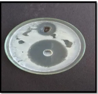
In these studies, the diameter of the zone of inhibition of optimized formulations was
compared with that of standard drug solution. It is evident that the zone of inhibition value of
[image:10.595.174.422.471.646.2]study indicated that Moxifloxacin retained its antimicrobial efficacy when formulated as an
[image:11.595.212.382.113.277.2]in- situ gelling system.
Fig. 6. Zone for drug soln and optimized batch of moxifloxacin
Table 6: Measurement of zone of inhibition
Code Zone of inhibition area (cm)
Drug solution 1.43
F7 0.61
Statistical Analysis
32 Full Factorial Design Batches
A 32 full factorial design was used in the present study. Two factors were evaluated, each at 3 levels, and experimental trials were performed at all 9 possible combinations . Carbopol 940
with HPMC E15LV. An optimized combination of these two able to achieve desired drug
release. Hence amount Carbopol 940 and amount of HPMC E15LV were assumed as
independent variables in a 32 full factorial design. The amount of carbopol 940 was taken as 0.4%, 0.5%, 0.6% and while that of HPMCE 15 LV was taken as 1%, 1.5%, 2% which
responded as -1, 0 or 1 levels respectively. The factorial design batches were evaluated for
Viscosity and % drug release study. The thickness, % cumulative drug release at 8 hrs and
drug diffusion were taken as dependent variables.
Viscosity (Y1)
Viscosity (Y1) = +93444.44+ (-3833.33*X1) + (+5916.67*X2) + (-3750.00X1X2) +
(-20666.67X2) + (-20666.67X2)………. (1)
%drug release at 8 hours (Y2)= +80.55+3.62* X1 -0.43* X2 -0.64* X1X2 +5.93* X12 +0.96
Response Surface and Contour Plot
The quadratic surface model obtained from the regression analyses was used to build up 3D
surface and 2D contour plots in which the responses were represented by curvature surface as
a function of independent variables. The relationship between the response and independent
variables can be directly visualized from the response surface plots.
[image:12.595.166.431.191.348.2]Fig.7- Different plots showing effect of independent variables on Viscosity of In-situ gelling eye drop.
Fig. 8- Different plots showing effect of independent variables on % drug release of In-situ gelling eye drop
CONCLUSION
The optimized formulation (F7) contained 0.6 % w/w carbopol 940 P and 1 %w/w HPMC
E15 LV wherein carbopol caused initial fast release of drug due to its hydrophilic nature later
on hydroxypropyl methylcellulose imparted sustained release property to the gel formed in
[image:12.595.167.430.417.606.2]and gradually erodes by dissolution of the gel, avoiding the need for removal. Hence, it can
be concluded that in-situ gels are a viable alternative to conventional eye drops by providing
sustained release of medicament resulting in decreased frequency of administration leading to
better patient acceptance.
ACKNOWLEDGEMENTS
Authors gratefully acknowledge to SAVA Pharmaceutical pvt. ltd. for providing gift sample
of Moxifloxacin.
REFERENCES
1. Thilekkumar M, Bharathi D, Balasubhramaniam J, Kant S, Pandit JK, PH-induced in-situ
gelling systems of indomethacin for sustained ocular delivery. Ind J Pharm Sci.2005; (3):
327-333.
2. Liu Z, Li J, Liu H, Ding P, Pan W, Study of an alginate/HPMC-based in situ gelling
ophthalmic delivery system for gatifloxacin. Int J Pharm 2006; 315: 12-17.
3. Lane SS, A fall update on the advantages of fourth generation fluoroquinolones.
www.eyeworld org/supplements/vigamox%206pg%2010- 1-4r%20to%20print.pdf
4. Sultana Y, Aqil M, Ali A, Ion-activated, gelrite-based in situ ophthalmic gels of
pefloxacinmesylate: comparison with conventional eye drops. Drug Delivery 2006; 13:
215-219.
5. Nandgude T, Bhise K, Characterization of Hydrochloride and Tannate Salts of
Diphenhydramine. Ind. J. Pharm. Sci. Aug. 2008; 70 (4): 482 – 486.
6. Nandgude T, Bhise K. Characterization of Drug and Polymers for Development of Colon
Specific Drug Delivery System. Asian Journal of Biomedical and Pharmaceutical
Sciences. 2011; 1(4): 17-21.
7. Srividya B, Cardaza RM, Amin PD, Sustained ophthalmic delivery of oflaxacin from a
pH triggered in situ gelling system. J Controlled Release 2001; 73: 205-211.
8. Nandgude TD, Bhise KS. Development of Controlled and Colon Specific Drug Delivery
System of Capecitabine. Inventi Rapid: NDDS. Vol. 2013, Issue 1, Published on Web
01/01/2013, www.inventi.in.
9. Jain SP, Shah SP, Rajadhyaksha NS, Singh PP, Amin PD, In situ ophthalmic gel of
ciprofloxacin hydrochloride for once a day sustained delivery. Drug DevInd Pharm 2008;
10.Nandgude T, Thube R, Jaiswal N,Deshmukh P, Chatap V and Hire N Formulation and
Evaluation of pH Induced In-Situ Nasal Gel of Salbutamol Sulphate. Int. J. of Pharm. Sci.
and Nanotech. July-Sept. 2008; 1(2): 177-183.
11.Doijad RC, Manvi FV, Malleswara VS, Alase P, Sustained ophthalmic delivery of
gatifloxacin from in situ gelling system. Ind J Pharm Sci 2006; 68(6): 814-818.
12.Rao KP, Bushetti SS, Phase transition systems of timolol maleate for glaucoma treatment:
Prolonged acting ocular drug delivery in glaucoma therapy. Indian Drugs 2006; 43(11):
909 913.
13.Sathe S, Bagade MY, NandgudeTD, et al. Formulation and Evaluation of Thermo
Reversible In-Situ Nasal Gel of Terbutaline Sulphate. Indo American Journal of
Pharmaceutical Research.2015; 5(09): 3680-3687.
14.Kulkarni MC, Damle AV, Development of ophthalmic in situ gelling formulation of
flurbiprofen sodium using gellan gum and antimicrobial activity of in-situ gel . Indian
Drugs 2007; 44(5): 373-377.
15.Jayarajkumar AS, Jayachandran EP, formulation and evaluation of in-situ gels containing
cicloprioxolamine for oral thrush. JITPS 2010; 1(5).
16.Dimitrova E, Bogdanova S, Mitcheva M, Tanev I, Minkov E, Development of model
aqueous ophthalmic solution of indomethacin. Drug DevInd Pharm 2000; 26(12): 1297-
1301.
17.Indian Pharmacopoeia. Government of India. Ministry of health and family welfare.