ULTRASOUND SCREENING FOR CONGENITAL UTERINE
ANOMALIES
Bushra Toman Oleiwi*1 and Angham Mohammed Ali Darweesh2
1
Ministry of Health, Baghdad, Iraq.
2
Ministry of Health - Baghdad Medical Office - Al-Karkh, Al-Aamel District of Primary Healthcare, Baghdad, Iraq.
ABSTRACT
The aim of this study was to investigate the prevalence of congenital uterine anomalies in 1046 women attending gynecological ultrasound clinics for a variety of indications. Using three‐dimensional ultrasound, anomalies were found in 55 women (5.4%), including 32 (3.1%) with an arcuate uterus and 23 (2.3%) with major anomalies. The prevalence of uterine anomalies was similar to the findings in women undergoing elective sterilization but lower than in studies of women with recurrent miscarriage. The true prevalence of congenital uterine anomalies in the general population is not known; previous studies reporting on uterineanomalies examined women with a history of infertility or recurrent miscarriage. One of the reasons for this pre‐selection of study populations was the invasive nature of the tests for the examination of the uterus including hysterosalpingography, hysteroscopy, and laparoscopy. Three‐dimensional ultrasound, that has recently been introduced into clinical practice, enables noninvasive and accurate diagnosis of congenital uterine anomalies.[1] We used this new technique to determine the prevalence of congenital anomalies in women attending gynecological ultrasound unit for a variety of indications. Women with a history of miscarriage or miscarriage and infertility have higher prevalence of congenital uterine anomalies compared with the unselected population.
KEYWORDS: Congenital uterine anomalies, prevalence, miscarriage, preterm, uterus.
INTRODUCTION
Congenital uterine anomalies result from abnormal formation, fusion or resorption of the Müllerian ducts during fetal life (Moore et al., 2008). These anomalies have been associated
Volume 8, Issue 8, 941-953. Research Article ISSN 2277– 7105
Article Received on 19 May 2019,
Revised on 09 June 2019, Accepted on 29 June 2019,
DOI: 10.20959/wjpr20198-15336
*Corresponding Author
Bushra Toman Oleiwi
Ministry of Health, Baghdad,
with an increased rate of miscarriage, preterm delivery and other adverse fetal outcomes (Green and Harris, 1976; Rock and Schlaff, 1985; Acien, 1993; Raga et al., 1997; Grimbizis et al., 2001; Tomazevic et al., 2007).
However, such associations might be artefactual. The true population prevalence of congenital uterine anomalies is difficult to assess partly because there are no universally agreed standardized classification systems and partly because the best diagnostic techniques are invasive and, therefore, rarely applied to low-risk study populations. As a result, reported population prevalence rates have varied between 0.06% and 38% (Simon et al., 1991; Makino et al., 1992a, b; Clifford et al., 1994; Acien, 1996; Homer et al., 2000; Guimaraes Filho et al., 2006a, b). This wide variation is likely to be linked to the assessment of different patient populations and the use of different diagnostic techniques with variable, and yet to be determined, test accuracy as well as reliance on non-standardized classification systems. Previous reviews have not considered these factors when investigating the prevalence of uterine anomalies (Acien, 1997; Nahum, 1998; Grimbizis et al., 2001; Troiano and McCarthy, 2004). Saravelos et al. (2008) carried out a critical review to determine the prevalence of congenital uterine anomalies. Their review has assessed the accuracy of different diagnostic procedures, but their search was limited to MEDLINE database and specifically limited to recurrent miscarriage, infertile and general population groups.
We conducted a systematic review of studies evaluating the prevalence of congenital uterine anomalies in the unselected population and in women with a history of infertility, including those undergoing IVF treatment, miscarriage, infertility and recurrent miscarriage combined, and preterm delivery and attempted to explore the inconsistencies present in the literature. This new systematic review is not only an update of the work by Saravelos et al. (2008) but also represents a different approach to the classification of optimal and suboptimal tests.
This was a prospective observational study involving pelvic ultrasound examination in 1046 women attending our gynecological ultrasound unit. The women were either self‐referred for ovarian cancer screening or they were referred for a scan by their consultant gynecologists or general practitioners because of suspected pelvic abnormalities (Table 1).
The prevalence of congenital uterine anomalies in high-risk women is unclear, as several different diagnostic approaches have been applied to different groups of patients.
This review aims to evaluate the prevalence of such anomalies in unselected populations and in women with infertility, including those undergoing IVF treatment, women with a history of miscarriage, women with infertility and recurrent miscarriage combined, and women with a history of preterm delivery.
Searches of MEDLINE, EMBASE, Web of Science and the Cochrane register were performed. Study selection and data extraction were conducted independently by two reviewers. Studies were grouped into those that used ‘optimal’ and ‘suboptimal’ tests for uterine anomalies. Meta-analyses were performed to establish the prevalence of uterine anomalies and their subtypes within the various populations.
RESULTS
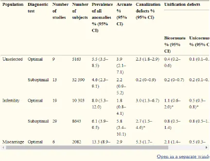
We identified 94 observational studies comprising 89 861 women. The prevalence of uterine anomalies diagnosed by optimal tests was 5.5% [95% confidence interval (CI), 3.5–8.5], In the unselected population, 8.0% (95% CI, 5.3–12) in infertile women, 13.3% (95% CI, 8.9– 20.0) in those with a history of miscarriage and 24.5% (95% CI, 18.3–32.8) in those with miscarriage and infertility.
Arcuate uterus is most common in the unselected population (3.9%; 95% CI, 2.1–7.1), and its prevalence is not increased in high-risk groups. In contrast, septate uterus is the most common anomaly in high-risk populations.
Methods
Search strategy
Articles were identified through the following electronic databases: MEDLINE (1950 to March 2011), EMBASE (1980 to March 2011), Web of Science (1990 to March 2011) and the Cochrane Central Register of Controlled Trials (The Cochrane Library until January 2011).
the electronic searches. Authors were contacted for additional details where required. The searches were conducted independently by two reviewers (Y.Y.C. and K.J.). see Table (1).
Search terms (Unless otherwise stated, search terms were free text terms; mp, term appears in title, original title, abstract, name of substance word, subject heading word; any character).
The search terms in Table I were designed specifically for MEDLINE. This search was modified for EMBASE, Web of Science and the Cochrane Library.
Selection criteria
Studies were selected if the incidence of any uterine anomaly was reported. Studies of all types of congenital uterine anomalies were included but limited to ‘Humans and Female’. Only cohort studies were included in the review. Studies were excluded when the population examined or the diagnostic methods used were not accurately defined. Only publications in English were considered in our selection.
displays a mild concave indentation or contour towards the uterine cavity (The American Fertility Society, 1988; Salim et al., 2003).
Many authors consider the arcuate uterus a normal variant rather than a true anatomical or developmental anomaly (Heinonen et al., 1982; Buttram et al., 1988), but this can only be properly evaluated if the true prevalence of the anomaly can be defined and appropriate associations with relevant outcome measures assessed. Neither can be assessed in the absence of an accurate test to identify the anomaly and differentiate it from more complex uterine anomalies and the normal uterus. In view of this, studies that failed to identify or record any arcuate uteri were excluded from the subtype analysis as we were unable to determine if these studies excluded arcuate uteri or if they failed to identify them because of the inaccuracy of the diagnostic tests employed.
Study Selection
Studies were selected in a two-stage process. First, the titles and abstracts from the electronic searches were examined independently by two reviewers (Y.Y.C. and K.J.) and full manuscripts of all citations that met the predefined selection criteria were then obtained.
Secondly, examinations of the full manuscripts were carried out to make final inclusion or exclusion decisions. In cases of duplicates, the most recent or the most complete publication was used. Any disagreements about inclusion were resolved by consensus or arbitration by a third reviewer (N.R.-F.).
All selected papers were assessed for the following: study design; adequate sampling (random or consecutive rather than convenience sampling); adequate description of population characteristics; completeness of information in the data sets; and use of a validated diagnostic method.
Data Collection and Extraction
Data collection and extraction were performed by the two reviewers (Y.Y.C. and K.J.) independently. Data were extracted on patients’ characteristics, study quality, inclusion and exclusion criteria, diagnostic tools used and anomaly occurrence rates.
methods used based on evidence from other studies (Scarsbrook and Moore, 2003; Saravelos et al., 2008; Olpin and Heilbrun, 2009). Diagnostic methods that were accepted as ‘optimal diagnostic tests’ included three-dimensional transvaginal ultrasound, laparoscopy or laparotomy performed in conjunction with hysteroscopy or hysterosalpingography (HSG), magnetic resonance imaging (MRI) and saline sonohysterography. Suboptimal tests, which could identify and differentiate most but not all anomalies, included two-dimensional transvaginal ultrasound, hysteroscopy performed in isolation, HSG and clinical assessment at the time of Caesarean section.
We devised this classification based on the ability of the test to demonstrate both the external contour of the uterus and the fungal aspect of the endometrial cavity.
Statistical Analysis
Meta-analyses were performed to establish the prevalence of uterine anomalies, and their subtypes, in each group of women. For meta-analyses, log rates were pooled, weighting each study by the inverse of its variance, and the summary estimates were exponentiated.
A random-effects model was used for analysis. Comparisons between the unselected population and the high-risk populations were carried out with the aid of meta-regression. Statistical analyses were performed using Stata 11.0 statistical software (Stata Corp, TX, USA).
RESULTS
All uterine anomalies
Table (2): Indications for ultrasound examination and prevalence of congenital uterine anomalies. Values are given as n or n (%). IUCD = intrauterine contraceptive device.
Indication n Arcuate uterus Major anomalies
Screening for ovarian cancer 253 6 (2.3) 4 (1.6) Pelvic pain 280 7 (2.5) 7 (2.5) Abnormal uterine bleeding 250 8 (3.2) 6 (2.4) Clinical suspicion of fibroids 126 6 (4.8) 4 (3.2) Clinical suspicion of ovarian cyst 103 4 (3.9) 2 (1.9) Localisation of IUCD 34 1 (2.9) 0 (0)
TOTAL 1046 32 (3.1) 23 (2.3)
Each woman first underwent a conventional B‐mode transvaginal ultrasound examination using a 7.5 MHz mechanical probe (Combison 530, 3D Voluson, Kretztechnik, Zipf, Austria).
The longitudinal axis of the uterus, from the isthmus to the fundus, was first defined and then a series of transverse sections were obtained. When there was any duplication or splitting of the endometrial echo, fusion anomalies were suspected and, in these women, a three‐dimensional ultrasound scan was used to determine the diagnosis. In the three‐dimensional ultrasound examination, the uterus was visualized in a longitudinal plane and a three‐dimensional volume was generated by the automatic rotation of the mechanical transducer through 360°.
The volumes, in the shape of truncated cones with a depth of 4.3–8.6 cm and a vertical angle of 90°, were analyzed on‐line using the technique of computer‐generated planar reformatted sections.
Although the images were similar to those of conventional two‐dimensional sonography, with this technique it is possible to obtain an unlimited number of sections through the uterus which cannot be seen on routine scans. The acquisition of three‐dimensional volumes is about 10 s, but complete analysis is achieved in 3 to 10 minutes.
Congenital uterine anomalies were classified according to the criteria of the American Fertility Society.[2]
there is a fundal cleft of at least 1 cm in depth. Cases with rudimentary uterine horns were classified as unicornuate uteri.
Demographic details and ultrasound findings were entered into a computer database at the time of examination and this database was used to obtain details on the age of the women, indications for the scan and the ultrasound findings on uterine morphology.
The significance of the association between the prevalence of congenital uterine abnormalities and both the indication for the ultrasound scan and the age of the women was examined using the χ2
test. Forage analysis, the following groups were used: 19‐29 years, 30‐39 years, 40‐49 years and > 49 years.
RESULTS
Successful examination of the uterus was achieved in 1022/1046 women (97.7%). The uterine cavity could not be clearly visualized due to the presence of large calcified anterior fibroids in 12 women, thin atrophic endometrium in nine women, an intrauterine contraceptive device in two women and previous endometrial resection in one woman. These 24 women were excluded from further analysis.
Congenital uterine anomalies were observed in 55 women (5.4%), including 32 (3.1%) with an arcuate uterus and 23 (2.3%) with major anomalies; there were 16 women with a subseptate uterus, four with a bicornuate uterus, two with an unicornuate uterus and one with uterine agenesis. There was no significant association between the prevalence of uterine anomalies and either the indication for referral (χ2
= 1.76; P > 0.05; 5 df; Table 2) or the women's age (χ2
Table (3): Results of SPSS.
DISCUSSION
In this study of women attending a gynecological ultrasound unit, the prevalence of all congenital uterine anomalies was 5.4%, while the prevalence of major anomalies was 2.3%.
Although our study population may not be representative of the general population, as most women attended for the scan because of a specific complaint, there was no significant association between the prevalence of anomalies and either the indication for the scan or the age of women.
[image:9.595.83.516.134.465.2]in 16(1.9%) of 840 hysterosalpingograms following transcervical tubal sterilization and Simon et al.[5] found major anomalies in 22 (3.2%) of 679 women that had a hysterosalpingogram five months after sterilization by laparoscopy or laparotomy.
Our findings are also compatible with the 1% to 7% prevalence of uterine anomalies in studies utilizing laparoscopy or hysterosalpingography to investigate women with infertility. In contrast, as expected, our prevalence of anomalies was lower than the 10% to 25% reported in studies of women with recurrent miscarriages.
The advantages of three‐dimensional ultrasound in the study of uterine morphology are first, the technique is noninvasive, secondly, it provides quantitative information on parameters such as fundal indentation or septum length, which is not possible with the endoscopic methods or hysterosalpingography, and thirdly, the stored ultrasound data may be re‐sliced in a way which provides standardized views of the uterus which is also impossible with the traditional techniques.
In addition, three‐dimensional ultrasound may improve our understanding of the pathophysiology of conditions such as recurrent miscarriage in the context of uterine anomalies and help to identify those women that may benefit from surgery; recent reports have shown that the prognosis in individuals with the same type of uterine anomaly is varied and have questioned the value of metroplasty in the treatment of multiple pregnancy loss.[6]
The disadvantage of three‐dimensional ultrasound is that it may not provide adequate examination in the presence of large uterine fibroids. Additionally, the technique is currently confined to a few specialized centers.
REFERENCES
1. Jurkovic, D, Geipel, A, Gruboeck, K, Jauniaux, E, Natucci, M, Campbell, S. Three‐dimensional ultrasound for the assessment of uterine anatomy and detection of congenital uterine anomalies. A comparison with hysterosalpingography and two‐dimensional sonography. Ultrasound Obstet Gynecol, 1995; 5: 233–237.
3. Cooper, JM, Houck, RM, Rigberg, HS. The incidence of intrauterine abnormalities found at hysteroscopy in patients undergoing elective hysteroscopic sterilization. J Reprod Med, 1983; 28: 659– 661.
4. Ashton, D, Amin, HK, Richart, RM, Neuwirth, RS. The incidence of asymptomatic uterine anomalies in women undergoing transcervical tubal sterilization. Obstet Gynecol, 1988; 72: 28– 30.
5. Simon, C, Martinez, L, Prado, F, Torajada, M, Pellicer, A. Mullerian defects in women with normal reproductive outcome. Fertil Steril, 1991; 56: 1192– 1193.
6. Kirk, EP, Choung, CJ, Coulam, CB, Williams, TJ. Pregnancy after metroplasty for uterine anomalies. Fertil Steril, 1993; 59: 1164– 1168.
7. Hinckley MD, Milki AA. 1000 office-based hysteroscopies prior to in vitro fertilization: feasibility and findings. JSLS., 2004; 8: 103–107.
8. Homer HA, Li TC, Cooke ID. The septate uterus: a review of management and reproductive outcome. Fertil Steril, 2000; 73: 1–14.
9. Jaipur de Sa Rosa e Silva AC, Rosa e Silva JC, Candido Dos Reis FJ, Nogueira AA, Ferriani RA. Routine office hysteroscopy in the investigation of infertile couples before assisted reproduction. J Reprod Med., 2005; 50: 501–506.
10.Jayaprakasan K, Chan YY, Sur S, Deb S, Clewes JS, Raine-Fenning NJ. The prevalence of uterine anomalies and their impact upon early pregnancy in women conceived following assisted reproduction treatment. Ultrasound Obstet Gynecol, 2011; 37: 727–732.
11.Jurkovic D, Geipel A, Gruboeck K, Jauniaux E, Natucci M, Campbell S. Three-dimensional ultrasound for the assessment of uterine anatomy and detection of congenital anomalies: a comparison with hysterosalpingography and two-dimensional sonography, 1995; 5: 233–237.
12.Jurkovic D, Gruboeck K, Tailor A, Nicolaides KH. Ultrasound screening for congenital uterine anomalies. Br J Obstet Gynecol, 1997; 104: 1320–1321.
13.Kupesic S. Clinical implications of sonographic detection of uterine anomalies for reproductive outcome. Ultrasound Obstet Gynecol, 2001; 18: 387–400.
14.Kupesic S. Three-dimensional ultrasound in reproductive medicine. Ultrasound Rev Obstet Gynecol, 2005; 5: 304–315.
16.Kupesic S, Kurjak A, Skenderovic S, Bjelos D. Screening for uterine abnormalities by three-dimensional ultrasound improves perinatal outcome. J Perinat Med., 2002; 30: 9–17.
17.Laifer-Narin S, Raghavendra N, Parmenter EK, Grant EG. False-normal appearance of the endometrium on conventional transvaginal sonography: comparison with saline hysterosonography. Am J Roentgenol, 2002; 178: 129–133.
18.Lazzarin N, Vaquero E, Exacoustos C, Romanini E, Amadio A, Arduini D. Midluteal phase Doppler assessment of uterine artery blood flow in nonpregnant women having a history of recurrent spontaneous abortions: correlation to different etiologies. Fertil Steril, 2007; 87: 1383–1387.
19.Leible S, Munoz H, Walton R, Sabaj V, Cumsille F, Sepulveda W. Uterine artery blood flow velocity waveforms in pregnant women with Mullerian duct anomaly: a biologic model for uteroplacental insufficiency. Am J Obstet Gynecol, 1998; 178: 1048–1053. 20.Li S, Qayyum A, Coakley FV, Hricak H. Association of renal agenesis and Mullerian
duct anomalies. J Comput Assist Tomogr, 2000; 24: 829–834.
21.Lin PC. Reproductive outcomes in women with uterine anomalies. J Womens Health (Larchmt), 2004; 13: 33–39.
22.Lindheim SR, Sauer MV. Upper genital-tract screening with hysterosonography in patients receiving donated oocytes. Int J Gynecol Obstet., 1998; 60: 47–50.
23.Makino T, Hara T, Oka C, Toyoshima K, Sugi T, Iwasaki K, Umeuchi M, Iizuka R. Survey of 1120 Japanese women with a history of recurrent spontaneous abortions. Eur J Obstet Gynecol Reprod Biol., 1992a; 44: 123–130.
24.Makino T, Umeuchi M, Nakada K, Nozawa S, Iizuka R. Incidence of congenital uterine anomalies in repeated reproductive wastage and prognosis for pregnancy after metroplasty. Int J Fertil., 1992b; 37: 167–170.
25.Malek KA, Hassan M, Soliman A, El-sawah H, Azab AO. A prospective comparative study to assess the accuracy of MRI versus HSG in tubouterine causes of female infertility. Middle East Fertil Soc J., 2005; 10: 250–257.
26.Malik E, Berg C, Sterzik K, Stolz F, Rossmanith WG. Reproductive outcome of 32 patients with primary or secondary infertility and uterine pathology. Arch Gynecol Obstet, 2000; 264: 24–26.
28.Maneschi F, Zupi E, Marconi D, Valli E, Romanini C, Mancuso S. Hysteroscopically detected asymptomatic mullerian anomalies. Prevalence and reproductive implications. J Reprod Med., 1995; 40: 684–688.
29.Momtaz MM, Ebrashy AN, Marzouk AA. Three-dimensional ultrasonography in the evaluation of the uterine cavity. Middle East Fertil Soc J., 2007; 12: 41–46.