Frontline
AMPK
α
1: A glucose sensor that controls CD8 T-cell
memory
Julia Rolf
1, Marouan Zarrouk
1, David K. Finlay
1,2, Marc Foretz
3,
Benoit Viollet
3and Doreen A. Cantrell
11Division of Cell Signalling and Immunology, University of Dundee, Dundee, Scotland, UK 2School of Biochemistry and Immunology, Trinity Biomedical Sciences Institute, Trinity
College, Dublin, Ireland
3Inserm, U1016, Institut Cochin, Cnrs, UMR8104, Universit´e Paris Descartes, Sorbonne Paris
Cit´e, France
The adenosine monophosphate-activated protein kinase (AMPK) is activated by antigen receptor signals and energy stress in T cells. In many cell types, AMPK can maintain energy homeostasis and can enforce quiescence to limit energy demands. We conse-quently evaluated the importance of AMPK for controlling the transition of metabolically active effector CD8 T lymphocytes to the metabolically quiescent catabolic memory T cells
during the contraction phase of the immune response. We show that AMPKα1 activates
rapidly in response to the metabolic stress caused by glucose deprivation of CD8
cyto-toxic T lymphocytes (CTLs). Moreover, AMPKα1 restrains mammalian target of rapamycin
complex 1 activity under conditions of glucose stress. AMPKα1 activity is dispensable for
proliferation and differentiation of CTLs. However, AMPKα1 is required for in vivo
sur-vival of CTLs following withdrawal of immune stimulation. AMPKα1nullT cells also show
a striking defect in their ability to generate memory CD8 T-cell responses duringListeria
monocytogenesinfection. These results show that AMPKα1 monitors energy stress in CTLs and controls CD8 T-cell memory.
Keywords: Cytotoxic T lymphocyte r Energy stress r Listeria monocytogenes r Memory r
Metabolism
See accompanying commentary by Araki and Ahmed
Introduction
T lymphocytes respond to antigen by proliferating and differen-tiating to effector cells that mediate adaptive immune responses. Na¨ıve and memory T lymphocytes are metabolically quiescent and have low rates of amino acid uptake and protein synthesis. They also have low rates of glucose uptake and use oxidative phos-phorylation to efficiently metabolize glucose to generate ATP [1]. However, effector T cells metabolically reprogram and
upregu-Correspondence:Dr. Doreen A. Cantrell e-mail: [email protected]
T cells during the contraction phase of the immune response. In this context, recent studies have shown that the mammalian tar-get of rapamycin complex 1 (mTORC1) controls the production of memory T cells [9]. Inhibition of mTORC1 with rapamycin thus increases the memory T-cell formation following virus chal-lenge. There is an increasing awareness that the control of T-cell metabolism has the potential to dictate T-cell fate.
One evolutionarily conserved molecule that controls cell metabolism is the serine/threonine kinase adenosine monophosphate-activated protein kinase (AMPK). AMPK is phos-phorylated and activated by liver kinase B1 when cells are energy stressed [10]. Triggering of TCR also activates AMPK via Ca2+– calmodulin-dependent protein kinase kinases [11]. The proposed role for active AMPK is to restore energy balance in a cell by inhibit-ing ATP-consuminhibit-ing processes and stimulatinhibit-ing ATP-generatinhibit-ing pathways [10]. In energy stressed cells, AMPK thus enforces quiescence to limit energy demands. T cells exclusively express the AMPKα1 catalytic isoform. In this context, germ-line deletion of AMPKα1 on a C57BL/6 background results in embryonic lethality (http://www.emmanet.org/mutant_types.php?keyword=0417). However, mixed genetic background AMPKα1 null mice are viable and appear fully immune-competent in vivo [12, 13]. These data indicate that AMPK is dispensable for T-cell effector function. However, a recent study has shown that metformin, a pharma-cological activator of AMPK promotes the production of memory T cells [14]. The caveat is that metformin indirectly activates AMPK because it inhibits respiratory chain complex I and thereby causes an increase in cellular adenosine monophosphate/ATP ratio. Metformin has many effects on cell metabolism that are not mediated by AMPK [15,16]. Moreover, even if activation of AMPK can promote the formation of memory T cells, this does not inform whether AMPK is essential for this key process. Metformin actions in vivo can thus be independent of AMPK or indeed could be T-cell extrinsic [15, 16]. Hence for a detailed analysis of the role of AMPKα1 in T cells, there is a requirement to examine the consequences of a T-cell-specific deletion of this kinase. Accordingly, the present study uses a CD4Cre transgene model to delete AMPKαl floxed alleles in thymocytes. We show that AMPKα1 activity is not required for CD8 T-cell proliferation or differentiation. However, AMPKα1 is shown to act as a critical sensor of energy status to control mTORC1 activity in CTLs and is required for CD8 T-cell memory.
Results
AMPKα1 is dispensable for proliferation, generation and function of CTLs
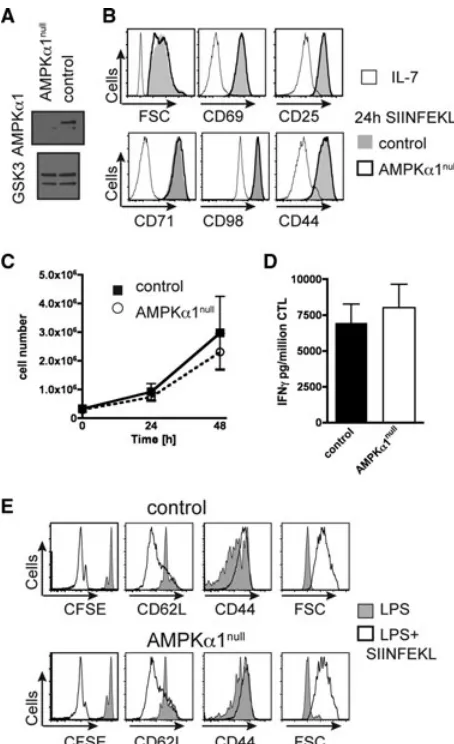
Mice with floxed AMPKα1 alleles were backcrossed to transgenic mice expressing Cre recombinase under the control of the CD4 promoter (CD4Cre) to delete the AMPKα1 gene in CD4/CD8 dou-ble positive thymocytes and hence in all subsequent T-cell pop-ulations (Fig. 1A). AMPKα1fl/fl(control) and CD4creAMPKα1fl/fl
[image:2.594.317.544.61.433.2](AMPKα1null) mice had a normal distribution of peripheralα/β
Figure 1. AMPKα1nullT cells activate, proliferate, and function
nor-mally. (A) Immunoblot analysis of total AMPKα1 and GSK3 in con-trol and AMPKα1nullCD4 thymocytes, two experiments. (B) FSC, CD69,
CD25, CD71, CD98, and CD44 expression by control (gray shade) and AMPKα1null(thick line) OT1 LN cells activated in vitro for 24 h with
SIINFEKL compared with IL-7 (thin line). (C) IL-2 maintained prolif-eration in vitro, control (filled squares) and AMPKα1null(open circles)
of OT1 cytotoxic T lymphocytes (CTLs), average±SD, three experi-ments. (D) IFN-γsecretion (pg/million CTLs) 3 h SIINFEKL restimulation of control and AMPKα1nullOT1 CTLs. Data are shown as mean+SEM of n=3 mice/genotype representing three experiments. (E) CFSE profile, CD62L, CD44, and FSC analysis of control (top panel) and AMPKα1null
(bottom panel) OT1 cells adoptively cotransferred into Ly5.1 recipient mice, 2 days after immunization with LPS+SIINFEKL (open) or LPS (gray shade), two experiments, two to three recipients.
T cells in the thymus, lymph nodes, and spleen (data not shown). TCR primed CD8 T cells cultured in IL-2 clonally expand and differ-entiate to cytotoxic T lymphocytes (CTLs) [17]. AMPKα1nullCD8
Figure 2. AMPKα1 is dispensable for generation of CD8 effector T cells during recombinantListeria monocytogenesOVA infection. Analysis day 7 after primary recombinantL.monocytogenesOVA infection showing (A) frequency transferred control and AMPKα1nullOT1 cells from recipient spleens.
(B) IL-7Rα, KLRG1, CD62L, and CD44 expression by transferred OT1 cells. (C) Frequency of ex vivo splenic IFN-γ-producing control and AMPKα1null
OT1 cells, 5 h SIINFEKL restimulation. Each symbol represents congenically marked (A) OT1 and (C) IFN-γ-producing cells among total CD8 cells from an individual recipient. (A–C) Data shown are representative of one out of two independent experiments (n=4–7 recipients/experiment), pairedt-test.
by the MHC class I molecule H-2Kb by APCs. AMPKα1null OT1
cells responded normally to TCR triggering in vivo and blasted and proliferated (Fig. 1E). We also determined the ability of AMPKα1null OT1 cells to differentiate into effector CD8 T cells
during an immune response against the attenuated bacterial strain
Listeria monocytogenes engineered to express the OVA-derived
peptide SIINFEKL, i.e. recombinant L. monocytogenes OVA (rLMOVA) [18]. Equal numbers of control and AMPKα1nullOT1
T cells were adoptively transferred into recipient mice that were challenged with rLMOVA. The frequency of OT1 cells in the pathogen-challenged animals was analyzed at day 7, the peak of the effector phase. At this time point, the relative frequency of AMPKα1nullOT1 T cells in the spleen was modestly increased
com-pared with control cells (Fig. 2A). Both control and AMPKα1null
OT1 cells had downregulated expression of IL-7Rαand CD62L and upregulated expression of CD44 and KLRG1: a cell surface phenotype characteristic of effector CD8 T cells (Fig. 2B). Control and AMPKα1nullcells were equally able to respond rapidly ex vivo
to produce high levels of IFN-γupon cognate antigen rechallenge (Fig. 2C). Collectively, these data reveal that AMPKα1 is dispens-able for CD8 T-cell differentiation into effector cells during an immune response.
AMPKα1 acts as a sensor of glucose metabolism in CTLs
Effector CD8 T cells are highly glycolytic and maintain high levels of glucose uptake [19]. CTLs treated with 2-deoxyglucose, an inhibitor of glycolysis, activated AMPK as judged by high levels of AMPKT172phosphorylation and also increased levels of
acetyl–CoA carboxylase phosphorylated on its AMPK substrate sequence Ser79 (pACCS79) (Fig. 3A). There was no detectable ACC
phosphorylation in AMPKα1nullCTLs treated with 2-deoxyglucose
(Fig. 3A). CTLs thus exclusively expressed the AMPKα1 catalytic subunit and do not compensate for AMPKα1 deletion by express-ing AMPKα2. Glucose deprivation also activated AMPKα1; even a brief 1 h switch of T cells into low glucose (1 mM) resulted in pAMPKT172stabilization (Fig. 3B). Moreover, the titratable effect
of different levels of exogenous glucose on AMPKα1 activity in CTLs demonstrated the ability of AMPKα1 to act as a quantita-tive sensor of glucose uptake in CTLs (Fig. 3B). Recent studies have revealed the importance of energy-generating glutaminoly-sis pathways in T cells [8]. However, glutamine deprivation did not cause AMPKα1 activation in T cells, indicating that AMPKα1 selectively monitors glucose metabolism (Fig. 3C).
One proposed function of AMPKα1 is to switch cells to a qui-escent catabolic state. In this context, one conserved mechanism used by AMPKα1 to restore energy balance in cells is inhibition of mTORC1 [20, 21]. Previous studies have shown that glucose deprivation inhibits mTORC1 in T cells [22] but whether this is mediated by AMPKα1 has not been explored. The present exper-iments address this issue by monitoring the impact of glucose deprivation on mTORC1 activity in control and AMPKα1nullCTLs.
In these experiments, mTORC1 activity was monitored by assess-ing the phosphorylation of mTORC1 substrate sequences in p70 S6-Kinase 1 (S6K1T389, T421/S424) and 4EBP-1T37/46.
Phosphory-lation of S6K substrate sequences in the S6 ribosomal subunit (pS6S235/6, S240/4) was also monitored. Figure 3D shows that in
control CTLs, the activity of mTORC1 was strictly dependent on cells sustaining high levels of glucose uptake as even a switch into 1 mM glucose inactivated mTORC1. Strikingly, glucose-deprived AMPKα1null CTLs maintained high levels of mTORC1 activity
(Fig. 3D). AMPKα1 is thus a dynamic sensor for glucose uptake and functions to terminate mTORC1 activity under conditions of energy stress in CTLs.
These data raise the possibility that AMPKα1 might have a role in supporting the switch of effector T cells to a quiescent catabolic state. Accordingly, to explore the capacity of AMPKα1 to modulate metabolic adaptation, fully differentiated control and AMPKα1null
Figure 3. Energy stress activates AMPKα1 and inhibits mTORC1 in an AMPK-dependent manner in cytotoxic T lymphocytes (CTLs). Immunoblot detection of (A) pAMPKT172 and pACCS79in control and AMPKα1nullpolyclonal CTLs incubated±50 mM 2-deoxyglucose for 20 min. Relative
pACC quantified as integrated intensity measured by the LICOR. (B) pAMPKT172and pACC1S79in control and AMPKα1nullpolyclonal CTLs cultured
in 0–5 mM glucose for 1 h. (C) OT1 CTLs pAMPKT172, pS6S235/6, and total GSK3-α/βafterL-glutamine deprivation or glucose deprivation for 2 h.
(D) pAMPKT172, total AMPKα1, pS6KT389, pS6KT421/S424, pS6S235/6, pS6S240/4, p4EBP1T37/46 and total S6K detection in control and AMPKα1nullCTLs
cultured in 0–5 mM glucose for 1 h. Representative data from three experiments (A and B) and two experiments (C and D) are shown.
state will be able to survive. The data show that 1 week after the adoptive transfer, a small subpopulation of the transferred CTLs could be recovered from secondary lymphoid organs. However, few transferred AMPKα1nullcells were recovered from secondary
lymphoid organs compared with control cells (Fig. 4B). These results argue that AMPKα1null effector T cells were impaired in
their ability to make the transition back to quiescence compared with control cells.
It was important to consider whether there was a general inability of AMPKα1nullT cells to compete in vivo for secondary
lymphoid tissue niches. We examined the ability of AMPKα1null
primary T cells to undergo lymphopenia-induced proliferation in
Figure 4. AMPK is required for cytotoxic T lymphocytes (CTLs) adaptation to metabolic stress in vivo but dispensable for lymphopenia-induced proliferation. (A) Control (gray shade) and AMPKα1null(thick line) polyclonal CTLs used for adoptive cotransfer gated on CD45.1 congenic marker,
expression of CD62L and CD44. (B) Frequency of cotransferred CFSEhiCD8 CD45 congenically marked CTLs 7 days after adoptive transfer. Each
symbol represents recovered cells from an individual Ly5.1 recipient among CD8 cells, LN left panel, spleen middle panel or Rag2-/-recipient
spleen among leukocytes, right panel. Data shown are pooled data from three experiments, pairedt-test. (C) Frequency of recovered control and AMPKα1nullna¨ıve CD8 T cells co-injected into Rag2-/-mice at day 14 after adoptive transfer. Data shown are pooled from two experiments, each
[image:4.594.99.484.420.654.2]competition with control T cells. In these experiments, control and AMPKα1nullT cells were adoptively transferred at a 1:1 ratio into
Rag2-/-mice. After 14 days, the recovery of control and AMPKα1null
cells from the recipient spleens showed that AMPKα1nullT cells
were equally efficient at undergoing proliferative expansion in a lymphopenic environment. There was no intrinsic problem with the ability of na¨ıve AMPKα1nullCD8 T cells to compete with control
T cells for the homeostatic antigen and cytokine signals (Fig. 4C). The inability to recover AMPKα1nulleffector cells after adoptive
transfer reflects that effector CD8 T cells need AMPKα1 to revert to a quiescent state.
CD8 T-cell memory immune responses depend on AMPKα1
In a response to infection with rLMOVA, AMPKα1null CD8 OT1
T cells undergo normal clonal expansion and contraction (Fig. 2 and 5A). Different CD8 effector subsets can be defined based on their relative expression of IL-7R-α and KLRG1 [23, 24]. It has been shown that IL-7RαhighKLRG1lowmemory precursor effector
cells have a greater potential to become long-lived memory cells and contribute to secondary responses than IL-7RαlowKLRG1high
short-lived effector cells. However, the memory precursor effector cells/short-lived effector cells distribution of OT1 T cells follow-ing rLMOVA infection was independent of AMPKα1 at various time-points after primary infection (Fig. 5B). AMPKα1 thus seems dispensable for CD8 T-cell immune responses during primary infection with rLMOVA. To address whether AMPKα1 contributes to secondary CD8 responses, control and AMPKα1null OT1 cells
were cotransferred and recipient mice rechallenged with rLMOVA 3 weeks following primary infection and assessed for a recall response after a further 6 days. These recall experiments revealed a striking decrease in the relative frequency of AMPKα1nullOT1
cells compared with control OT1 cells both at the sites of
Listeria infection: spleen and liver and in the bone marrow
(Fig. 5C). Taken together, AMPKα1nullOT1 cells were strikingly
defective in their ability to generate a secondary immune response to rLMOVA (Fig. 5C). To further confirm whether AMPKα1 may play a role in primary infection, we analyzed the frequency and absolute numbers of polyclonal ova-specific CD8 T cells defined by MHC class I+SIINFEKL pentamer flow cytometry staining. Also in a polyclonal setting, in CD4creAMPKα1fl/flmice, AMPKα1 appears
dispensable for primary CD8 T-cell responses (Fig. 5D). Figure 5E shows that CD4CreAMPKα1fl/flmice undergoing a secondary
chal-lenge to rLMOVA had reduced frequencies of ova-reactive CD8 T cells in the spleen, liver, and bone marrow compared with control mice.
AMPKα1null CD8 T cells are thus impaired in their ability to
make effector T cells in a secondary immune response.
Discussion
The control of T-cell metabolism seems to be important in the effector/memory transition of T cells. The cytokine IL-2 that drives
effector CTLs differentiation at the expense of memory cell forma-tion is a strong activator of mTORC1 signaling whereas IL-15, which supports memory T-cell formation, only weakly initiates mTORC1 activity [25]. CTLs have high rates of glucose and amino acid uptake to meet the energy demands associated with rapid proliferation and abundant effector cytokine production. In con-trast, memory T cells are relatively metabolically quiescent cells. Effector CD8 T cells would thus need to revert from a metaboli-cally active state to a quiescent catabolic state to produce memory T cells [26]. The strength of mTORC1 activation appears to be a pivotal control switch that determines effector versus memory CD8 T-cell differentiation [9, 27]. Previous studies have shown that glucose deprivation inhibits mTORC1 activity in T cells [22] but the glucose sensor that mediates this response was unknown. The present article demonstrates that AMPKα1 acts as a critical glucose sensor in effector CTLs and is activated by glucose depri-vation and functions to restrain mTORC1 activity under conditions of metabolic stress.
TCR triggering activates AMPKα1 [11], but AMPKα1 seems dis-pensable for the initial antigen receptor-induced metabolic switch that accompanies effector T-cell differentiation. AMPKα1 is not required for T-cell proliferation or the induction of CD8 effector T cells in a primary immune response toL. monocytogenes infec-tion. However, AMPKα1nullCD8 T cells are impaired in their
abil-ity to make effector T cells in a secondary immune response. The developmental processes that regulate effector T cells progression to form memory cells must include mechanisms that allow CD8 T cells to revert from the metabolically active state characteristic of effectors to the quiescent catabolic state of memory cells. The present data are consistent with a model whereby AMPKα1 regu-lates the metabolic adaptations that allow secondary effector cell generation. AMPKα1 thus monitors energy stress in CD8 T cells and is a key regulator of CD8 T-cell memory to secondary effector transition during an immune response to infection.
Materials and methods
Mouse models
AMPKα1fl/fl mice were obtained from Benoit Viollet and Marc
Foretz and bred to CD4cre transgenic and OT1 TCR transgenic [28] mice. Mice were maintained in the Biological Resource unit at the University of Dundee in compliance with UK Home Office Animals (Scientific Procedures) Act 1996 guidelines.
Cell culture
Figure 5. AMPK is dispensable for CD8 cells during primary infection but essential during secondary infection. (A) Frequency OT1 cells among total CD8 cells, average±SD, eight Ly5.1 recipients, control (filled squares), and AMPKα1null(open circles) in blood day 7, 10, 14, and 35 after
recombinantListeria monocytogenesOVA (rLMOVA) infection. (B) IL-7Rαand KLRG1 expression by OT1 cells in the blood, control (upper panel) and AMPKα1null(lower panel) day 7, 14, and 39 after primary rLMOVA infection. Data shown are representative from two experiments. (C) Frequency
of cotransferred control and AMPKα1nullTCR transgenic OT1 cells day 6 post secondary challenge in the spleen, liver, and bone marrow. Each
symbol represents CD45 congenically marked OT1 cells among total CD8 cells in individual recipients. Data shown are representative from two experiments, (n=4–6 recipients), pairedt-test. (D) Frequency (left panel) and absolute number (right panel) of MHC class H-2Kb+SIINFEKL
pentamer+cells among total CD8 cells in the spleen. Data are shown as mean±SD (n=3–5 mice/genotype/time-point). Control AMPKα1fl/flmice
(black) and AMPKα1nullCD4creAMPKα1fl/fl(open) mice days 7, 14, and 28 after primary rLMOVA infection. (E) Frequency of pentamer+cells among
CD8 cells day 6 post secondary rLMOVA challenge in control AMPKα1fl/flmice (black square) compared with AMPKα1nullCD4creAMPKα1fl/flmice
(open circle) in spleen, liver, and bone marrow. Each symbol represents one mouse, Mann–Whitney test.
approximately 0.3×10⁶cells/mL daily. The in vitro generated
CTLs were used day 5–7 after activation. For glucose/glutamine deprivation, CTLs were incubated 1 h in glucose-free RPMI with dialyzed FCS (GIBCO) or 2 h inL-glutamine-free RPMI (GIBCO). Treatment with 50 mM 2-deoxyglucose (Sigma) for 20 min was used as a positive control.
Western blot
CTLs were gently centrifuged and then lyzed at a density of 1–2 × 10⁷/mL in Tris lysis buffer containing 10 mM Tris pH
inhibitors (Roche), and Calyculin A phosphatase inhibitor (Cal-biochem). Proteins were resolved on SDS 4–12% polyacrylamide gel, transferred to nitrocellulose membranes, blocked in 5% milk, and incubated with primary antibody and developed using HRP-coupled secondary antibodies and ECL. Antibodies to detect pAMPKT172, pS6KT389, S6KT421/S424, pS6S235/6, p4EBP1T37/46and
total S6K were from Cell Signalling Technologies. pACCS79
was detected by anti-sheep AlexaFluor 680 and Streptavidin-IRDye800 using LICOR system (Odyssey). Antibodies recognizing pACCS79and AMPKα1 were kindly provided by Professor Grahame
Hardie.
Flow cytometry
Cells (1–2×10⁶/sample) were incubated in Fc-block (2.4G2) at
4◦C in PBS with 10% complete RPMI. Fluorescent-labeled anti-bodies to detect: CD8-α (53−6.7), CD25 (7D4), CD44 (IM7), CD45.1 (A20), CD45.2 (104), CD62L (MEL-14), CD69 (H1.2F3), CD71 (C2), CD98 (RL388), IL-7Rα(SB/199), KLRG1 (2F1), Vα2 (B20.1), and Vβ5.1/2 (MR9–4) were obtained from BD Bio-science or eBioBio-science. To detect SIINFEKL-specific cells, MHC class I H-2Kb coupled to SIINFEKL pentamers labeled with
allo-phycocyanin were used according to manufacturer’s instructions (Proimmune).
For cytokine staining afterL. monocytogenesinfection, spleno-cytes were restimulated with SIINFEKL 1μM for 5 h, Brefeldin A was added for 4 h. Intracellular cytokine staining to detect IFN-γ (XMG1.2) was performed using eBioscience reagents according to manufacturer’s instructions. Flow cytometry used BD Calibur or BD LSR Fortessa and the data were processed using FlowJo software (Treestar).
ELISA
Day 7 after initial activation OT1 CTLs were restimulated with 1μM SIINFEKL for 3 h and the supernatant was harvested. IFN-γ ELISA was done according to manufacturer’s instructions (eBio-science).
Adoptive transfer
For in vivo proliferation, CD45 congenically differently marked OT1 cells from AMPKα1fl/fl (control) and CD4creAMPKα1fl/fl
(AMPKα1null) mice were purified using CD8 T-cell isolation kit
(Miltenyi) according to manufacturer’s instructions. The OT1 cells were mixed at an equal ratio and were labeled with 5 μM CFSE for 10 min at 37◦C. Approximately 4 × 10⁶ OT1 cells
were co-injected iv into recipient C57BL/6 Ly5.1 mice. The mice were immunized with 40 μg SIINFEKL and 25 μg LPS ip the next day. The CFSE dilution was analyzed day 2 and 4 after immunization. For adoptive cotransfer of CTLs, day 7 fully dif-ferentiated CD4creAMPKα1fl/+ (control) and CD4creAMPKα1fl/fl
(AMPKα1null) CTLs with different CD45 congenic markers were
mixed at equal ratio, CFSE labeled and injected intravenously into C57BL/6 Ly5.1 or Rag2−/−recipient mice. The CTLs recovery was analyzed 1 week later by flow cytometry. To assess lymphopenia-induced proliferation, T cells were purified using pan-T-cell kit (Miltenyi) and 2–3×10⁶polyclonal CD8 T cells were mixed at an
equal ratio and adoptively transferred intravenously into Rag2−/− recipient mice. The recovered cells were analyzed by flow cytom-etry 14 days later. To exclude the possibility of CD4cre-dependent rejection, CD4creAMPKα1fl/+ mice were used as controls in Figure 4.
Listeria monocytogenesinfection
In total 104OT1 cells mixed at a 1:1 ratio from CD45 congenically
differently marked AMPKα1fl/fl(control) and CD4creAMPKα1fl/fl
(AMPKα1null) mice were coinjected ip into C57BL/6 Ly5.1
recipi-ent mice. Alternatively, AMPKα1fl/fland CD4creAMPKα1fl/flmice
were infected to determine polyclonal responses. The mice were infected with attenuated ActA-deleted OVA-expressingL.
monocy-togenes(kindly provided by Professor Hao Shen)[18]. For primary
infection 1–5×10⁶and for secondary challenge 10×106colony
forming units were injected iv.
Statistical analysis
Statistical analysis was done using Graph Pad Prism,t-test was performed on paired values, and Mann–Whitney nonparametric test was used for all other data.*p<0.05, **p<0.01, ***p<
0.001.
Acknowledgements: We thank biological service unit staff, Eliz-abeth Emslie, members of the Cantrell group, and Grahame Hardie. We are grateful to Hao Shen, Verity Pearce, and Klaus Okkenhaug for theListeria monocytogenesmodel. This work was funded by Wellcome Trust Principal Research Fellowship grant 065975/Z/01/A and 097418/Z/11Z to DAC.
Conflict of interest: The authors have declared no commercial or financial conflict of interest.
References
1 Krauss, S.,Brand, M. D. and Buttgereit, F., Signaling takes a breath– new quantitative perspectives on bioenergetics and signal transduction.
Immunity2001.15:497–502.
3Frauwirth, K. A.,Riley, J. L.,Harris, M. H.,Parry, R. V.,Rathmell, J. C.,
Plas, D. R.,Elstrom, R. L. et al., The, CD28 signaling pathway regulates glucose metabolism.Immunity2002.16:769–777.
4Rathmell, J. C.,Vander Heiden, M. G.,Harris, M. H.,Frauwirth, K. A. and Thompson, C. B., In the absence of extrinsic signals, nutrient utilization by lymphocytes is insufficient to maintain either cell size or viability.
Mol. Cell.2000.6:683–692.
5Michalek, R. D.,Gerriets, V. A.,Jacobs, S. R.,Macintyre, A. N.,MacIver N. J.,Mason, E. F.,Sullivan, S. A. et al., Cutting edge: distinct glycolytic and lipid oxidative metabolic programs are essential for effector and regulatory CD4+T-cell subsets.J. Immunol.2011.186:3299–3303.
6Jacobs, S. R.,Herman, C. E.,MacIver, N. J.,Wofford, J. A.,Wieman, H. L.,Hammen, J. J. and Rathmell, J. C., Glucose uptake is limiting in T-cell activation and requires CD28-mediated Akt-dependent and inde-pendent pathways.J. Immunol.2008.180:4476–4486.
7van der Windt, G. J. W.,Everts, B.,Chang, C.-H.,Curtis, J. D.,Freitas, T. C.,Amiel, E.,Pearce, E. J. et al., Mitochondrial respiratory capacity is a critical regulator of CD8+T-cell memory development.Immunity2012.
36:68–78.
8Wang, R.,Dillon, C. P.,Shi, L. Z.,Milasta, S.,Carter, R.,Finkelstein, D.,
McCormick L. L. et al., The transcription factor Myc controls metabolic reprogramming upon T lymphocyte activation. Immunity 2011. 35:
871–882.
9Araki, K.,Turner, A. P.,Shaffer, V. O.,Gangappa, S.,Keller, S. A., Bach-mann, M. F.,Larsen, C. P. et al., mTOR regulates memory CD8 T-cell differentiation.Nature2009.460:108–112.
10Hardie, D. G.,Ross, F. A. and Hawley, S. A., AMPK: a nutrient and energy sensor that maintains energy homeostasis.Nat. Rev. Mol. Cell, Biol.2012.
13:251–262.
11Tam ´as, P.,Hawley, S. A.,Clarke, R. G.,Mustard, K. J.,Green, K.,Hardie, D. G. and Cantrell, D. A., Regulation of the energy sensor AMP-activated protein kinase by antigen receptor and Ca2+in T lymphocytes.J. Exp. Med.2006.203:1665–1670.
12Mayer, A.,Denanglaire, S.,Viollet, B.,Leo, O. and Andris, F., AMP-activated protein kinase regulates lymphocyte responses to metabolic stress but is largely dispensable for immune cell development and func-tion.Eur. J. Immunol.2008.38:948–956.
13MacIver, N. J.,Blagih, J.,Saucillo, D. C.,Tonelli, L.,Griss, T.,Rathmell, J. C. and Jones, R. G., The liver kinase B1 is a central regulator of T-cell development, activation, and metabolism. J. Immunol. 2011. 187:
4187–4198.
14Pearce, E. L.,Walsh, M. C.,Cejas, P. J.,Harms, G. M.,Shen, H.,Wang, L.-S.,
Jones, R. G. et al., Enhancing, CD8 T-cell memory by modulating fatty acid metabolism.Nature2009.460:103–107.
15Foretz, M.,H ´ebrard, S.,Leclerc, J.,Zarrinpashneh, E.,Soty, M.,Mithieux, G.,Sakamoto, K. et al., Metformin inhibits hepatic gluconeogenesis in mice independently of the LKB1/AMPK pathway via a decrease in hepatic energy state.J. Clin. Invest.2010.120:2355–2369.
16Kalender, A.,Selvaraj, A.,Kim, S. Y.,Gulati, P.,Br ˆul ´e, S.,Viollet, B.,Kemp, B. E. et al., Metformin, independent of AMPK, inhibits mTORC1 in a rag GTPase-dependent manner.Cell Metab.2010.11:390–401.
17Cantrell, D. A. and Smith, K. A., Transient expression of interleukin 2 receptors. Consequences for T-cell growth.J. Exp. Med. 1983.158:
1895–1911.
18Pearce, E. L. and Shen, H., Generation of CD8 T-cell memory is regulated by IL-12.J. Immunol.2007.179:2074–2081.
19Macintyre, A. N.,Finlay, D.,Preston, G.,Sinclair, L. V.,Waugh, C. M.,
Tam ´as, P.,Feijoo, C. et al., Protein kinase B controls transcriptional programs that direct cytotoxic T-cell fate but is dispensable for T-cell metabolism.Immunity2011.34:224–236.
20Gwinn, D. M., Shackelford, D. B., Egan, D. F., Mihaylova, M. M.,
Mery, A., Vasquez, D. S., Turk, B. E. et al., AMPK phosphoryla-tion of raptor mediates a metabolic checkpoint. Mol. Cell 2008. 30:
214–226.
21Inoki, K., Zhu, T. and Guan, K.-L., TSC2 mediates cellular energy response to control cell growth and survival. Cell 2003. 115:
577–590.
22Cham, C. M. and Gajewski, T. F., Glucose availability regulates IFN-gamma production and p70S6 kinase activation in CD8+effector T cells.
J. Immunol.2005.174:4670–4677.
23Sarkar, S.,Kalia, V.,Haining, W. N.,Konieczny, B. T.,Subramaniam, S. and Ahmed, R., Functional and genomic profiling of effector CD8 T-cell subsets with distinct memory fates. J. Exp. Med. 2008. 205:
625–640.
24Obar, J. J., Jellison, E. R., Sheridan, B. S., Blair, D. A., Pham, Q.-M.,Zickovich, J. M. and Lefranc¸ois, L., Pathogen-induced inflamma-tory environment controls effector and memory CD8+T-cell differenti-ation.J. Immunol.2011.187:4967–4978.
25Cornish, G. H., Sinclair, L. V. and Cantrell, D. A., Differential regulation of T-cell growth by IL-2 and IL-15. Blood 2006. 108:
600–608.
26D’Cruz, L. M.,Rubinstein, M. P. and Goldrath, A. W., Surviving the crash: transitioning from effector to memory CD8+T cell.Semin. Immunol.2009.
21:92–98.
27Rao, R. R.,Li, Q.,Odunsi, K. and Shrikant, P. A., The mTOR kinase deter-mines effector versus memory CD8+T-cell fate by regulating the expres-sion of transcription factors T-bet and Eomesodermin.Immunity2010.32:
67–78.
28Hogquist, K. A.,Jameson, S. C.,Heath, W. R.,Howard, J. L.,Bevan, M. J. and Carbone, F. R., T-cell receptor antagonist peptides induce positive selection.Cell1994.76:17–27.
29Waugh, C., Sinclair, L., Finlay, D., Bayascas, J. R. and Cantrell, D., Phosphoinositide (3,4,5)-triphosphate binding to phosphoinositide-dependent kinase 1 regulates a protein kinase B/Akt signaling threshold that dictates T-cell migration, not proliferation.Mol. Cell. Biol.2009.29:
5952–5962.
Abbreviations:AMPK: AMP-activated protein kinase·CTL: cytotoxic T lymphocyte·mTORC1: mammalian target of rapamycin complex 1·
rLMOVA: recombinantListeria monocytogenesOVA
Full correspondence: Dr. Doreen A. Cantrell, Division of Cell Signaling and Immunology, University of Dundee, Dow Street, Dundee, UK, DD15EH
Fax:+44-0-1382-38-5047
e-mail: [email protected]
See accompanying commentary: http://dx.doi.org/10.1002/eji.201343483
Received: 20/9/2012 Revised: 10/12/2012 Accepted: 7/1/2013