SYNTHESIZED SILVER NANOPARTICLES FROM PSEUDARTHRIA
VISCIDA
EXERT HIGH ANTI-MICROBIAL ACTIVITY AGAINST
PATHOGENIC BACTERIA
Kunal Naik1 and R. Krishnamurthy2*
1
Ph.D. Student at C. G. Bhakta Institute of Biotechnology, Uka Tarsadia University.
2
Director at C. G. Bhakta Institute of Biotechnology, Uka Tarsadia University, Bardoli,
(Gujarat) India.
ABSTRACT
In recent decades the use of plant based antimicrobial formulation is on
increase due to microbial resistance to chemical drugs. The present
study attempted to synthesize and evaluate Silver Nanoparticles
(AgNPs) from methanolic extract of P. viscida under diverse
temperature against pathogenic bacteria. AgNPs were prepared from a
mixture of 1mM to5mM silver nitrate and 1-5% extract of P. viscida.
Optimum proportion and concentration of extract and silver nitrate was
determined and used for production of stable AgNPs. The
characterization of stable AgNPs was done by UV-vis spectroscopy
and Transmission Electron Microscope (TEM). Loss of organic
content and specific characteristic of AgNPs were measured by
Differential Scanning Calorimetry (DSC). The spectroscopy showed presence of AgNPs at
430 nm absorbance peak. The TEM analysis revealed AgNPs of good quality, dominant
spherical in shape and particle size range from 59-95 nm. AgNPs shows similar inhibitory
activity against 4 (67%) out of 6 bacteria tested. 1mM silver nitrate and 2% extract mixture
prepared at 40˚c and pH 12-13 was found to be optimum for production of stable AgNPs with induced inhibitory activity that crude extract can’t gives alone. The study concludes that
AgNPs of good quality, stability and antimicrobial activity can be obtained from methanolic
extract of P. viscida.
KEYWORDS: Silver Nanoparticles, Pseudarthria viscida, pathogenic bacteria,
anti-microbial activity.
Volume 7, Issue 11, 1203-1215. Research Article ISSN 2277– 7105
Article Received on 18 April 2018,
Revised on 08 May 2018, Accepted on 29 May 2018,
DOI: 10.20959/wjpr201811-12470
*Corresponding Author
R. Krishnamurthy
Director at C. G. Bhakta
Institute of Biotechnology,
Uka Tarsadia University,
INTRODUCTION
P. viscida belongs to fabaceae family and it exhibits wide range of medicinal properties. The
decoction is used in pain, hysteria, rheumatism, asthma, heart diseases, paralysis and urinary
stones. In India, it is commonly called as Salaparni and it is a keen stimulant for digestive
system and is used in digestive ailments like anorexia, flatulence, diarrhea, vomiting and
piles. It is good as a pain-killer in general body ache. It also possesses anti-inflammatory and
wound healing activity.[1] Indian forests are major and cheap source for medicinal plants and
its product. Many medicinal plants are gaining importance due to their unique
phyto-constituent and versatile application in field of development.[2] The use of environment
friendly material like plant extract for synthesis of AgNPs offers several advantages which
include eco friendliness, pharmaceutical capability and biomedical application as this
synthesis is free of any toxic chemicals.[3] It is a well-known fact that silver ions and
silver-based compounds are highly toxic to micro-organisms which include sixteen major species of
bacteria.[4] Hence, investigation on synthesis of plant based silver nano particles has regained
more importance due to overuse of antibiotics which have caused resistance in many
microbes. In this study we have formulated AgNPs with the help of methanolic extract of P.
viscida by reduction of Ag+ to Ag- from silver nitrate solution under optimized condition.
Due to wide range of its application, numbers of synthetic methods have been developed.[5]
Many literatures show different methods for synthesis of gold and silver particles of different
size and shape in polar and non-polar organic solvents. The nano particles synthesized this
way shows excellent stability for long period. The smaller particles have the greater effect.[6]
MATERIALS
Silver nitrate (AgNO3) and Nutrient agar were purchased from Hi-Media, India. Mature and
healthy entire plant of Pseudarthria viscida was collected from Waghai Botanical Garden,
NH360, Ambapada, Gujarat-394730, India, and was taxonomically identified and
authenticated by Dr. Bimal Desai, Assistant professor, Forestry department, Navsari
Agricultural University, Navsari, Gujarat, India. A voucher specimen (voucher number: -
UTU/CGBIBT/16-17/01) were kept in Botany lab, Biotech department, UTU, Bardoli,
Gujarat for future utilization. All the microbes used in this study were obtained from MTCC,
METHODS
Preparation of plant extract
Whole plant of P. viscida was washed for three times with distilled water. Whole plant
materials were shed dry and fine powder was obtained using grinding mill, approx 200 gm
whole plant powders of P. viscida was used for extraction process by Soxhlet apparatus using
methanol as a solvent. Then these methanolic extract was concentrated by rotary evaporator.
These concentrated methanolic extract of P. viscida was stored at 4˚c and used for this study.
Optimization of various parameters for synthesis of eco-friendly silver nano particles
Concentration of plant extract
To synthesize AgNPs from whole plant extract of P. viscida, different concentration of
methanolic extract were prepared ranges 1% to 5%.
Concentration of silver nitrate
For optimization of silver ion concentration, different concentration of silver nitrate (range-
1mM to 5 mM) were mixed with fixed concentration of plant extract and incubated in dark
condition at different temperature.
Temperature
Mixture of plant extract and silver nitrate were kept at different temperature ranges 12˚c,
28˚c, 37˚c and 60˚c under shaking condition keeping other parameter constant.
Reaction time
Time interval was optimized in terms of properties and production by examine the sample at
various time interval (0, 15, 30, 60, 90 and 120 minutes) using spectrophotometer.Different
concentration of methanolic extract of plant, concentration of silver nitrate, temperature and
reaction time was optimized for the synthesis of AgNPs under controlled pH (12.5).
Characterization of AgNPs
TEM (Transmission Electron Microscopy) analysis
TEM of the methanolic extract of P. viscida was carried out at Sardar Patel Center for
Science and Technology, CharutarVidyaMandal, VallabhVidyanagar, Anand. Analysis report
was given by Sophisticated Instrumentation Centre for Applied Research and Testing, Govt.
silver nitrated solution was used to prepare mixture of 2%. Then this mixture was used for
TEM analysis for confirmation of formation of silver nanoparticles.
DSC analysis
Thermal decomposition behavior of the AgNPs has been studied using Netzsch (STA 449C)
DSC/TG. The DSC/TG patterns were collected as a function of temperature up to 900˚c
under N2 atmosphere. The heating rate was 10˚c /min. in N2. Alpha alumina was used as
reference material.
Zeta potential
The size distribution or average size of the synthesized AgNPs were determined by dynamic
light scattering (DLS) and zeta potential measurements were carried out using DLS (Malvern,
UK). For DLS analysis the samples were diluted 10 folds by 0.15M phosphate buffer (pH
7.4) and the measurements were taken in the range between 0.1 and 10,000 nm. Dynamic
light scattering (DLS) which is based on the laser diffraction method with multiple scattering
techniques was employed to study the average particle size of silver nanoparticles. The
prepared sample was dispersed in de-ionized water and then for ultra-sonication. Then
solution was filtered and centrifuged for 15 min. at 25˚c with 5000 rpm and the supernatant
was collected. The supernatant was diluted for 4 to 5 times and then the particle distribution
in liquid was studied in a computer controlled particle size analyzer.
Antimicrobial study
The comparative antimicrobial activity of the both plant extracts and AgNPs synthesized
from plant extract was effectively tested against Gram (+) ve and Gram (-) ve bacteria. The
antibacterial activity testing was determined by disc diffusion method. Sterile discs of
uniform size were soaked in distilled water, bulk silver, crude extract, AgNPs including
standard drug separately. Then the discs were air dried under sterile condition. The plates of
Mueller Hinton agar media were prepared and then the soaked discs were placed on
respective place. Plates were incubated at 27˚c for 24-48 hrs. Streptomycin was used
(10µg/ml) as a standard antibacterial drug.
RESULT
Preparation of extract
Soxhlet extraction process gave 18.92 gm (12.61%) yield from 150 gm powder of whole
Synthesis and optimization of AgNPs
As a result of simple distinctive process of AgNPs preparation, when silver nitrate is mixed
with crude methanolic extract of plant, color started to change from colorless to yellow
brown and then dark brown over the time. Change in color was characterized for all five tests
by spectrophotometer at 430nm on interval of 15 min as shown in Graph-I.
Graph-I: Highest peak of silver nitrate at 430 nm with different concentration of plant
extract 1% to 5%.
Pure silver nitrate and methanol were used as blank to nullify its interference in test sample.
Effect of different parameters like different temperature, concentration of plant extract and
concentration of silver nitrate are shown in Graph II on interval of 30 minutes.
Graph-II: Effect of (a) Temperature, (b) extract concentration and (c) silver nitrate
concentration on synthesis of AgNPs.
As shown in Graph-II (a), 1% and 2% concentration of plant extract produced stable AgNPs,
plant extracts were getting precipitated after 120 minutes. 2% plant extract produced higher
amount of AgNPs compare to 1%, hence the optimized concentration for stable AgNPs
production was found to be 2%. Same way as shown in Graph-II (b), minimum 2% silver
nitrate was found to be adequate for stable AgNPs production. Both of these parameters were
optimized under different temperature and 28˚c was found to be the best optimum
temperature for synthesis of such stable AgNPs as shown in Graph-II (c).
Result of TEM
Morphology and size of produced AgNPs were determined by TEM analysis. Figures I show
the obtained TEM images which confirmed that AgNPs were produced from methanolic
extract of P. viscida.
Image I: TEM images of AgNPs from extract of P. viscida.
It was also observed that the produced AgNPs were predominantly spherical in shape. The
overall morphology of the produced AgNPs was composed of almost uniform nanoparticles
which can be clearly seen in Image 1.[7] which was produced by reaction between 2%
methanolic extract of P. viscida and 2mM silver nitrate mixture.
DSC analysis
DSC study shows the nature of the plant extract compound loaded on the AgNPs. Different
compound shows their particular characteristic endodermic peaks in DSC. The endodermic
peak of AgNPs was found at 102.02˚c, range is 93.96˚c to 105.54˚c as shown in Graph-III,
Graph-III: DSC analysis of AgNPs synthesized from extract of P. viscida.
These findings are in agreement with the literature report of.[8] The plot showed a steady
weight loss due to moisture loss, between 100-750˚c the weight loss was due to degradation
of organic compounds then after no degradation and remaining content accounts for silver
weight.
Zeta sizer
The particle size range of AgNPs synthesized from P. viscida monitored by using particle
size analyzer Mastsizer 2000. The result showed that AgNPs average size range was found to
be 59.50 nm as shown in Graph-IV.
Graph IV: Particle size distribution of AgNPs from extract of P. viscida.
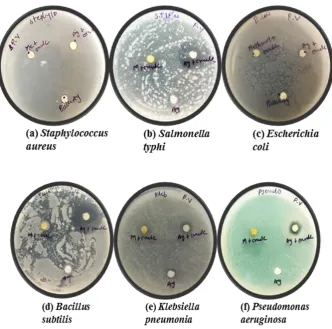
Anti bacterial activity
The silver nano particles with narrow size distribution obtained in this study were tested as
against different pathogenic bacterial strains. Standard antibiotic drug showed zone of
inhibition in almost all the bacterial strains as showed in Images-II.
Image II: Zone of inhibition of standard drug in all selected bacterial strains.
Crude extract alone shows no inhibitory activity against all selected bacterial strains as shown
in Images-III. While, combined with silver nitrate and crude extract of P. viscidai.e. AgNPs,
Images III: Zone of inhibition of AgNPs.
These AgNPs showed greater synergy and was found to be superior against Salmonella typhi
and Bacillus subtilis as it showed greater of zone of inhibition. These AgNPs showed
comparatively low inhibitory activity against Klebsiella pneumonia and Pseudomonas
aeruginosa and no activity against Escherichia coli and Staphylococcus aureus as small and
no zone of inhibition was observed respectively. The diameter of zone of inhibition of all
tested sample were measured and fold increase in inhibition were also calculated, data are
[image:9.595.132.465.68.397.2]exert in Table- II.
Table II: Diameter of zone of inhibition and fold increase in inhibition by AgNPs.
Crude (a) AgNPs (b) Fold increase (c)
Salmonella typhi 2 19 89.3
Bacillus subtilis 2 20 99.0
Klebsiella pneumonia 2 5 5.3
Pseudomonas aeruginosa 2 6 8.0
Escherichia coli 2 2 0.0
DISCUSSION
In this study we have successfully verified and confirm the quick and proficient route for
synthesis of silver nanoparticles using extract of P. viscida. The progression of intense
yellowish brown colour confirms the synthesis of silver nano particles which is completely
due to surface plasmon resonance. According to literature P. viscida is in rich source of
flavanoids and phenolic acid derivatives.[9,10] Flavanoids are widely known for reduction
reaction for successful synthesis of nano particles.[11] Hence, according to result of
phytochemical analysis, presence of high flavanoids and phenolic content in P. viscida
extract rationally increase the potential of P. viscida extract for bioreduction of Ag+ to Ag0.
Many literatures revealed P. viscida contains ascorbic acid and citric acid in notable
amount.[9,10] In addition to that ascorbic acid and citric acid are widely used in synthesis of
AgNPs, it is a strong justification to use P. viscida extract for synthesis and shaping of silver
nano particles.[12] Presence of Diosgenin as a dominant saponins in P. viscida is also reported
which might contribute to surfactant properties of P. viscida extract.[13,14] Plant surfactants are
already been in use for synthesis of silver nano particles. similarly the starch content reflects
the capping properties of the extract and starch is widely used in various synthetic process for
capping and stabilizing AgNPs.[15,16] Thus phytochemical studies support the use of P. viscida
extract as a suitable agent to facilitate the synthesis of AgNPs. The complete reduction of
Ag+ with in 4 hrs indicates that synthesis is much faster compared with other plants, which
takes more than 24 hrs for complete synthesis of AgNPs to occur. Optimization studies
showed that the sharpness of the peak increased with an increase in temperature. Most likely
this occurs due to an increase in the reaction rate of conversion of metal ions to nanoparticles
at higher temperature.[17] P. viscida is also known to contain various bioactive compounds
like flavanoids, terpenoids, amino acid, protein and glycosides. It is speculated that the
alcohol groups are oxidize to carbonyl groups in the course of reduction, which support the
suggestion that polyol groups are primarily responsible for the bioreduction process. Thus the
water soluble fraction of P. viscida played key roles in the bioreduction of the precursors and
evolution in shape in the AgNPs. Silver ion and silver based compounds are highly toxic to
micro-organisms, showing a strong biocidal effect against microbial species because they are
highly reactive species with larger surface area.[18,19,20] AgNPs produced using microbes and
plant extract are known to exhibit potent anti microbial activity. Anti bacterial activity
determined using disc diffusion method confirms that combining crude extract with silver
nano particles resulted in higher bactericidal effect on the test pathogen than the either crude
between crude extract and silver nitrate. It is very interesting observation that some pathogens
are resistant to almost all the antibiotic and medicinal plants. Therefore this combinational
approach of crude extract and silver nitrate would provide a strategy for effective control on
such pathogens. From the result of our study we can conclude that the antibacterial effect of
silver nitrate is the key contributor of the synergistic effect observed with a combination of
silver nitrate and crude extract.
Mode of action of all known antibiotics have been recognized by the scientist.[21,22] Silver has
been in use since century for burn wounds but still there is not any reliable explanation on
mechanism of silver.[23] Silver at low concentration cannot penetrate into the cells but it can
be adsorb on bacterial cell wall. Respiration in bacteria occurs across the cell membrane
hence adsorption of silver in cell wall prevent the dehydrogenation. According many
hypotheses, the catalytic oxidation of silver nitrate reacts with bacterial cell membrane which
causes cell death. As discussed above, the killing mechanism of crude extract and silver
nitrate is different. Therefore synergistic effect can be a powerful alternative against drug
resistant micro-organism. Silver nitrate shows the selective approach towards phospholipids
and glycoproteins which are present in cell membrane, consequently concentration of anti
bacterial agents at specific point on the cell membrane will increase due to reaction between
crude extract and silver nitrate. In addition we can say that silver nitrate aid in transportation
of bio-active compounds to the cell surface. Silver nitrate is also specific towards sulfur in
protein of bacterial cell and as a result it increases membrane permeability and enhanced
infiltration of the bio active compounds. Due to the larger size of AgNPs in comparison to
silver ion, more molecules can react with them which will directly increase antibacterial
activity.
CONCLUSION
From this study we conclude that, synthesis of plant based silver nano particles is speedy and
economical approach as it produced numbers of highly stable nano particles. P. viscida is rich
in source of flavanoids and phenols, which may play important role for in synthesis of
nanoparticles. These synthesized nanoparticles are capped with phenolic compound and can
ACKNOWLEDGMENT
All the authors would like to show their gratitude to, Sophisticated Instrumentation Centre for
Applied Research and Testing, Govt. of India at Anand, Gujarat, India.
REFERENCES
1. Mittal, A. K., Kaler, A. & Banerjee, U. C. Free Radical Scavenging and Antioxidant
Activity of Silver Nanoparticles Synthesized from Flower Extract of Rhododendron
dauricum. Nano Biomed. Eng. 2012; 4: 118–124.
2. Banerjee, P., Satapathy, M., Mukhopahayay, A. & Das, P. Leaf extract mediated green
synthesis of silver nanoparticles from widely available Indian plants: synthesis,
characterization, antimicrobial property and toxicity analysis. Bioresour. Bioprocess.
2014; 1: 3.
3. Lakshmi, Y. S., Banu, F. & Gopalakrishnan, S. Green Synthesis , And Antimicrobial
Activity Of Silver Nanoparticles From The Medicinal Plant Vernonia Amygdalina.
Indian J. Nanosci. 2013; 1: 3–6.
4. Azad, B. & Banerjee, A. Formulation of silver nanoparticles using methanolic extract of
stem of plant Desmodium gangeticum , their characterization and antibacterial and
anti-oxidant evaluation. Pharma Innov. J., 2014; 3: 77–81.
5. Kapinus, D., Gopalakrishnan, S., Lakshmi, S. Y. & Banu, F. Comparison of antimicrobial
activities of silver nanoparticles synthesized from Bridelia minutiflora Hook . f . through
Boiling method and Microwave irradiation method. Indian J. drugs Dis., 2015; 4: 1–5.
6. Kvı, L. & Vec, R. Silver Colloid Nanoparticles : Synthesis, Characterization, and Their
Antibacterial Activity. J Phys. Chem. B. 2006; 110: 16248–16253.
7. KUMARASAMYRAJA, D. & JEGANATHAN, N. S. Green Synthesis of Silver
Nanoparticles Using Aqueous. Int J Pharm Bio Sci. 2013; 4: 469–476.
8. Pol, V. G. et al. Sonochemical Deposition of Silver Nanoparticles on Silica Spheres.
Langmuir. 2002; 18: 3352–3357.
9. Hemlal, H. & Ravi, S. GC-MS, HPTLC and Antimicrobial analysis of Root extracts of
Pseudarthria viscida Wight and Arn and Desmodium gangeticum (Linn) DC. I. Res. J.
Biol. Sci. 2012; 1: 57–65.
10. Deepa, M. A., Narmatha Bai, V. & Basker, S. Antifungal properties of Pseudarthria
viscida. Fitoterapia. 2004; 75: 581–584.
11. Shankar, S. S., Rai, A., Ahmad, A. & Sastry, M. Rapid synthesis of Au, Ag, and
Colloid Interface Sci. 2004; 275: 496–502.
12. Qin, Y. et al. Size control over spherical silver nanoparticles by ascorbic acid reduction.
Colloids Surfaces A Physicochem. Eng. Asp. 2010; 372: 172–176.
13. Guclu-Ustundag, Ö. & Mazza, G. Saponins: Properties, applications and processing. Crit.
Rev. Food Sci. Nutr. 2007; 47: 231–258.
14. Wang, Z. W., Gu, M. Y. & Li, G. Z. Surface properties of Gleditsia saponin and
synergisms of its binary system. J. Dispers. Sci. Technol. 2005; 26: 341–347.
15. Nadagouda, M. N., Hoag, G., Collins, J. & Varma, R. S. Green synthesis of Au
nanostructures at room temperature using biodegradable plant surfactants. Cryst. Growth
Des. 2009; 9: 4979–4983.
16. Sharma, V. K., Yngard, R. A. & Lin, Y. Silver nanoparticles: Green synthesis and their
antimicrobial activities. Adv. Colloid Interface Sci. 2009; 145: 83–96.
17. Song, J. Y. & Kim, B. S. Rapid biological synthesis of silver nanoparticles using plant
leaf extracts. Bioprocess Biosyst. Eng. 2009; 32: 79–84.
18. Suvorova, E. I., Klechkovskaya, V. V., Kopeikin, V. V. & Buffat, P. A. Stability of Ag
nanoparticles dispersed in amphiphilic organic matrix. J. Cryst. Growth. 2005; 275:
2351–2356.
19. Dibrov, P., Dzioba, J., Gosink, K. K., Häse, C. C. & Ha, C. C. Chemiosmotic Mechanism
of Antimicrobial Activity of Ag + in Vibrio cholerae Chemiosmotic Mechanism of
Antimicrobial Activity of Ag ϩ in Vibrio cholerae. Antimicrob. Agents Chemother. 2002;
46: 2668–2670.
20. Shahverdi, A. R., Fakhimi, A., Shahverdi, H. R. & Minaian, S. Synthesis and effect of
silver nanoparticles on the antibacterial activity of different antibiotics against
Staphylococcus aureus and Escherichia coli. Nanomedicine Nanotechnology, Biol. Med.,
2007; 3: 168–171.
21. Kaur, M., Rai, J. & Randhawa, G. Recent advances in antibacterial drugs. Int. J. Appl.
Basic Med. Res., 2013; 3: 3.
22. Thangamani, S. et al. Antibacterial activity and mechanism of action of auranofin against
multi-drug resistant bacterial pathogens. Sci. Rep., 2016; 6: 22571.
23. Vijayabaskaran, M., Sajeer, P. & Perumal, P. Wound Healing Activity of Ethanol Extract