Received 29 July 2019 Accepted 13 January 2020
Edited by D. Chopra, Indian Institute of Science Education and Research Bhopal, India
Keywords:substituted arenes; pentafluorothio; functionalized aromatic rings; organometallic synthesis; crystal structure.
CCDC reference:1943767
Supporting information:this article has supporting information at journals.iucr.org/e
Structural characterization and Hirshfeld surface
analysis of 2-iodo-4-(pentafluoro-
k
6-sulfanyl)-benzonitrile
Jean C. Gonza´lez Espiet,aJuan A. Cintro´n Cruzaand Dalice M. Pin˜ero Cruzb*
a
Department of Chemistry, University of Puerto Rico-Rio Piedras Campus, PO Box 23346, San Juan, 00931-3346, Puerto Rico, andbDepartment of Chemistry and the Molecular Sciences Research Center, University of Puerto Rico-Rio Piedras Campus, PO Box 23346, San Juan, 00931-3346, Puerto Rico. *Correspondence e-mail: [email protected]
The title compound, C7H3F5INS, a pentafluorosulfanyl (SF5) containing arene,
was synthesized from 4-(pentafluorosulfanyl)benzonitrile and lithium tetra-methylpiperidide following a variation to the standard approach, which features simple and mild conditions that allow direct access to tri-substituted SF5
intermediates that have not been demonstrated using previous methods. The molecule displays a planar geometry with the benzene ring in the same plane as its three substituents. It lies on a mirror plane perpendicular to [010] with the iodo, cyano, and the sulfur and axial fluorine atoms of the pentafluorosulfanyl substituent in the plane of the molecule. The equatorial F atoms have symmetry-related counterparts generated by the mirror plane. The pentafluorosulfanyl group exhibits a staggered fashion relative to the ring and the two hydrogen atoms orthoto the substituent. S—F bond lengths of the pentafluorosulfanyl group are unequal: the equatorial bond facing the iodo moiety has a longer distance [1.572 (3) A˚ ] and wider angle compared to that facing the side of the molecules with two hydrogen atoms [1.561 (4) A˚ ]. As expected, the axial S—F bond is the longest [1.582 (5) A˚ ]. In the crystal, in-plane C—H F and N I interactions as well as out-of-plane F C interactions are observed. According to the Hirshfeld analysis, the principal intermolecular contacts for the title compound are F H (29.4%), F I (15.8%), F N (11.4%), F F (6.0%), N I (5.6%) and F C (4.5%).
1. Chemical context
Organic compounds containing the trifluoromethyl (CF3) or
pentafluorothio (or pentafluoro-6
-sulfanyl, SF5) groups play
an important role in organofluorine chemistry because of their special properties including low surface energy, hydro-phobicity, high chemical resistance, high thermal stability and high electronegativity (Kirsch et al., 1999, 2014; Iida et al., 2015; Beier et al., 2011). SF5, coined as the
‘super-trifluoro-methyl’ group, is often preferred to CF3 as it is more
elec-tronegative, lipophilic and chemically stable, and possesses a higher steric effect (Bowdenet al., 2000). The current interest in the field of drug discovery of fluorinated substituents is based on the possibility of improving both the metabolic stability and bioavailability of receptor binders upon the incorporation of susbtituents with one or more fluorine atoms (Altomonteet al., 2014; Savoie & Welch, 2015; Sowailehet al., 2017). In fact, several blockbuster drugs include such a group, demonstrating the prominent role of the trifluoromethyl group in the area of drug discovery (O’Hagan, 2010; Mu¨lleret al., 2007; Purseret al., 2008). New molecules incorporating the SF5group are thus potential alternatives to already existing
biologically active molecules containing the CF3substitution.
Additionally, the chemical robustness of SF5 has been
explored in other areas such as polymer chemistry (Zhouet al., 2016). Despite the popularity of the title compound, an important precursor in organofluorine chemistry, its crystal-lographic characterization, which is an important milestone in the synthesis of next-generation materials containing this motif, has not been reported. Herein, we describe a variation to the synthetic approach and give details of its simple crys-tallization through slow evaporation methods, yielding X-ray diffraction-quality single crystals.
The title compound was obtained as part of our studies toward the synthesis of functionalized arenes containing the SF5moiety. Its synthesis involves a one-pot reaction in which
the interaction of the cyano group in 4-(pentafluorosulfan-yl)benzonitrile to the Lewis acidic lithium cation in lithium tetramethylpiperidide (LiTMP) allows deprotonation from the nearest ortho-H atom on the arene. The SF5-containing
organolithium species is then quenched with iodine to yield the title compound. This reaction pathway was proposed by Iida et al. (2015) for the synthesis of SF5-substituted zinc
phthalocyanines. We modified the synthesis by adding tetra-methylethylenediamine (TMEDA), an amine additive that serves to break up the lithiated base aggregates, allowing for accelerated reactivity because of the increased basicity. This variation improves the total yield of the title compound by 8%.
[image:2.610.312.566.94.163.2]2. Structural commentary
Fig. 1 shows the molecular structure of the title compound, which crystallizes in the space group Pnma. Its asymmetric unit comprises a single molecule lying on a mirror plane perpendicular to [010] with the iodo, cyano, and the sulfur and
axial fluorine atoms of the pentafluorosulfanyl substituent in the plane of the molecule. The fluorine atoms of the penta-fluorosulfanyl group in the equatorial positions lie above and below the plane in a staggered fashion relative to the two hydrogen atomsorthoto the substituent; of those, two of the four fluorine atoms are generated symmetrically by the mirror plane. The S1—F(eq) bond distances differ from each other
depending on which side of the molecule the bond is located (Table 1). The S1—F2(eq) bond and its symmetry equivalent
S1—F2i(eq)[symmetry code: (i)x,y+32,z] are on the same
side as the iodine atom and exhibit a longer bond distance of 1.572 (3) A˚ in comparison to S1—F1(eq)and S1—F1
i
(eq), which
are further away from the iodine and have a shorter bond length distance of 1.561 (4) A˚ . The S1—F3(ax)bond length of
1.582 (5) A˚ is the longest and is consistent with those in similar structures [1.588 (2) and 1.573 (3) A˚ ; Duet al., 2016].
3. Supramolecular features
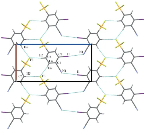
The packing of the title compound is consolidated through a series of intermolecular interactions, which can be classified as being in-plane and out-of-plane (Table 2). Each molecule acts as a C—H donor through themeta- andpara-hydrogen atoms of the phenyl ring counter to the iodine atom. Two C–H F
232
Gonza´lez Espietet al. C7H3F5INS Acta Cryst.(2020). E76, 231–234 [image:2.610.48.293.198.243.2]research communications
Figure 1
Molecular structure of the title compound, including atom labelling. Displacement ellipsoids are drawn at the 50% probability level. Atoms generated by the mirror plane [symmetry code: (i) x, y+ 3
2, z] are
depicted in dark green.
Table 1
Selected bond lengths and angles.
S1—F1(eq)and S1—–F1i(eq) 1.561 (4)
S1—F2(eq)and S1—F2i(eq) 1.572 (3)
S1—F3(ax) 1.582 (5)
C4—S1—F2(eq) 92.0 (2)
C4—S1—F1(eq) 92.2 (2)
Symmetry code: (i)x,y+3 2,z.
Figure 2
[image:2.610.313.564.490.720.2] [image:2.610.47.299.552.702.2]hydrogen bonds, C5—H5 F3 and C6—H6 F3 with H F distances of 2.5 and 2.6 A˚ , respectively, create an in-plane network (Table 2 and Fig. 2). Both the H5 and H6 atoms are highly acidic because of the electron-withdrawing effects of the –SF5 and –CN substituents. Additionally, significant
in-plane halogen-bonding interactions [N1 I1(1 2+x,
3 2y,
1 2z)
= 3.408 (10) A˚ ] are observed (Metrangoloet al., 2005). Out-of-plane intermolecular interactions arise primarily from F
ring interactions at one of the ‘corners’ of the ring (Fig. 3) with an F2 C3(2x,12+y, 1z) distance of 3.124 (5) A˚ .
4. Hirshfeld surface analysis
The Hirshfeld surface (Spackman & Jayatilaka, 2009) for the title compound mapped over dnorm is shown in Fig. 4 while
Fig. 5 shows the associated two-dimensional fingerprint plots (McKinnon et al., 2007), both generated with Crystal-Explorer17(Turneret al., 2017). Red spots on the Hirshfeld surface mapped over dnorm in the colour range 0.4869 to
1.4157 arbitrary units confirm the previously mentioned main intermolecular contacts. The fingerprint plots are given for all contacts and those delineated into F H/H F (29.4%; Fig. 5b), F I/I F (15.8%; Fig. 5c), F N/N F (11.4%; Fig. 5d), H N/N H (6.3%; Fig. 5e), I N/N I (5.6%; Fig. 5f), C F/F C (4.5%; Fig. 5g), C H/H C (4.5%; Fig. 5h), I H/H I (3.3%; Fig. 5i), C N/N C (1.6%; Fig. 5j), C C (9.5%; Fig. 5k), F F (6.0%; Fig. 5l) and I I (2.2%; Fig. 5m) interactions. Thus, the Hirshfeld surface analysis indicates that the most significant contributions arise from F H and F I contacts.
5. Database survey
A search of the Cambridge Structural Database (Version 5.39, updated May 2017; Groomet al., 2016) revealed no matching compounds with the title compound substructure and the
three substituents. However, a search for SF5aryl compounds
fragment revealed about 85 hits: 77 of these structures were reported in the last 10 years, which shows the increasing interest in the SF5group. Most of these compounds are used as
reagents in the synthesis and modification of pharmaceuticals, such as the antimalarial agent mefloquine (Wipfet al., 2009) and the anti-obesity drug fenfluramine (Welchet al., 2007).
6. Synthesis and crystallization
All solvents and reagents were purified prior to being used. 4-(Pentafluorosulfanyl)benzonitrile was obtained commer-cially and used without further purification. A solution of 2.5 M n-butyl lithium in hexanes was used. Column chroma-tography was carried out on a column packed with silica gel 70–230 mesh.
The synthesis of the title compound was performed through the regioselective ortho-lithiation of 4-(pentafluorosulfan-yl)benzonitrile with lithium tetramethylpiperidide (LiTMP) in THF as solvent, favouring the formation of theorthoproduct (1,2,4-substituted arene) over themetaproduct (1,3,4-substi-tuted arene). Theortho-metalated product was subsequently quenched with I2to afford the iodinated trisubstituted arene.
A dry 50 mL Schlenk tube was charged with 4 mL of dry THF and 300mL of 2,2,6,6-tetramethyl piperidine (1.75 mmol, 2 eq.) and 262mL of N,N,N,N-tetramethylethylendiamine (1.75 mmol) were added under an inert atmosphere. The
Figure 3
Out-of-plane contacts. Partial packing diagram for the the title compound viewed along theaaxis. F interactions are shown as dashed lines.
Figure 4
A view of the Hirshfeld surface of the title compound mapped overdnorm
with the four main intermolecular contacts in the crystal lattice.
Figure 5
Full (a) and individual (b)–(m) two-dimensional fingerprint plots showing the 12 intermolecular contacts present in the crystal structure.
Table 2
Hydrogen-bond geometry (A˚ ,).
D—H A D—H H A D A D—H A
C5—H5 F3i 0.93 2.57 3.501 (1) 174
C6—H6 F3ii 0.93 2.56 3.476 (1) 169
Symmetry codes: (i)x1 2;yþ
3 2;zþ
3
solution was cooled to 273 K and 700mL of 2.5 M n-butyl lithium in hexane (1.75 mmol, 2 eq.) were added slowly. The reaction mixture was stirred at 273 K for 30 minutes and then cooled to 195 K. A solution containing 200 mg of 4-(penta-fluorosulfanyl)benzonitrile (0.872 mmol, 1 eq.) in 4 mL THF was added dropwise: the solution changed from pale yellow to dark brown upon formation of the metalated intermediary. After stirring for 1 h at 195 K, a solution of 244 mg I2
(0.960 mmol, 1.2 eq.) in 4 mL THF was added dropwise and stirred for 2 h. The mixture was then warmed to room temperature and stirred for 1 h.
The reaction was quenched with water and THF was removed under reduced pressure, followed by extraction with diethyl ether. The combined organic phase was washed with aqueous 0.1MHCl, 0.1MNa2S2O3and brine, then dried over
MgSO4. The crude product was purified by column
chroma-tography (9:1, hexane:ethyl acetate) to yield 71 mg (46%) of the pure arene product as a yellow solid (m.p. 367–369 K). Block-like yellow crystals suitable for X-ray diffraction were obtained by slow evaporation of a saturated CH2Cl2solution
of the 2-iodo-4-(pentafluoro-6
-sulfanyl)benzonitrile at room temperature over a period of four days. NMR analyses were performed on a Bruker AV-500 spectrometer using chloro-form-d as solvent (CDCl3). The solvent signals at 7.26 and
77.00 ppm were used as internal standards for proton and carbon, respectively. 1H NMR (500 MHz, Chloroform-d)
8.31 (d,J= 2.1 Hz, 1H), 7.89 (dd,J= 8.6, 2.1 Hz, 1H), 7.75 (d,J = 8.6 Hz, 1H).13C NMR (125 MHz, CDCl3) , 98.22, 117.83,
124.10, 126.16, 134.39, 136.82, 156.15.
7. Refinement
Data collection, crystal data and structure refinement para-meters are summarized in Table 3. H atoms were included in geometrically calculated positions and refined as riding atoms with C—H = 0.93 A˚ andUiso(H) = 1.2Ueq(C).
Funding information
The authors acknowledge financial support by the NSF– CREST Center for Innovation, Research and Education in Environmental Nanotechnology (CIRE2N) grant No. HRD-1736093. The single crystal x-ray diffractometer was acquired through the support of the National Science Foundation under the Major Research Instrumentation Award No. CHE-1626103.
References
Altomonte, S., Baillie, G. L., Ross, R. A., Riley, J. & Zanda, M. (2014).
RSC Adv.4, 20164–20176.
Beier, P., Pasty´rˇı´kova´, T. & Iakobson, G. (2011).J. Org. Chem.76, 4781–4786.
Bowden, R. D., Comina, P. J., Greenhall, M. P., Kariuki, B. M., Loveday, A. & Philp, D. (2000).Tetrahedron,56, 3399–3408. Dolomanov, O. V., Bourhis, L. J., Gildea, R. J., Howard, J. A. K. &
Puschmann, H. (2009).J. Appl. Cryst.42, 339–341.
Du, J., Hua, G., Beier, P., Slawin, A. M. Z. & Woollins, J. D. (2016).
Struct. Chem. 28, 723–733.
Groom, C. R., Bruno, I. J., Lightfoot, M. P. & Ward, S. C. (2016).Acta Cryst.B72, 171–179.
Iida, N., Tanaka, K., Tokunaga, E., Mori, S., Saito, N. & Shibata, N. (2015).Chem. Open.4, 698–702.
Kirsch, P. & Bremer, M. (2014).Chimia,68, 363–370.
Kirsch, P., Bremer, M., Heckmeier, M. & Tarumi, K. (1999).Angew. Chem. Int. Ed.38, 1989–1992.
McKinnon, J. J., Jayatilaka, D. & Spackman, M. A. (2007).Chem. Commun.pp. 3814–3816.
Metrangolo, P., Neukirch, H., Pilati, T. & Resnati, G. (2005).Acc. Chem. Res.38, 386–395.
Mu¨ller, K., Faeh, C. & Diederich, F. (2007).Science,317, 1881–1886. O’Hagan, D. (2010).J. Fluor. Chem.131, 1071–1081.
Purser, S., Moore, P. R., Swallow, S. & Gouverneur, V. (2008).Chem. Soc. Rev.37, 320–330.
Rigaku OD (2018). CrysAlis PRO. Rigaku Oxford Diffraction, Yarnton, England.
Savoie, P. R. & Welch, J. T. (2015).Chem. Rev.115, 1130–1190. Sheldrick, G. M. (2015a).Acta Cryst.A71, 3–8.
Sheldrick, G. M. (2015b).Acta Cryst.C71, 3–8.
Sowaileh, M. F., Hazlitt, R. A. & Colby, D. A. (2017).Med. Chem. 12, 1481–1490.
Spackman, M. A. & Jayatilaka, D. (2009).CrystEngComm,11, 19– 32.
Turner, M. J., McKinnon, J. J., Wolff, S. K., Grimwood, D. J., Spackman, P. R., Jayatilaka, D. & Spackman, M. A. (2017).
CrystalExplorer17. University of Western Australia. http://hirsh-feldsurface. net
Welch, J. T. & Lim, D. S. (2007).Bioorg. Med. Chem.15, 6659–6666. Wipf, P., Mo, T., Geib, S. J., Caridha, D., Dow, G. S., Gerena, L., Roncal, N. & Milner, E. E. (2009).Org. Biomol. Chem.7, 4163– 4165.
Zhou, Y., Wang, J., Gu, Z., Wang, S., Zhu, W., Acen˜a, J. L., Soloshonok, V. A., Izawa, K. & Liu, H. (2016).Chem. Rev.116, 422–518.
234
Gonza´lez Espietet al. C7H3F5INS Acta Cryst.(2020). E76, 231–234 [image:4.610.311.563.90.358.2]research communications
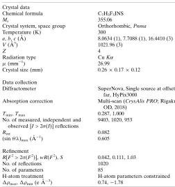
Table 3
Experimental details.
Crystal data
Chemical formula C7H3F5INS
Mr 355.06
Crystal system, space group Orthorhombic,Pnma
Temperature (K) 300
a,b,c(A˚ ) 8.0634 (1), 7.7088 (1), 16.4410 (3)
V(A˚3) 1021.96 (3)
Z 4
Radiation type CuK
(mm1) 26.99
Crystal size (mm) 0.260.170.12
Data collection
Diffractometer SuperNova, Single source at offset/ far, HyPix3000
Absorption correction Multi-scan (CrysAlis PRO; Rigaku OD, 2018)
Tmin,Tmax 0.287, 1.000
No. of measured, independent and observed [I> 2(I)] reflections
9403, 1020, 953
Rint 0.082
(sin/)max(A˚
1) 0.605
Refinement
R[F2> 2(F2)],wR(F2),S 0.042, 0.111, 1.03
No. of reflections 1020
No. of parameters 85
H-atom treatment H-atom parameters constrained
max, min(e A˚3) 0.74,1.78
sup-1
Acta Cryst. (2020). E76, 231-234
supporting information
Acta Cryst. (2020). E76, 231-234 [https://doi.org/10.1107/S2056989020000365]
Structural characterization and Hirshfeld surface analysis of
2-iodo-4-(penta-fluoro-
λ
6-sulfanyl)benzonitrile
Jean C. Gonz
á
lez Espiet, Juan A. Cintr
ó
n Cruz and Dalice M. Pi
ñ
ero Cruz
Computing details
Data collection: CrysAlis PRO (Rigaku OD, 2018); cell refinement: CrysAlis PRO (Rigaku OD, 2018); data reduction:
CrysAlis PRO (Rigaku OD, 2018); program(s) used to solve structure: ShelXT (Sheldrick, 2015a); program(s) used to
refine structure: SHELXL (Sheldrick, 2015b); molecular graphics: OLEX2 (Dolomanov et al., 2009); software used to
prepare material for publication: OLEX2 (Dolomanov et al., 2009).
2-Iodo-4-(pentafluoro-λ6-sulfanyl)benzonitrile
Crystal data
C7H3F5INS
Mr = 355.06
Orthorhombic, Pnma
a = 8.0634 (1) Å
b = 7.7088 (1) Å
c = 16.4410 (3) Å
V = 1021.96 (3) Å3
Z = 4
F(000) = 664
Dx = 2.308 Mg m−3
Cu Kα radiation, λ = 1.54184 Å
Cell parameters from 6922 reflections
θ = 2.7–68.4°
µ = 26.99 mm−1
T = 300 K
Irregular, clear light yellow 0.26 × 0.17 × 0.12 mm
Data collection
SuperNova, Single source at offset/far, HyPix3000
diffractometer
ω scans
Absorption correction: multi-scan (CrysAlisPro; Rigaku OD, 2018)
Tmin = 0.287, Tmax = 1.000
9403 measured reflections
1020 independent reflections 953 reflections with I > 2σ(I)
Rint = 0.082
θmax = 68.8°, θmin = 5.4°
h = −9→9
k = −9→9
l = −19→19
Refinement
Refinement on F2
Least-squares matrix: full
R[F2 > 2σ(F2)] = 0.042
wR(F2) = 0.111
S = 1.03
1020 reflections 85 parameters 0 restraints
Hydrogen site location: inferred from neighbouring sites
H-atom parameters constrained
w = 1/[σ2(F
o2) + (0.071P)2 + 1.8371P]
where P = (Fo2 + 2Fc2)/3 (Δ/σ)max < 0.001
Δρmax = 0.74 e Å−3
supporting information
sup-2
Acta Cryst. (2020). E76, 231-234
Special details
Geometry. All esds (except the esd in the dihedral angle between two l.s. planes) are estimated using the full covariance matrix. The cell esds are taken into account individually in the estimation of esds in distances, angles and torsion angles; correlations between esds in cell parameters are only used when they are defined by crystal symmetry. An approximate (isotropic) treatment of cell esds is used for estimating esds involving l.s. planes.
Refinement. ShelXL
Fractional atomic coordinates and isotropic or equivalent isotropic displacement parameters (Å2)
x y z Uiso*/Ueq
I1 0.67725 (6) 0.750000 0.31142 (3) 0.0540 (3)
S1 0.97572 (19) 0.750000 0.62917 (9) 0.0398 (4)
F3 1.1407 (6) 0.750000 0.6812 (3) 0.0641 (14)
F2 1.0576 (4) 0.6065 (5) 0.5743 (2) 0.0641 (9)
F1 0.9063 (5) 0.8925 (6) 0.68738 (19) 0.0819 (13)
C4 0.7869 (8) 0.750000 0.5682 (4) 0.0356 (13)
C3 0.8020 (7) 0.750000 0.4841 (4) 0.0335 (13)
H3 0.905856 0.750000 0.459508 0.040*
C2 0.6572 (8) 0.750000 0.4373 (4) 0.0352 (13)
C1 0.5034 (8) 0.750000 0.4752 (4) 0.0467 (16)
C7 0.3514 (9) 0.750000 0.4290 (5) 0.056 (2)
C5 0.6348 (10) 0.750000 0.6059 (5) 0.065 (3)
H5 0.628167 0.750000 0.662401 0.078*
N1 0.2295 (10) 0.750000 0.3943 (6) 0.080 (2)
C6 0.4948 (10) 0.750000 0.5606 (5) 0.072 (3)
H6 0.391926 0.750000 0.586180 0.087*
Atomic displacement parameters (Å2)
U11 U22 U33 U12 U13 U23
I1 0.0575 (4) 0.0786 (4) 0.0259 (3) 0.000 −0.00566 (16) 0.000
S1 0.0409 (8) 0.0534 (9) 0.0250 (8) 0.000 −0.0044 (6) 0.000
F3 0.055 (3) 0.097 (4) 0.040 (3) 0.000 −0.020 (2) 0.000
F2 0.0622 (18) 0.0697 (19) 0.0605 (18) 0.0256 (16) −0.0188 (15) −0.0192 (17)
F1 0.081 (3) 0.110 (3) 0.055 (2) 0.022 (2) −0.0146 (16) −0.044 (2)
C4 0.038 (3) 0.045 (3) 0.024 (3) 0.000 −0.002 (3) 0.000
C3 0.037 (3) 0.038 (3) 0.026 (3) 0.000 0.000 (2) 0.000
C2 0.047 (4) 0.033 (3) 0.025 (3) 0.000 −0.003 (2) 0.000
C1 0.038 (3) 0.066 (4) 0.036 (4) 0.000 −0.003 (3) 0.000
C7 0.046 (4) 0.081 (6) 0.042 (5) 0.000 −0.004 (3) 0.000
C5 0.047 (4) 0.121 (8) 0.027 (4) 0.000 0.002 (3) 0.000
N1 0.049 (4) 0.121 (7) 0.071 (5) 0.000 −0.016 (4) 0.000
C6 0.036 (4) 0.139 (9) 0.042 (4) 0.000 0.014 (3) 0.000
Geometric parameters (Å, º)
I1—C2 2.076 (6) C3—H3 0.9300
sup-3
Acta Cryst. (2020). E76, 231-234
S1—F2 1.572 (3) C2—C1 1.388 (9)
S1—F2i 1.572 (3) C1—C7 1.442 (10)
S1—F1i 1.561 (4) C1—C6 1.406 (11)
S1—F1 1.561 (4) C7—N1 1.136 (10)
S1—C4 1.823 (7) C5—H5 0.9300
C4—C3 1.388 (9) C5—C6 1.353 (11)
C4—C5 1.374 (10) C6—H6 0.9300
F3—S1—C4 179.4 (3) C5—C4—C3 121.8 (6)
F2i—S1—F3 87.52 (18) C4—C3—H3 120.8
F2—S1—F3 87.52 (18) C4—C3—C2 118.4 (6)
F2i—S1—F2 89.4 (3) C2—C3—H3 120.8
F2—S1—C4 92.04 (19) C3—C2—I1 118.9 (5)
F2i—S1—C4 92.04 (19) C1—C2—I1 121.2 (5)
F1i—S1—F3 88.3 (2) C1—C2—C3 119.9 (6)
F1—S1—F3 88.3 (2) C2—C1—C7 121.6 (6)
F1—S1—F2i 90.4 (2) C2—C1—C6 119.5 (6)
F1—S1—F2 175.8 (2) C6—C1—C7 118.9 (7)
F1i—S1—F2 90.4 (2) N1—C7—C1 178.4 (9)
F1i—S1—F2i 175.8 (2) C4—C5—H5 120.1
F1i—S1—F1 89.5 (4) C6—C5—C4 119.8 (7)
F1i—S1—C4 92.2 (2) C6—C5—H5 120.1
F1—S1—C4 92.2 (2) C1—C6—H6 119.7
C3—C4—S1 118.3 (5) C5—C6—C1 120.6 (7)
C5—C4—S1 119.8 (5) C5—C6—H6 119.7
I1—C2—C1—C7 0.000 (2) F1i—S1—C4—C5 44.79 (18)
I1—C2—C1—C6 180.000 (2) C4—C3—C2—I1 180.000 (1)
S1—C4—C3—C2 180.000 (2) C4—C3—C2—C1 0.000 (2)
S1—C4—C5—C6 180.000 (2) C4—C5—C6—C1 0.000 (3)
F2—S1—C4—C3 −44.74 (15) C3—C4—C5—C6 0.000 (3)
F2i—S1—C4—C3 44.74 (15) C3—C2—C1—C7 180.000 (2)
F2i—S1—C4—C5 −135.26 (15) C3—C2—C1—C6 0.000 (2)
F2—S1—C4—C5 135.26 (15) C2—C1—C6—C5 0.000 (3)
F1i—S1—C4—C3 −135.21 (18) C7—C1—C6—C5 180.000 (2)
F1—S1—C4—C3 135.21 (18) C5—C4—C3—C2 0.000 (2)
F1—S1—C4—C5 −44.79 (18)
Symmetry code: (i) x, −y+3/2, z.
Hydrogen-bond geometry (Å, º)
D—H···A D—H H···A D···A D—H···A
C5—H5···F3ii 0.93 2.57 3.501 (1) 174
C6—H6···F3iii 0.93 2.56 3.476 (1) 169
supporting information
sup-4
Acta Cryst. (2020). E76, 231-234
Non-covalent intermolecular interactions (Å)
N1—I1 3.408 F2—C3′ 3.408
F3—H5 3.123 F3—H6 2.573, 2.558
Hydrogen-bond and short-contact geometry (Å, °)
D—H···A/D···A D—H H···A D···A D—H···A
C5—H5···F3 0.93 2.57 3.501 (1) 174
F2···C3 – – 3.123 (1) –
C6—H6···F3 0.93 2.56 3.476 (1) 169
N1···I1 – – 3.408 (1)
-Percentage contributions of interatomic contacts to the Hirshfeld surface
Contact % contribution Contact % contribution
F···H/H···F 29.4 C···C 9.5
F···I/I···F 15.8 F···F 6.0
F···N/N···F 11.4 I···I 2.2
H···N/N···H 6.3
I···N/N···I 5.6
C···F/F···C 4.5
C···H/H···C 4.5
I···H/H···I 3.3
![Crystal structure, Hirshfeld surface analysis and physicochemical characterization of bis[4 (dimethylamino)pyridinium] di μ chlorido bis[dichloridomercurate(II)]](data:image/gif;base64,R0lGODlhAQABAIAAAP///wAAACH5BAEAAAAALAAAAAABAAEAAAICRAEAOw==)