Crystal structure of
1-(2-chloroacetyl)-3,3-dimethyl-2,6-di-
p
-tolylpiperidin-4-one
S. Jothivel,aJibon Kotokyband S. Kabilana*
aDrug Discovery Lab., Department of Chemistry, Annamalai University, Annamalai Nagar, Tamil Nadu 608 002, India, andbDivision of Life Sciences, Central Instrumentation Facility, Institute of Advanced Study in Science & Technology (IASST), Guwahati 781 035, Assam, India. *Correspondence e-mail: kabilanchem60@rediffmail.com
Received 7 January 2015; accepted 7 February 2015
Edited by H. Stoeckli-Evans, University of Neuchaˆtel, Switzerland
In the title compound, C23H26ClNO2, the piperidin-4-one ring adopts a distorted boat conformation. The two p-tolyl rings are nearly normal to each other, making a dihedral angle of 83.33 (10). They are inclined to the mean plane of the
piperidine ring by 73.2 (1) and 87.22 (9). In the crystal, there
are no significant intermolecular interactions present.
Keywords:crystal structure; piperidones; piperidin-4-one;p-tolyl.
CCDC reference:1030980
1. Related literature
For some biological properties of piperidones, see: Dimmock
et al.(2001); Perumalet al.(2001). For the synthesis of the title compound, see: Aridosset al.(2007). For further literature on piperidones and the crystal structures of similar compounds, see: Parthibanet al.(2009); Ravindranet al.(1991); Krishna-kumar & Krishnapillay (1996).
2. Experimental
2.1. Crystal data
C23H26ClNO2
Mr= 383.90 Monoclinic,C2=c a= 18.7923 (6) A˚
b= 18.8289 (5) A˚
c= 11.6689 (3) A˚
= 93.162 (2)
V= 4122.6 (2) A˚3
Z= 8
MoKradiation
= 0.20 mm 1
T= 296 K
0.350.300.25 mm
2.2. Data collection
Bruker Kappa APEXII CCD diffractometer
Absorption correction: multi-scan (SADABS; Bruker, 2004)
Tmin= 0.931,Tmax= 0.959
29055 measured reflections 3989 independent reflections 3097 reflections withI> 2(I)
Rint= 0.028
2.3. Refinement
R[F2> 2(F2)] = 0.041
wR(F2) = 0.124
S= 1.03 3989 reflections
244 parameters
H-atom parameters constrained
max= 0.30 e A˚ 3
min= 0.22 e A˚ 3
Data collection:APEX2(Bruker, 2004); cell refinement:APEX2and
SAINT(Bruker, 2004); data reduction:SAINTandXPREP(Bruker, 2004); program(s) used to solve structure:SIR92 (Altomareet al., 1993); program(s) used to refine structure:SHELXL97(Sheldrick, 2008); molecular graphics:ORTEP-3 for Windows(Farrugia, 2012); software used to prepare material for publication:SHELXL97and
PLATON(Spek, 2009).
Acknowledgements
SJ is thankful to the CSIR, New Delhi, for the award of a Senior Research Fellowship through research grant No. 01/ 2454/11/EMR-II, and is also grateful to the UGC for the award of a UGC–BSR fellowship through a Research Fellowship in Science for Meritorious Students (RFSMS). The authors acknowledge the SAIF, IIT Madras, for the data collection.
Supporting information for this paper is available from the IUCr electronic archives (Reference: SU5059).
data reports
Acta Cryst.(2015).E71, o173–o174 doi:10.1107/S2056989015002613 Jothivelet al.
o173
References
Altomare, A., Cascarano, G., Giacovazzo, C. & Guagliardi, A. (1993).J. Appl. Cryst.26, 343–350.
Aridoss, G., Balasubramanian, S., Parthiban, P. & Kabilan, S. (2007).
Spectrochim. Acta Part A,68, 1153–1163.
Bruker (2004).APEX2,SAINT,XPREPandSADABS. Bruker AXS Inc., Madison, Wisconsin, USA.
Dimmock, J. R., Padmanilayam, M. P., Puthucode, R. N., Nazarali, A. J., Motaganahalli, N. L., Zello, G. A., Quail, J. W., Oloo, E. O., Kraatz, H. B., Prisciak, J. S., Allen, T. M., Santos, C. L., Balzarini, J., De Clercq, E. & Manavathu, E. K. (2001).J. Med. Chem.44, 586–593.
Farrugia, L. J. (2012).J. Appl. Cryst.45, 849–854.
Krishnakumar, R. & Krishnapillay, M. (1996).Indian J. Chem. Sect. B,35, 418– 425.
Parthiban, P., Aridoss, G., Rathika, P., Ramkumar, V. & Kabilan, S. (2009).
Bioorg. Med. Chem. Lett.19, 2981–2985.
Perumal, R. V., Agiraj, M. & Shanmugapandiyan, P. (2001).Indian Drugs,38, 156–159.
Ravindran, T., Jeyaraman, R., Murray, R. W. & Singh, M. J. (1991).J. Org. Chem.56, 4833–4840.
supporting information
sup-1
Acta Cryst. (2015). E71, o173–o174supporting information
Acta Cryst. (2015). E71, o173–o174 [doi:10.1107/S2056989015002613]
Crystal structure of 1-(2-chloroacetyl)-3,3-dimethyl-2,6-di-
p
-tolylpiperidin-4-one
S. Jothivel, Jibon Kotoky and S. Kabilan
S1. Comment
Piperidones are an important group of heterocyclic compounds in the field of medicinal chemistry due to their biological activities, including cytotoxic properties (Dimmock et al., 2001). They were also reported to possess analgesic, anti-inflammatory, central nervous system (CNS), local anaesthetic, anticancer and antimicrobial activities (Perumal et al., 2001). The present investigation was undertaken to establish the structure, conformation of the heterocyclic ring and orientation of the 4-tolyl groups in the title compound.
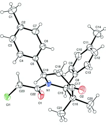
The molecular structure of the title compound is illustrated in Fig. 1. The sum of the bond angles around atom N1 is 359.39° indicating sp2 hybridization. The N1—C22 [1.349 (2) Å] and C22—O1[1.218 (2) Å] bond distances indicate
electron delocalization. The six membered piperidine ring (N1/C15-C19) adopts a distorted boat conformation. The two
p-tolyl rings are nearly orthogonal to each other with a dihedral angle of 83.33 (10)°. The methyl substituents are oriented equatorially [N1—C15—C16—C20 = 175 (16)°] and axially [N1—C15—C16—C21 = 56.52 (19)°] at the C3 position. The two p-tolyl (C2-C7 and C8-C13) are inclined to the mean plane of the piperidine ring by 73.2 (1) and 87.22 (9) °, respectively.
In the crystal, there are no significant intermolecular interactions present.
S2. Experimental
The title compound was synthesized according to a published procedure (Aridoss et al., 2007). To a well stirred solution of 3, 3-dimethyl-2, 6-di-p-tolyl piperidin-4-one (5 mmol), and triethylamine (5 mmol) in 20 ml of benzene, dichloro-acetylchloride (5 mmol) in 20 ml of benzene was added drop wise through the additional funnel over ca. 30 min. Stirring was continued with mild heating using a magnetic stirrer for 7 h. The progress of the reaction was monitored by TLC. After completion of reaction, the mixture was poured into water and extracted with ether. The collected ether extracts were then washed well with 3% sodium bicarbonate solution and dried over anhydrous Na2SO4. The pasty mass obtained
was purified by crystallization from distilled ethanol giving the compound in pure form as colourless block-like crystals.
S3. Refinement
H atoms were positioned geometrically and refined using a riding model: C—H = 0.93–0.98 Å with Uiso(H) = 1.5Ueq(C)
Figure 1
The molecular structure of the title compound, with atom labelling. Displacement ellipsoids are drawn at the 30% probability level.
1-(2-Chloroacetyl)-3,3-dimethyl-2,6-di-p-tolylpiperidin-4-one
Crystal data
C23H26ClNO2
Mr = 383.90 Monoclinic, C2/c
Hall symbol: -C 2yc
a = 18.7923 (6) Å
b = 18.8289 (5) Å
c = 11.6689 (3) Å
β = 93.162 (2)°
V = 4122.6 (2) Å3
Z = 8
F(000) = 1632
Dx = 1.237 Mg m−3
Mo Kα radiation, λ = 0.71073 Å Cell parameters from 8523 reflections
θ = 2.3–25.5°
µ = 0.20 mm−1
supporting information
sup-3
Acta Cryst. (2015). E71, o173–o174Data collection
Bruker Kappa APEXII CCD diffractometer
Radiation source: fine-focus sealed tube Graphite monochromator
ω and φ scan
Absorption correction: multi-scan
(SADABS; Bruker, 2004)
Tmin = 0.931, Tmax = 0.959
29055 measured reflections 3989 independent reflections 3097 reflections with I > 2σ(I)
Rint = 0.028
θmax = 25.8°, θmin = 2.2°
h = −23→22
k = −23→23
l = −14→14
Refinement
Refinement on F2
Least-squares matrix: full
R[F2 > 2σ(F2)] = 0.041
wR(F2) = 0.124
S = 1.03 3989 reflections 244 parameters 0 restraints
Primary atom site location: structure-invariant direct methods
Secondary atom site location: difference Fourier map
Hydrogen site location: inferred from neighbouring sites
H-atom parameters constrained
w = 1/[σ2(F
o2) + (0.0577P)2 + 3.5027P]
where P = (Fo2 + 2Fc2)/3
(Δ/σ)max < 0.001
Δρmax = 0.30 e Å−3
Δρmin = −0.22 e Å−3
Special details
Geometry. All e.s.d.'s (except the e.s.d. in the dihedral angle between two l.s. planes) are estimated using the full covariance matrix. The cell e.s.d.'s are taken into account individually in the estimation of e.s.d.'s in distances, angles and torsion angles; correlations between e.s.d.'s in cell parameters are only used when they are defined by crystal symmetry. An approximate (isotropic) treatment of cell e.s.d.'s is used for estimating e.s.d.'s involving l.s. planes.
Refinement. Refinement of F2 against ALL reflections. The weighted R-factor wR and goodness of fit S are based on F2,
conventional R-factors R are based on F, with F set to zero for negative F2. The threshold expression of F2 > σ(F2) is used
only for calculating R-factors(gt) etc. and is not relevant to the choice of reflections for refinement. R-factors based on F2
are statistically about twice as large as those based on F, and R- factors based on ALL data will be even larger.
Fractional atomic coordinates and isotropic or equivalent isotropic displacement parameters (Å2)
x y z Uiso*/Ueq
C1 0.99934 (14) 0.15212 (14) 0.4159 (3) 0.0785 (8) H1A 0.9762 0.1118 0.3802 0.118* H1B 1.0097 0.1423 0.4959 0.118* H1C 1.0429 0.1616 0.3795 0.118* C2 0.95102 (11) 0.21596 (11) 0.40374 (19) 0.0516 (5) C3 0.97183 (11) 0.28078 (11) 0.44787 (18) 0.0532 (5)
H3 1.0169 0.2854 0.4841 0.064*
C4 0.92751 (10) 0.33922 (10) 0.43974 (17) 0.0466 (5)
H4 0.9431 0.3823 0.4711 0.056*
C5 0.86037 (9) 0.33477 (9) 0.38582 (15) 0.0372 (4) C6 0.83961 (11) 0.26980 (10) 0.34030 (19) 0.0519 (5)
H6 0.7949 0.2652 0.3029 0.062*
C7 0.88421 (12) 0.21166 (11) 0.3495 (2) 0.0603 (6)
H7 0.8688 0.1685 0.3183 0.072*
H9 0.7118 0.3326 0.0669 0.060* C10 0.66551 (12) 0.23935 (12) 0.1004 (2) 0.0580 (6) H10 0.6770 0.2177 0.0322 0.070* C11 0.62763 (11) 0.20178 (11) 0.1777 (2) 0.0540 (5) C12 0.61085 (12) 0.23649 (12) 0.27655 (19) 0.0607 (6) H12 0.5842 0.2129 0.3296 0.073* C13 0.63251 (11) 0.30541 (12) 0.29906 (18) 0.0529 (5) H13 0.6203 0.3271 0.3668 0.064* C14 0.60485 (15) 0.12615 (13) 0.1559 (3) 0.0794 (8) H14A 0.5792 0.1093 0.2194 0.119* H14B 0.6462 0.0970 0.1475 0.119* H14C 0.5747 0.1239 0.0869 0.119* C15 0.70211 (9) 0.41716 (10) 0.24177 (16) 0.0388 (4) H15 0.6940 0.4422 0.1686 0.047* C16 0.66973 (10) 0.46453 (10) 0.33298 (16) 0.0433 (4) C17 0.68764 (10) 0.43549 (10) 0.45226 (16) 0.0426 (4) C18 0.75270 (10) 0.38867 (10) 0.46625 (16) 0.0449 (4) H18A 0.7370 0.3396 0.4611 0.054* H18B 0.7738 0.3958 0.5432 0.054* C19 0.81131 (9) 0.39888 (9) 0.38170 (15) 0.0369 (4) H19 0.8397 0.4402 0.4071 0.044* C20 0.58947 (11) 0.47386 (14) 0.3074 (2) 0.0619 (6) H20A 0.5810 0.4923 0.2312 0.093* H20B 0.5710 0.5064 0.3618 0.093* H20C 0.5661 0.4288 0.3134 0.093* C21 0.70482 (13) 0.53857 (11) 0.33111 (19) 0.0565 (5) H21A 0.6953 0.5600 0.2572 0.085* H21B 0.7554 0.5339 0.3459 0.085* H21C 0.6856 0.5680 0.3891 0.085* C22 0.82141 (10) 0.43472 (10) 0.17806 (16) 0.0419 (4) C23 0.90131 (10) 0.43746 (11) 0.20559 (18) 0.0494 (5) H23A 0.9206 0.3897 0.2052 0.059* H23B 0.9106 0.4570 0.2819 0.059* N1 0.78074 (7) 0.41404 (8) 0.26376 (12) 0.0364 (3) O1 0.79731 (8) 0.44923 (10) 0.08186 (12) 0.0640 (4) O2 0.65195 (8) 0.44814 (8) 0.53268 (12) 0.0578 (4) Cl1 0.94435 (3) 0.48999 (3) 0.10507 (5) 0.05769 (18)
Atomic displacement parameters (Å2)
U11 U22 U33 U12 U13 U23
supporting information
sup-5
Acta Cryst. (2015). E71, o173–o174C8 0.0325 (9) 0.0502 (11) 0.0366 (9) −0.0013 (8) −0.0010 (7) −0.0024 (8) C9 0.0458 (11) 0.0631 (13) 0.0421 (11) −0.0107 (9) 0.0082 (8) −0.0081 (9) C10 0.0530 (12) 0.0657 (14) 0.0558 (13) −0.0064 (10) 0.0087 (10) −0.0222 (11) C11 0.0475 (11) 0.0507 (12) 0.0632 (14) −0.0044 (9) −0.0033 (10) −0.0054 (10) C12 0.0657 (14) 0.0617 (14) 0.0556 (13) −0.0186 (11) 0.0106 (11) 0.0026 (11) C13 0.0562 (12) 0.0604 (13) 0.0432 (11) −0.0132 (10) 0.0111 (9) −0.0082 (9) C14 0.0800 (17) 0.0545 (14) 0.103 (2) −0.0098 (13) −0.0022 (15) −0.0091 (14) C15 0.0326 (9) 0.0460 (10) 0.0373 (9) 0.0011 (7) −0.0022 (7) 0.0009 (8) C16 0.0405 (10) 0.0446 (10) 0.0442 (11) 0.0056 (8) −0.0030 (8) −0.0038 (8) C17 0.0431 (10) 0.0423 (10) 0.0427 (10) −0.0013 (8) 0.0033 (8) −0.0077 (8) C18 0.0504 (11) 0.0487 (11) 0.0355 (10) 0.0053 (9) 0.0011 (8) 0.0014 (8) C19 0.0391 (9) 0.0370 (9) 0.0339 (9) 0.0012 (7) −0.0044 (7) 0.0012 (7) C20 0.0449 (12) 0.0752 (15) 0.0650 (14) 0.0169 (11) −0.0015 (10) −0.0091 (12) C21 0.0664 (14) 0.0447 (11) 0.0574 (13) 0.0061 (10) −0.0057 (11) −0.0001 (10) C22 0.0396 (10) 0.0468 (10) 0.0392 (10) −0.0015 (8) −0.0002 (8) 0.0064 (8) C23 0.0396 (10) 0.0571 (12) 0.0516 (12) −0.0056 (9) 0.0025 (8) 0.0161 (10) N1 0.0324 (7) 0.0433 (8) 0.0331 (8) −0.0007 (6) −0.0022 (6) 0.0033 (6) O1 0.0448 (8) 0.1056 (13) 0.0410 (8) −0.0026 (8) −0.0019 (6) 0.0212 (8) O2 0.0571 (9) 0.0680 (10) 0.0495 (8) 0.0061 (7) 0.0138 (7) −0.0085 (7) Cl1 0.0489 (3) 0.0632 (3) 0.0617 (3) −0.0087 (2) 0.0092 (2) 0.0175 (3)
Geometric parameters (Å, º)
C1—C2 1.508 (3) C14—H14B 0.9600
C1—H1A 0.9600 C14—H14C 0.9600
C1—H1B 0.9600 C15—N1 1.487 (2)
C1—H1C 0.9600 C15—C16 1.540 (3)
C2—C3 1.373 (3) C15—H15 0.9800
C2—C7 1.377 (3) C16—C17 1.516 (3)
C3—C4 1.380 (3) C16—C20 1.531 (3)
C3—H3 0.9300 C16—C21 1.543 (3)
C4—C5 1.381 (3) C17—O2 1.207 (2)
C4—H4 0.9300 C17—C18 1.509 (3)
C5—C6 1.381 (3) C18—C19 1.531 (3)
C5—C19 1.518 (2) C18—H18A 0.9700
C6—C7 1.379 (3) C18—H18B 0.9700
C6—H6 0.9300 C19—N1 1.489 (2)
C7—H7 0.9300 C19—H19 0.9800
C8—C9 1.385 (3) C20—H20A 0.9600
C8—C13 1.386 (3) C20—H20B 0.9600
C8—C15 1.520 (3) C20—H20C 0.9600
C9—C10 1.381 (3) C21—H21A 0.9600
C9—H9 0.9300 C21—H21B 0.9600
C10—C11 1.376 (3) C21—H21C 0.9600
C10—H10 0.9300 C22—O1 1.218 (2)
C11—C12 1.377 (3) C22—N1 1.349 (2)
C12—H12 0.9300 C23—H23A 0.9700
C13—H13 0.9300 C23—H23B 0.9700
C14—H14A 0.9600
C2—C1—H1A 109.5 C8—C15—C16 118.50 (15)
C2—C1—H1B 109.5 N1—C15—H15 106.0
H1A—C1—H1B 109.5 C8—C15—H15 106.0
C2—C1—H1C 109.5 C16—C15—H15 106.0
H1A—C1—H1C 109.5 C17—C16—C20 112.73 (17) H1B—C1—H1C 109.5 C17—C16—C15 110.46 (15) C3—C2—C7 117.28 (18) C20—C16—C15 110.77 (16) C3—C2—C1 121.0 (2) C17—C16—C21 105.40 (15) C7—C2—C1 121.7 (2) C20—C16—C21 108.11 (17) C2—C3—C4 121.64 (19) C15—C16—C21 109.16 (16) C2—C3—H3 119.2 O2—C17—C18 120.85 (18) C4—C3—H3 119.2 O2—C17—C16 122.55 (18) C3—C4—C5 121.03 (18) C18—C17—C16 116.60 (16) C3—C4—H4 119.5 C17—C18—C19 117.67 (16)
C5—C4—H4 119.5 C17—C18—H18A 107.9
C4—C5—C6 117.42 (17) C19—C18—H18A 107.9 C4—C5—C19 120.27 (16) C17—C18—H18B 107.9 C6—C5—C19 122.27 (16) C19—C18—H18B 107.9 C7—C6—C5 121.02 (19) H18A—C18—H18B 107.2 C7—C6—H6 119.5 N1—C19—C5 112.60 (14) C5—C6—H6 119.5 N1—C19—C18 111.41 (14) C2—C7—C6 121.6 (2) C5—C19—C18 109.69 (14)
C2—C7—H7 119.2 N1—C19—H19 107.6
C6—C7—H7 119.2 C5—C19—H19 107.6
supporting information
sup-7
Acta Cryst. (2015). E71, o173–o174H14A—C14—H14B 109.5 Cl1—C23—H23B 109.4 C11—C14—H14C 109.5 H23A—C23—H23B 108.0 H14A—C14—H14C 109.5 C22—N1—C15 117.32 (14) H14B—C14—H14C 109.5 C22—N1—C19 122.34 (14) N1—C15—C8 110.26 (14) C15—N1—C19 119.73 (14) N1—C15—C16 109.30 (14)