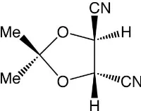
(4
S
,5
S
)-2,2-Dimethyl-1,3-dioxolane-4,5-dicarbonitrile
Alan H. Haines* and David L. Hughes*
School of Chemistry, University of East Anglia, Norwich NR4 7TJ, England Correspondence e-mail: a.haines@uea.ac.uk, d.l.hughes@uea.ac.uk
Received 7 May 2013; accepted 7 June 2013
Key indicators: single-crystal X-ray study;T= 140 K; mean(C–C) = 0.001 A˚;
Rfactor = 0.030;wRfactor = 0.076; data-to-parameter ratio = 16.9.
The title compound, C7H8N2O2, formed by dehydration of the corresponding dicarboxamide, crystallizes as rectangular prisms. The molecules have a C2 axis of symmetry through the C atom bearing the methyl groups and the mid-point of the ring C—C bond, and the 1,3-dioxolane ring adopts the extreme twist conformation of the two possible with this symmetry. This brings the two nitrile groups nearest to a linear arrangement when the molecule is viewed along the ring C—C bond. The correct absolute configuration of the molecule was defined by that of the original starting material, (2R,3R )-tartaric acid. The packing is largely controlled by a number of C—H N interactions.
Related literature
For the first syntheses of the title compound, see: Briggset al.
(1985). For determination of the absolute configuration of (+)-tartaric acid, see: Bijvoetet al.(1951). For related structures, see: (4R,5R)-2,2-dimethyl-1,3-dioxolane-4,5-dicarboxamide, Shainyan et al. (2002); (2R,3S )-2,3-dihydroxy-2,3-dicyano-ethane and (2R,3S)-2,3-dibenzoyloxy-2,3-dicyanoethane, Rychlewska et al. (2008); and (2S,3S )-2,3-dibenzoyloxy-2,3-dicyanoethane, Gawron´ski et al. (2007). For the Flack x
parameter, see: Flack (1983).
Crystal data
C7H8N2O2 Mr= 152.15 Tetragonal,P41212 a= 8.7740 (2) A˚ c= 10.0282 (3) A˚ V= 772.00 (3) A˚3
Z= 4
MoKradiation
= 0.10 mm1 T= 140 K
0.080.070.07 mm
Data collection
Oxford Diffraction Xcalibur 3/ Sapphire3 CCD diffractometer Absorption correction: multi-scan
(CrysAlis PRO REDRED; Oxford Diffraction, 2010) Tmin= 0.886,Tmax= 1.000
14922 measured reflections 1133 independent reflections 1073 reflections withI> 2(I) Rint= 0.032
Refinement
R[F2> 2(F2)] = 0.030 wR(F2) = 0.076 S= 1.09 1133 reflections
67 parameters
All H-atom parameters refined
max= 0.28 e A˚
3
min=0.11 e A˚
[image:1.610.120.220.592.671.2]3
Table 1
Hydrogen-bond geometry (A˚ ,).
D—H A D—H H A D A D—H A
C2—H2 N21i
0.946 (13) 2.450 (13) 3.2530 (14) 142.6 (10)
Symmetry code: (i)xþ1 2;yþ
3 2;zþ
3 4.
Data collection:CrysAlis PRO CCD(Oxford Diffraction, 2010); cell refinement:CrysAlis PRO RED(Oxford Diffraction, 2010); data reduction:CrysAlis PRO RED; program(s) used to solve structure: SHELXS97(Sheldrick, 2008); program(s) used to refine structure: SHELXL97 (Sheldrick, 2008); molecular graphics: ORTEPII (Johnson, 1976) andORTEP-3 for Windows(Farrugia, 2012); soft-ware used to prepare material for publication: SHELXL97 and WinGX(Farrugia, 2012).
Supplementary data and figures for this paper are available from the IUCr electronic archives (Reference: SJ5320).
References
Bijvoet, J. M., Peerdeman, A. F. & van Bommel, A. J. (1951).Nature,168, 271. Briggs, M. A., Haines, A. H. & Jones, H. F. (1985).J. Chem. Soc. Perkin Trans.
1, pp. 795–798.
Farrugia, L. J. (2012).J. Appl. Cryst.45, 849–854. Flack, H. D. (1983).Acta Cryst.A39, 876–881.
Gawron´ski, J., Gawron´ska, K., Was´cinska, N., Plutecka, A. & Rychlewska, U. (2007).Pol. J. Chem.81, 1917–1925.
Johnson, C. K. (1976).ORTEPII. Report ORNL-5138. Oak Ridge National Laboratory, Tennessee, USA.
Oxford Diffraction (2010).CrysAlis PRO. Oxford Diffraction Ltd, Yarnton, England.
Rychlewska, U., Was´cinska, N., Warz˙ajtis, B. & Gawron´ski, J. (2008).Acta Cryst.B64, 497–503.
Shainyan, B. A., Ustinov, M. V., Bel’skii, B. K. & Nindakova, L. O. (2002). Russ. J. Org. Chem.38, 104–110.
Sheldrick, G. M. (2008).Acta Cryst.A64, 112–122.
Structure Reports
Online
supporting information
Acta Cryst. (2013). E69, o1104 [https://doi.org/10.1107/S1600536813015973]
(4
S
,5
S
)-2,2-Dimethyl-1,3-dioxolane-4,5-dicarbonitrile
Alan H. Haines and David L. Hughes
S1. Comment
The stereoisomeric forms of tartaric acid have played a central role in determining the absolute and relative
stereochemistries of chiral carbon compounds, and our knowledge of the absolute configurations of such organic
compounds stems from determination of the absolute configuration of the sodium rubidium salt of (+)-tartaric acid
(Bijvoet et al., 1951). Since that time, many structural determinations by X-ray crystallography have been performed on derivatives of the three isomeric forms of tartaric acid, the chiral (R,R)- and (S,S)-isomers and the meso (R,S)-isomer. Relevant to our structural determination of the title compound are reports on the crystal structures of: (i) its precursor
(4R,5R)-2,2-dimethyl-1,3-dioxolane-4,5-dicarboxamide (Shainyan et al., 2002) - note: the structural diagram in this paper recording the compound's crystallographic data depicts, erroneously, the (4S,5S)-isomer despite the fact that the stated synthesis is from (2R,3R)-tartaric acid); (ii) (2R,3S)-2,3-dihydroxy-2,3-dicyanoethane (Rychlewska et al., 2008); (iii) (2S,3S)-2,3-dibenzoyloxy-2,3-dicyanoethane (Gawroński et al., 2007); and (iv) (2R,3S )-2,3-dibenzoyloxy-2,3-dicyano-ethane (Rychlewska et al., 2008).
We previously synthesized (4S,5S)-2,2-dimethyl-1,3-dioxolane-4,5-dicarbonitrile in 80.5% yield as a highly crystalline solid m.p. 163–164 °C by treatment of (4R,5R)-2,2-dimethyl-1,3-dioxolane-4,5-dicarboxamide with benzenesulfonyl chloride in pyridine (Briggs et al., 1985). [N.B. In the paper by Briggs et al., 1985, the stereochemical descriptors for positions 4 and 5 of the dioxolane ring were incorrectly assigned as R; it should be noted that conversion of an amide function into nitrile lowers the order of preference according to the sequence-rules.] Two noteworthy properties of the
dicarbonitrile were (i) its resistance to hydrolysis by trifluoroacetic acid-water, an acidic medium which normally brings
about ready hydrolysis of the type of acetal group present in this compound, and (ii) the lack of absorptions attributable
to a nitrile group in its IR spectrum although an expected absorption was present in its Raman spectrum. Interestingly,
(S,S)-2,3-dihydroxy-2,3-dicyanoethane, which was the desired hydrolysis product of the dicarbonitrile, also proved difficult to synthesize from the unprotected (R,R)-tartaric acid diamide, was only obtained in low yield, and is not stable at room temperature (Rychlewska et al., 2008). In contrast, (R,S)-1,2-dihydroxy-1,2-dicyanoethane has been isolated as a stable crystalline solid from the mixture of (R,R), (S,S) and (R,S)-dinitriles obtained in the reaction of glyoxal with potassium cyanide and hydrochloric acid (Rychlewska et al., 2008).
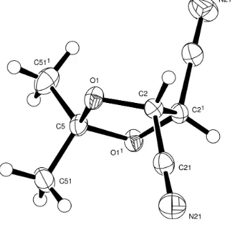
Our molecule has a 5-membered ring with a twist conformation, Figure 1. It lies about a twofold symmetry axis though
the Me2-carbon atom C5 and the mid-point of the ring C—C bond. Of the two possible extreme twist conformations
which may be so defined that one is adopted which brings the two nitrile groups nearest to linearity with the torsional
angle C21—C2—C21—C211 at -155.70 (11)°; the alternative twist conformation would set the nitrile groups with a
torsional angle near -90°.
leading to a very weak or absent overall absorption for the CN groups. However, the nitrile groups in our structure are not
parallel but related by a twofold screw (21) symmetry axis, and the angle between two C–N vectors is 64.35 (8)°.
The absolute configuration of the molecule cannot be determined unequivocally from the X-ray data, but is known from
that of the precursor, (2R,3R)-tartaric acid, and is that shown in Figure 1. We note that the Flack x parameter (Flack, 1983) is 0.8 (11), the value of which suggests that we should invert the structure; however, the large s.d. on this value
indicates that this parameter has not been reliably determined from the diffraction data.
In contrast to the C2 symmetry possessed by the dinitrile, the dicarboxamide precursor lacks similar symmetry and the
shape of its 1,3-dioxolane ring lies close to an envelope conformation with one of the ring O atoms, O2 in the original
publication (Shainyan et al., 2002), 0.503 Å out of the mean plane of the remaining ring atoms; extensive inter- and intra-molecular hydrogen bonding occurs involving the amide groups. In our structure, the packing is largely controlled by a
number of C–H···N contacts.
The parent (S,S)-1,2-dihydroxy-1,2-dicyanoethane is non-crystalline but the crystal structure of the corresponding (R,S)-1,2-dihydroxy-1,2-dicyanoethane has been reported (Rychlewska et al., 2008) and it has a perfectly staggered conformation about the central C—C bond suggesting that an anti-parallel arrangement of vicinal nitrile groups may be a
strong driving force influencing conformational preference and thus influencing the choice of twist conformations in the
title compound.
S2. Experimental
The preparation of the title compound, m.p. 163–164 °C (sublimes above 120 °C), [α]D -83 (c, 0.6), together with its IR,
Raman, and 1H and 13C NMR spectra has been described (Briggs et al., 1985).
S3. Refinement
The non-hydrogen atoms were refined with anisotropic thermal parameters. All the hydrogen atoms were located in a
Figure 1
Figure 2
View down the crystallographic a axis showing the apparent alignment of nitrile groups along a twofold screw axis.
(4S,5S)-2,2-Dimethyl-1,3-dioxolane-4,5-dicarbonitrile
Crystal data
C7H8N2O2
Mr = 152.15
Tetragonal, P41212
Hall symbol: P 4abw 2nw
a = 8.7740 (2) Å
c = 10.0282 (3) Å
V = 772.00 (3) Å3
Z = 4
F(000) = 320
Dx = 1.309 Mg m−3
Mo Kα radiation, λ = 0.71073 Å Cell parameters from 5971 reflections
θ = 3.1–32.7°
µ = 0.10 mm−1
T = 140 K Cube, colourless 0.08 × 0.07 × 0.07 mm
Data collection
Oxford Diffraction Xcalibur 3/Sapphire3 CCD diffractometer
Radiation source: Enhance (Mo) X-ray Source Graphite monochromator
Detector resolution: 16.0050 pixels mm-1
Thin–slice φ and ω scans
Absorption correction: multi-scan
(CrysAlis PRO RED; Oxford Diffraction, 2010)
Tmin = 0.886, Tmax = 1.000
14922 measured reflections 1133 independent reflections 1073 reflections with I > 2σ(I)
Rint = 0.032
θmax = 30.0°, θmin = 3.1°
h = −12→12
k = −12→12
Refinement
Refinement on F2
Least-squares matrix: full
R[F2 > 2σ(F2)] = 0.030
wR(F2) = 0.076
S = 1.09 1133 reflections 67 parameters 0 restraints
Primary atom site location: structure-invariant direct methods
Secondary atom site location: difference Fourier map
Hydrogen site location: difference Fourier map All H-atom parameters refined
w = 1/[σ2(F
o2) + (0.0398P)2 + 0.0831P]
where P = (Fo2 + 2Fc2)/3
(Δ/σ)max < 0.001
Δρmax = 0.28 e Å−3
Δρmin = −0.11 e Å−3
Special details
Geometry. All s.u.'s (except the s.u. in the dihedral angle between two l.s. planes) are estimated using the full covariance matrix. The cell s.u.'s are taken into account individually in the estimation of s.u.'s in distances, angles and torsion angles; correlations between s.u.'s in cell parameters are only used when they are defined by crystal symmetry. An approximate (isotropic) treatment of cell s.u.'s is used for estimating s.u.'s involving l.s. planes.
Refinement. Refinement of F2 against ALL reflections. The weighted R-factor wR and goodness of fit S are based on F2,
conventional R-factors R are based on F, with F set to zero for negative F2. The threshold expression of F2 > 2σ(F2) is
used only for calculating R-factors(gt) etc. and is not relevant to the choice of reflections for refinement. R-factors based on F2 are statistically about twice as large as those based on F, and R- factors based on ALL data will be even larger.
Fractional atomic coordinates and isotropic or equivalent isotropic displacement parameters (Å2)
x y z Uiso*/Ueq
O1 0.46094 (8) 0.42363 (7) 0.38782 (6) 0.02318 (17) C2 0.53777 (10) 0.55849 (10) 0.42482 (9) 0.02062 (18) C21 0.44612 (12) 0.69757 (11) 0.39872 (10) 0.0267 (2) N21 0.37695 (12) 0.80532 (11) 0.37960 (11) 0.0416 (3)
C5 0.37130 (10) 0.37130 (10) 0.5000 0.0224 (3)
C51 0.20468 (13) 0.40426 (15) 0.47877 (13) 0.0359 (3) H2 0.6296 (15) 0.5652 (14) 0.3757 (12) 0.026 (3)* H51A 0.1892 (16) 0.5121 (18) 0.4539 (15) 0.045 (4)* H51B 0.151 (2) 0.3837 (19) 0.5586 (17) 0.052 (4)* H51C 0.1670 (19) 0.339 (2) 0.4073 (19) 0.057 (5)*
Atomic displacement parameters (Å2)
U11 U22 U33 U12 U13 U23
O1 0.0289 (3) 0.0237 (3) 0.0169 (3) −0.0052 (3) 0.0036 (2) −0.0023 (2) C2 0.0204 (4) 0.0208 (4) 0.0206 (4) −0.0010 (3) 0.0000 (3) 0.0025 (3) C21 0.0264 (5) 0.0264 (4) 0.0272 (4) −0.0018 (4) −0.0049 (4) 0.0038 (4) N21 0.0405 (6) 0.0330 (5) 0.0513 (7) 0.0068 (4) −0.0120 (5) 0.0053 (5) C5 0.0237 (4) 0.0237 (4) 0.0196 (6) −0.0054 (5) 0.0034 (3) −0.0034 (3) C51 0.0230 (5) 0.0461 (7) 0.0385 (6) −0.0066 (5) −0.0005 (4) −0.0117 (5)
Geometric parameters (Å, º)
C2—C2i 1.5297 (17) C51—H51A 0.988 (15)
C2—H2 0.946 (13) C51—H51B 0.946 (18)
C21—N21 1.1397 (13) C51—H51C 0.975 (19)
C2—O1—C5 108.73 (7) O1—C5—C51i 108.28 (5)
O1—C2—C21 112.59 (7) O1i—C5—C51 108.28 (5)
O1—C2—C2i 102.49 (5) O1—C5—C51 110.91 (6)
C21—C2—C2i 109.62 (9) C51i—C5—C51 113.15 (13)
O1—C2—H2 108.8 (7) C5—C51—H51A 110.7 (9)
C21—C2—H2 108.6 (7) C5—C51—H51B 109.1 (10)
C2i—C2—H2 114.8 (8) H51A—C51—H51B 109.1 (12)
N21—C21—C2 179.18 (12) C5—C51—H51C 108.7 (9)
O1i—C5—O1 105.03 (10) H51A—C51—H51C 109.3 (15)
O1i—C5—C51i 110.91 (6) H51B—C51—H51C 109.9 (14)
C5—O1—C2—C21 −88.69 (8) C2—O1—C5—C51 104.75 (9)
C5—O1—C2—C2i 28.99 (9) O1—C2—C2i—O1i −35.25 (11)
C2—O1—C5—O1i −12.00 (4) O1—C2—C2i—C21i 84.52 (7)
C2—O1—C5—C51i −130.54 (9) C21—C2—C2i—C21i −155.70 (11)
Symmetry code: (i) y, x, −z+1.
Hydrogen-bond geometry (Å, º)
D—H···A D—H H···A D···A D—H···A
C2—H2···N21ii 0.946 (13) 2.450 (13) 3.2530 (14) 142.6 (10)