Low Temperature Swelling in Beta SiC Associated with Point Defect Accumulation
5
0
0
Full text
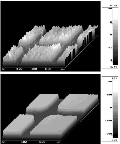
(2) Low Temperature Swelling in Beta-SiC. 613. by the damage range thickness (Rd ); S s = ∆z s /Rd. 2.5. 0.0025. 2.0. 0.0020. 1.5. 0.0015. dpa 1.0. 0.0010. 0.5. 0.0005. ion deposition 0.0 0. 500 1000 1500 2000 2500 Distance from Irradiated Surface, z / nm. Deposited Ni Concentration, CNi / apa. Displacement, H t / dpa. Fig. 1 Surface oblique plots of beta-SiC irradiated with 4 MeV nickel ions at 333 K to 0.01 dpa (upper) and 1.0 dpa (lower), respectively, through 0.254 mm-pitch molybdenum grid meshes following a surface finish with diamond paste.. 0.0000 3000. Fig. 2 Profiles of displacement damage and deposited Ni in irradiated monolithic SiC at a nominal fluence level of 1dpa. Calculation was performed assuming a sublattice-averaged displacement threshold energy of 35 eV and a target mass density of 3.21 × 10³ kg/m3 .. The practical resolution in the profiling is determined by the surface smoothness of the object. Examples of surface profilometry for the cases of 0.01 and 1.0 dpa at 333 K are presented in Fig. 1 as oblique plots. Figure 2 shows the calculated depth-profiles of displacement damage (dpa; displacement-per-atom) and stopped ions in silicon carbide for the case of 4 MeV nickel irradiation. A nominal damage level was defined as the dpa averaged over the damage range. The single-beam irradiation-induced swelling (S s ) was determined as the step-height (∆z s ) divided. (1). According to the calculation in Fig. 2, estimated contribution from ion deposition to the surface height increase is 0.45 nm/dpa, assuming unchanged atomic density due to the chemical effect of implantation. On the other hand, an estimated sputtering rate of the irradiated surface is ∼ 0.1 nm/dpa. Since these contributions are sufficiently small compared to the observed swelling-induced height changes, which were more than 20 nm at 1 dpa in the irradiation conditions in this study, we conclude that the effects of ion deposition and surface sputtering are negligible. However, because of the potential phase instability and atomic density change associated with ion implantation, the influences of ion deposition should carefully by examined in case the irradiation effects to very high fluence levels are of interest. In the case of dual-beam irradiation, additional swelling due to the co-implantation of helium was calculated by dividing the additional step height change (∆z d − ∆z s ) by the thickness of helium implantation range (RHe ), based on the fact that a partial damage range had been co-implanted with helium in the employed experimental set-up. The dual-beam irradiated swelling was derived as a sum of single-beam irradiated swelling and the added swelling due to helium coimplantation; S d = ∆z s /Rd + (∆z d − ∆z s )/RHe. (2). For a microstructural examination by cross-sectional transmission electron microscopy (XTEM), the irradiated specimens were subjected to a thin-foil processing using a focused ion beam (FIB) micro-processing device.8) The microstructural investigation was performed with JEOL JEM-2010 conventional TEM. Optical microscopy was supplementary utilized for surface feature characterization. 3. Results and Discussion Charged particle bombardment causes a roughening of the incident surface in scales ranging from inter-atomic distance to crystal grain size. For the surface height determination using optical profilometry, roughening in scales of 1 to 10 µm most likely imposes a quantitative uncertainty. The root-mean-squared (RMS) surface roughness as a function of displacement damage level at 333 K is presented in Fig. 3, where the roughness seems to be cumulatively increasing approximately in proportion to the ion fluence. An examination by Nomarski interferometric optical microscopy revealed that the morphology of height variations in the irradiated area was corresponding to crystal grain structure. The degree of the roughness increase was close to or less than 1% of the swelling in any case. Since it is well known that the irradiation-induced swelling of cubic silicon carbide is isotropic in stress-free conditions, the variation in crystallographic orientation probably caused a relatively minor grainto-grain height variation through interactive constraints in a polycrystalline system in the presence of irradiation-induced plastic flow. The fluence dependence of swelling was taken within a fluence range of 0.0026–3 dpa at 333 to 873 K as plotted in.
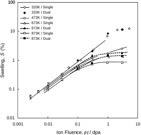
(3) Y. Katoh, H. Kishimoto and A. Kohyama Surface roughness, R / nm-rms. 614 6. CVD-SiC Irradiated by 4MeV Ni T = 333K. 5 4 3 2 1 0 0. 1. 2. 3. 4. Disp lacement Damage, t / dpa. Irradiated(3dpa @333K) Fig. 3 RMS surface roughness of CVD-SiC as a function of irradiation fluence (upper) and a Nomarski interferometric optical image of the irradiated sample (lower). The dotted line is a least-square linear fit.. 100. Swelling, S. 10. 333K / Single 333K / Dual 473K / Single 673K / Single 673K / Dual 873K / Single 873K / Dual. 1. 0.1. 0.01 0.001. 0.01. 0.1. 1. 10. Ion Fluence, t / dpa. Fig. 4 Fluence-dependence of irradiation-induced swelling at various temperatures.. Fig. 4. The surface roughness was the primary factor that imposed an uncertainty in the swelling measurement. The maximum uncertainty corresponded to 0.02% of swelling. At 333 K, the swelling increased apparently in proportion to the 0.81 power of the fluence until it approached the saturated value. Physical significance of such dependency may not be able to be clarified, since a very complex defect accumulation and recovery processes, which may incorporate cascade overlapping and/or interactions, Frankel defect recombination, anti-site production, stable cluster production, structural relaxation and dissociation of unstable clusters, etc., are oper-. ating in addition to an uneven damage profile in the irradiated volume. However, if one assumes a linear defect accumulation up to 0.0026 dpa and that each atomic displacement results in a swelling corresponding to one atomic volume for perfect silicon carbide by then, as a very rough estimation, a surviving defect fraction to NRT-dpa can be derived to be 21%.9) Since the fluences for apparent cascade overlapping onset in facecentered cubic metals are generally estimated to be within a lower milli-dpa range,10) cascade overlapping should not impose a large error to this estimation with a power law index of 0.81. The production and recovery behavior of individual defect specie will be studied in more details in separate works. A rather rapid swelling at near 1dpa at 333 K (Fig. 4) indicates a progressive crystalline-to-amorphous phase transition that initiated at the damage peak zone. The swelling due to full amorphization in the single-beam irradiated volume at 5 dpa at 333 K appeared to be 12.6%. This number falls in a typical range of swelling of originally full-dense silicon carbide associated with neutron-induced amorphization, which is reported to be 11–15%.11) At temperatures up to 873 K, the apparent defect accumulation rate decreased with the increasing temperature, as shown in Fig. 4. The swelling exhibited a tendency toward saturation at these temperatures. The estimated saturation fluence was smaller at a higher temperature. Hereafter, the swelling at 3dpa is regarded as a saturated swelling, since a significant swelling increase was not observed after 1 dpa at temperatures > 673 K. The influence of displacement damage rate on swelling behavior was not very significant but detectable in a consistent way as presented in Fig. 5. At temperature of 333 K, the reduction in displacement rate by one order decreased swelling by approximately 20% at a given fluence level, however, did not significantly change the swelling exponent. This indicates that thermally activated migration of self interstitial atoms is very likely even at temperature where irradiation-induced amorphization is allowed. The effect of displacement rate within the power-law swelling regime at higher temperatures appeared similar to that at 333 K. However, swelling after deviation from the power-law regime exhibits a reduced sensitivity to displacement rate until it becomes nearly independent of displacement rate at over about 0.3 dpa at both 473 K and 873 K. Because vacancy migration is almost prohibited in this temperature range,12) a volumetric swelling should be close to vacancy concentration and, on the other hand, a saturated vacancy concentration is determined by an equilibrium of vacancy production and Frenkel defect recombination through migration of self interstitial atoms (SIA). Therefore, the experimental observation of saturated swelling that is independent of displacement rate suggests that the equilibrium SIA concentration in a saturated swelling regime is proportional to the displacement rate. Such a situation is enabled only when the Frenkel defect recombination rate is much higher than the SIA annihilation rate at other sinks and the sink conditions are not strongly affected by the displacement rate. In Fig. 6, the swelling at 3dpa obtained in this work is plotted as a function of irradiation temperature, along with reported neutron irradiation data.4) All the ion irradiation data.
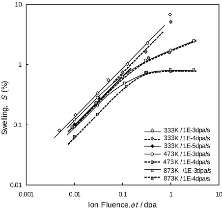
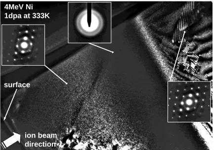
(4) Low Temperature Swelling in Beta-SiC 10. Swelling, S ( ). 1. 0.1. 0.01 0.001. 333K / 1E-3dpa/s 333K / 1E-4dpa/s 333K / 1E-5dpa/s 473K / 1E-3dpa/s 473 K / 1E-4dpa/s 873K /1E-3dpa/s 873K / 1E-4dpa/s 0.01. 0.1. 1. 10. Ion Fluence, t / dpa. Fig. 5 The influence of displacement damage rate on fluence-dependent swelling behavior of beta-SiC.. 100. Present work (single-beam) Present work (dual-beam) Price(1969,1973) Blackstone(1971) Snead(1998). Swelling, S. 10. 1. 0.1 200. 600. 1000. 1400. 1800. Irradiation Temperature, T / K. Fig. 6 Temperature-dependence of saturated swelling. Neutron data are from compilation in Ref. 11).. points fall close to the bottom of a neutron data band. Considering the outstanding controllability of irradiation conditions in the present ion irradiation experiment, we suspect that majority of neutron data have been affected by irradiation temperature uncertainty and/or unintentional transient low temperature neutron exposure that is peculiar to the irradiation in fission reactor cores.13) In addition, some of the neutron data might have been affected by transmutation of boron, that had been added as a sintering aid to produce dense materials. An XTEM image, taken at a {111} reflection in a dark-field condition, of the specimen irradiated to 1dpa at 333 K is provided in Fig. 7. An amorphous region was identified at around the damage peak depth. An amorphization threshold dose can accurately be determined from the XTEM by overlaying a calculated damage profile. The threshold dose of 1.07 dpa was obtained from this examination in the irradiation condition of 333 K and 1 × 10⁻³ dpa/s. The estimated accuracy of threshold dose determination in this procedure is within a few per-. 615. cent, although the conditions for calculation by SRIM may exert a more significant uncertainty. This data appeared to fall strictly on the reported line of temperature dependence of amorphization threshold dose for cases both hexagonal and cubic silicon carbide irradiated with heavy ion (Si and Fe) in an MeV energy range.14) In the damaged but still crystalline region, very fine defect clusters with unclear images were observed at high density. The types and configurations of these clusters were not identified. The density of dislocations in the substrate, or the region beyond the ion range, associated with a plastic deformation was not high, considering the extent of swelling in the adjacent amorphized band. This observation supports that the plastic flow associated with the irradiationinduced swelling occurred mostly toward the constraint-free direction and consequently the degree of linear deformation in this experiment is corresponding to that of the isotropic deformation in unconstrained conditions. 4. Conclusions An experimental technique to study the irradiation-induced volume changes and microstructural evolution in silicon carbide in accurately controlled irradiation conditions was successfully developed. The absolute accuracy in volume change measurement for the current technique is approximately 0.02%, which is determined by the specimens’ surface roughness. Surface roughness of the irradiated silicon carbide appeared to increase approximately in proportion to the ion fluence. However, the irradiation-induced roughening was not significantly disturbing the measurement, since it was minor and seemed to be related with the degree of volume changes. All the irradiated samples exhibited swelling that tended to saturate at certain fluences. The swelling increased with the increased ion fluence and the swelling rate and saturated volume decreased as temperature was elevated. The obtained temperature dependence of saturated swelling fell at the bottom of the reported neutron data band. A low fluence irradiation data suggested about 20% of surviving defect fraction to NRT-dpa at 333 K. Variations in defect production rate caused only minor change in swelling rates and almost no effect on a saturated swelling value. Amorphization was observed only at 333 K. The threshold dose was determined to be 1.07 dpa through an XTEM microstructural examination. The irradiated microstructure was high-density defect clusters with unclear images in TEM at 333 K. Acknowledgements This work is supported by Laboratory for Complex Energy Processes, Institute of Advanced Energy, Kyoto University and Core Research for Evolutional Science and Technology (CREST), Japan Science and Technology Corporation (JST). REFERENCES 1) A. Kohyama, Y. Katoh, T. Hinoki, W. Zhang and M. Kotani: Proceedings of the Eighth European Conference on Composite Materials, Vol. 4 (1998) 15–22..
(5) 616. Y. Katoh, H. Kishimoto and A. Kohyama. 4MeV Ni 1dpa at 333K. surface. ion beam direction Fig. 7 Cross-sectional transmission electron micrograph of cubic silicon carbide irradiated with 4 MeV nickel to 1 dpa at 333 K. Z ∼ 110 , g = {111}.. 2) P. Fenici, A. J. Frias Rebelo, R. H. Jones, A. Kohyama and L. L. Snead: J. Nucl. Mater. 258–263 (1998) 215–225. 3) S. J. Zinkle and C. Kinoshita: J. Nucl. Mater. 251 (1997) 200–217. 4) S. J. Zinkle and L. L. Snead: Fusion Materials, DOE/ER-0313/24 (1998) 93–114. 5) A. Kohyama, Y. Katoh, M. Ando and K. Jimbo: Fusion Engineering and Design 51–52 (2000) 789–795. 6) J. F. Ziegler: Ion Beam Interactions with Matter, http://www.research.ibm.com/ionbeams/ 7) H. L. Heinisch: Proc. of 2nd IEA/JUPITER Joint Int. Workshop on SiC/SiC Ceramic Composites for Fusion Application (1997) 156–161. 8) Y. Katoh, A. Kohyama and T. Hinoki: Proceedings of the Eighth European Conference on Composite Materials, Vol. 4 (1998) 351–357.. 9) B. N. Singh and S. J. Zinkle: J. Nucl. Mater. 206 (1993) 212–229. 10) K. Kitagawa, K. Yamakawa, H. Fukushima, T. Yoshiie, Y. Hayashi, H. Yoshida, Y. Shimomura and M. Kiritani: J. Nucl. Mater. 133&134 (1985) 395–399. 11) C. J. McHargue and J. M. Williams: Nucl. Instrum. Meth. B80/81 (1993) 889–894. 12) H. Itoh, M. Yoshikawa, I. Nashiyama, H. Okamura, S. Misawa and S. Yoshida: J. Appl. Phys. 77 (1995) 837–842. 13) M. Kiritani: J. Nucl. Mater. 160 (1988) 135–141. 14) L. L. Snead and S. J. Zinkle: Mater. Res. Soc. Symp. Proc. 439 (1997) 595–606..
(6)
Figure
Related documents