Received 21 July 2017 Accepted 29 August 2017
Edited by M. Weil, Vienna University of Technology, Austria
Keywords:crystal structure; dimethyl sulfoxide; van der Waals interaction; geometric para-meters; crystal packing.
CCDC reference:1571260
Supporting information:this article has supporting information at journals.iucr.org/e
Structural parameters of dimethyl sulfoxide, DMSO,
at 100 K, based on a redetermination by use of
high-quality single-crystal X-ray data
Hans Reuter*
Institute of Chemistry of New Materials, University of Osnabru¨ck, Barbarastrasse 7, 49069 Osnabru¨ck, Germany. *Correspondence e-mail: [email protected]
The title compound, C2H6OS, is a high melting, polar and aprotic solvent widely used in organic and inorganic chemistry. It serves as a H-atom acceptor in hydrogen bonding and is used as an ambidentate ligand in coordination chemistry. The evaluation of the influence of intermolecular interactions on the internal structural parameters of the chemically bonded DMSO molecules affords precise structural data of the free molecule as a point of reference. So far, valid data have been obtained only by use of neutron powder diffraction [Ibberson (2005).Acta Cryst.C61, o571–o573]. In the present redetermination, structural data have been obtained from a single-crystal X-ray diffraction experiment at 100 K, revealing a better comparison with DMSO molecules in other crystal structures. In the solid state, the pyramidal molecule exhibits a nearly perfectCssymmetry [including H atoms, which are eclipsed with respect to the C C axis], with a C—S—C bond angle of 97.73 (7)and an S—O bond
length of 1.5040 (10) A˚ , corresponding very well with an S O double bond, and with almost equal S—C bond lengths [mean value = 1.783 (4) A˚ ] and O—S—C bond angles [mean value = 106.57 (4)]. The crystal packing is influenced by C—
H O interactions (2.42–2.47 A˚ ) between all three H atoms of only one methyl group with the O atoms of three neighbouring DMSO molecules. The interactions of the O atom with H atoms (or Lewis acids, or hydrogen-donor groups) of adjacent molecules in relation to the orientation of the complete DMSO molecule are described in terms of the angle!and the distancednorm;!
is the angle between the pseudo-mirror plane of the molecule and the plane defined through the S O bond and the interacting atom, and dnorm is the distance of the interacting atom from the plane perpendicular to the S O bond.
1. Chemical context
Dimethyl sulfoxide (DMSO), (CH3)2SO, is a colourless polar aprotic solvent with high melting (291 K) and boiling points (462 K), miscible with a wide range of organic solvents and water. It is commonly used in organic and inorganic chemistry because of its capability to dissolve numerous polar or nonpolar compounds. In addition to its solvation properties, the molecule may act as a H-atom acceptor in hydrogen bonding, as well as an ambidentate Lewis base in coordination compounds. In the latter case, DMSO reactivity follows the HSAB principle (Pearson, 1963) which means that in combi-nation with ‘hard’ acids like tin(IV), DMSO coordinates via
the ‘hard’ O atom [e.g. iPrSnCl3(DMSO-O)2; Kastner & Reuter, 1999] and in combination with ‘soft’ acids like plati-num(II) via the ‘soft’ S atom [e.g. cis-PtCl2(DMSO-S)2; Melanson & Rochon, 1975], while with acids at the ‘hard–soft’ borderline like ruthenium(II), both coordination modes can be realized [cis-RuCl2(DMSO-O)1(DMSO-S)3; Tarighi &
Abbasi, 2007]. DMSO is also used in pharmacology in trans-dermal drug delivery applications and in veterinary medicine.
Both hydrogen-bond formation and formation of coordi-nation bonds will change the structural parameters of the DMSO molecule, as was shown by Calligaris (2004) for DMSO and other sulfoxides. For the evaluation of the influence of these additional intermolecular bonds on the internal struc-tural parameters of the coordinating or hydrogen-bonded DMSO ligands, precise data on bond lengths and angles within the free molecule are required as a point of reference. The available data, however, in the case of single-crystal X-ray structure determinations, are from the late 1960s (Viswamitra & Kannan, 1966; Thomas et al., 1966) when precession and Weissenberg photographs were state of the art. Therefore,
these data are of less accuracy compared with modern X-ray data obtained with CCD area detectors. More recently, Ibberson (2005) published results on neutron powder diffraction studies of fully deuterated dime-thyl sulfoxide at 2 and 100 K. Although, the data obtained are of higher precision than those of the forgoing single-crystal X-ray measurements, they suffer from the limita-tions of powder diffraction techniques.
In the current study, the results of a redetermination of the crystal structure of DMSO based on single-crystal X-ray data at 100 K are presented. The results are comparatively discussed with the previous structure determinations.
2. Structural commentary
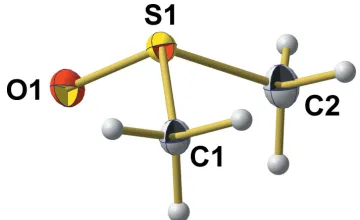
Unit-cell parameters of the current 100 K single-crystal X-ray measurement (SCXD) are consistent with those of the neutron powder diffraction (NPD) data of Ibberson (2005), but structural parameters of the DMSO molecule differ consid-erably between the two refinements (Table 1). In the pyra-midal molecule of crystallographic point group symmetryC1 (Fig. 1, atom positions and atom labelling according to NPD), the S atom lies 0.6994 (9) A˚ above the triangular base formed by the O and C atoms. The S—O bond length of 1.5040 (10) A˚ is slightly longer than the value [1.496 (2) A˚ ] determined by Ibberson at 100 K, but corresponds very well with a S O double bond in sulfoxides [1.497 (13) A˚ ; Allenet al., 1987].
Other differences between the single-crystal X-ray and neutron powder diffraction data, however, are strongly expressed with respect to S—C bond lengths and even more with respect to O—S—C bond angles (Table 1). In the case of the neutron data, the difference between both S—C bonds is 0.05 A˚ [S—C1 = 1.838 (3) A˚ and S—C2 = 1.788 (3) A˚], while in the case of the X-ray data, the difference between both bonds is reduced by a factor of about 10 to 0.006 A˚ [S—C1 = 1.7801 (14) A˚ and S—C2 = 1.7861 (15) A˚]. Moreover, the bond to atom C1 is shorter than the bond to C2, in contrast to the bond-length distribution observed by Ibberson. This is of
1406
Hans Reuter C2H6OS Acta Cryst.(2017). E73, 1405–1408
[image:2.610.119.218.363.434.2]research communications
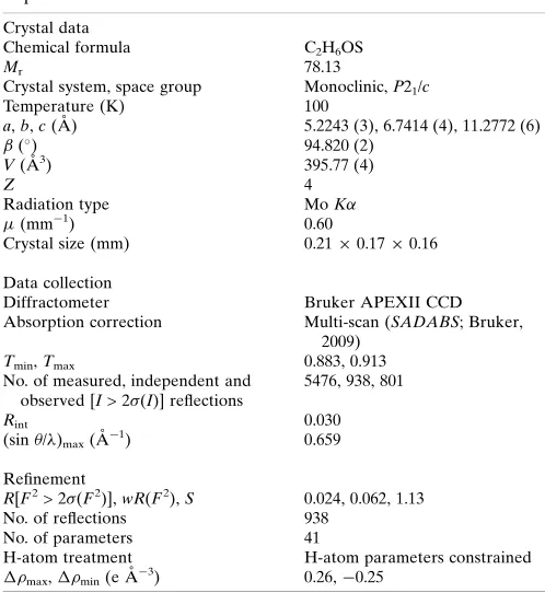
Table 1
Experimental details of previous crystal structure determinations of DMSO and their comparison with the present study.
Thomaset al.(1966) Ibberson (2005) This work
Space group,Z P21/c, 4 P21/c, 4 P21/c, 4
a(A˚ ) 5.303 (5) 5.2390 (1) 5.2243 (3)
b(A˚ ) 6.829 (3) 6.7581 (1) 6.7414 (4)
c(A˚ ) 11.693 (3) 11.2696 (1) 11.2772 (6)
() 94.5 (3) 94.8053 (3) 94.820 (2)
V(A˚3) 422.2 397.60 (1) 395.77 (4)
T(K) 278 100 100
Sample single-crystal powder single-crystal
Radiation MoK neutron MoK
Technique precession photographs HRPD CDC
Rvalue 7.4% 3.77% 2.4%
Number of reflections 777 not given 938
Number of parameters not given 93 41
H(D) atoms constrained refined constrained
d(S1—O1) (A˚ ) 1.531 (5) 1.496 (2) 1.5040 (10)
d(S1—C1) (A˚ ) 1.775 (8) 1.838 (3) 1.7801 (14)
d(S1—C2) (A˚ ) 1.821 (11) 1.788 (3) 1.7861 (15)
O1—S1—C1 (
) 106.7 (4) 105.2 (2) 106.54 (6)
O1—S1—C2 (
) 106.8 (4) 108.3 (2) 106.60 (7)
C1—S1—C2 (
[image:2.610.77.257.604.714.2]) 97.4 (4) 96.4 (1) 97.73 (7)
Figure 1
The molecular structure of the title compound, showing the atom-labelling scheme and displacement ellipsoids for the non-H atoms at the 50% probability level.
Figure 2
[image:2.610.314.566.605.722.2]special interest in view of the C—H O interactions discussed below. With respect to the C—S—O bond angles, structural differences between the NPD and SCXD model are enor-mous: the difference between both bond angles of 3.09[O—
S—C1 = 105.21 (16)and O—S—C2 = 108.30 (15)] found by
Ibberson at 100 K can be compared with a difference of only 0.06[O—S—C1 = 106.54 (6)and O—S—C2 = 106.60 (7)] in the case of the present work. All in all, the idealCspoint group
symmetry of the gaseous and liquid DMSO molecule is much better approached in the crystalline state, even at 100 K, than originally assumed from neutron powder data.
Although this symmetry consideration is not affected by the bond angle between the S atom and the methyl groups, it is important – on the background of coordination that seems to have a great influence on this bond angle – to emphasize that in the SCXD model [C—S—C = 97.73 (3)], this angle is about
1.4larger than in the NPD model [C—S—C = 96.37 (12)at
100 K]. With respect to the hydrogen/deuterium positions, no differences occur, as both methyl groups show an eclipsed orientation with respect to C C, thus fulfilling the nearly idealCssymmetry, too.
For the sake of completeness, structural data of the previous single-crystal X-ray structure determination by Thomaset al.
(1966) are also compiled in Table 1.
3. Supramolecular features
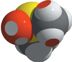
C—H O contacts are the most prominent intermolecular interactions responsible for the three-dimensional arrange-ment of the DMSO molecules in the solid state (Fig. 2). In order to compare our results with the results of the neutron powder diffraction experiment, one must take into account the different validity and refinement strategies for the H/D atoms in both methods. Under consideration of the van der Waals radii of H (1.10 A˚ ) and O (1.52 A˚) supplied by Mantinaet al.
(2009), relevant H O distances should be shorter than 2.62 A˚ . From the H atoms attached to C2, only one (H22) shows an interatomic distance below this threshold. With an H22 O1ii (for symmetry code, see Table 2) distance of 2.61 A˚ , a binding C—H O interaction other than a van der Waals interaction can be excluded. Just the opposite is observed in case of the H atoms attached to C1: all three H atoms show an intermolecular contact to one O atom of three different DMSO molecules in the range 2.42–2.47 A˚ (Table 2). In this case, these contacts fall below the van der Waals distance by 7.6–5.7% (0.20–0.15 A˚ ) which justifies the assumption of binding C—H O interactions. The corre-sponding intermolecular donor–acceptor distances are in the
range 3.318 (2)–3.445 (2) A˚ , while the C—H O angles are in the range 152.0–173.0 (Table 2). In summary, each DMSO molecule participates in six C—H O contacts to five neighbouring molecules (Fig. 3). The extent of the van der Waals and hydrogen-bonding interactions on the overlapping of the molecules is visualized in Fig. 4. Obviously, there is no weakening influence of these interactions on the S—C bond length. Quite the opposite, the S1—C1 bond is somewhat shorter than the S1—C2 bond (see above).
With respect to the O atom as an acceptor atom, bond angles (S O H) of the van der Waals contacts come to 114.0 for H132
(2 = 1 +x, y, z), 152.1 for H121
(1= x, 0.5 +y, 0.5z), and 103.4 for H113
(3= x,y,z). The geometrical aspects of these van der Waals interactions (or of coordinatively or hydrogen-bonded DMSO molecules) are described only incompletely with the foregoing used distances and angles as they disregard the orientation of the complete DMSO molecule in relation to the interactions described. In order to unambiguously account for this specific relationship, indexation by two additional values,! anddnorm, using two planes as a reference (Fig. 5) is suggested. The first plane is identical, with the pseudo-mirror planem0defined by O1, S1
and the mid-point between both C atoms. The second plane,
plO, is perpendicular to the S O bond and located in O1.
Table 2
Hydrogen-bond geometry (A˚ ,).
D—H A D—H H A D A D—H A
C1—H11 O1i 0.98 2.42 3.3318 (18) 155
C1—H12 O1ii 0.98 2.42 3.3184 (17) 152
C1—H13 O1iii 0.98 2.47 3.4450 (19) 173
[image:3.610.317.565.72.230.2]C2—H22 O1ii 0.98 2.61 3.4618 (18) 146
Figure 3
C—H O contacts (grey brocken sticks) between the DMSO molecule and its neighbours. [Symmetry codes used to generate equivalent atoms: (i) 1x,1
2+y, 1
2z; (ii)1 +x,y,z; (iii)x,y,z; (iv) 1 +x,y,z; (v)
x,1 2+y,
[image:3.610.375.500.604.712.2]1 2z.]
Figure 4
While dnorm represents the distance between the interacting atom (viaa van der Waals interaction, a hydrogen bond or a coordinative bond) and plO, the angle ! marks the angle
between m0 and the plane pl
H defined by O1, S1 and the
interacting atom. Values of!can stretch from 0 to 360when
looking down the O S bond as in a Newman projection. In the case of the van der Waals interactions discussed here, the corresponding !/dnorm values are: H12
1
= 98.2/2.139 A˚ ,
H132= 178.3/1.006 A˚ and H113
= 335.1/0.560 A˚ .
4. Synthesis and crystallization
Single crystals were grown from a commercial available sample (Sigma–Aldrich) within a 0.3 mm thick Lindemann capillary using the Kryoflex low-temperature device of the diffractometer.
5. Refinement
Crystal data, data collection and structure refinement details are summarized in Table 3. All six H atoms were found in a difference-Fourier map. They could be refined without any restraints in meaningful positions [C—H range = 0.91 (2)– 0.97 (2) A˚ ; H—C—H range = 107.6 (11)–112.2 (15)] with
individual isotropic displacement parameters [range = 0.018 (4)–0.036 (5) A˚2]. In order to obtain a structure model comparable to typical refinement techniques of DMSO mol-ecules in the structures of coordination compounds or with hydrogen bonds, conventional constraints [AFIX 137, C—H = 0.99 A˚ , H—C—H = 109.6 in SHELXL (Sheldrick, 2015)] with two common isotropic displacement parameters, one for each methyl group, have been applied. In summary, these restraints only slightly affected the final results: the final R
value increased from 2.37 to 2.42%, while the bond lengths and bond angles remained unchanged. All data have been approved by a second independently grown crystal.
Acknowledgements
We thank the Deutsche Forschungsgemeinschaft and the Government of Lower-Saxony for funding the diffractometer and acknowledge support by Deutsche Forschungsge-meinschaft (DFG) and Open Access Publishing Fund of Osnabru¨ck University.
References
Allen, F. H., Kennard, O., Watson, D. G., Brammer, L., Orpen, A. G. & Taylor, R. (1987).J. Chem. Soc. Perkin Trans. 2, pp. S1–19. Brandenburg, K. (2006).DIAMOND. Crystal Impact GbR, Bonn,
Germany.
Bruker (2009).APEX2,SADABS,SAINTandSHELXTL. Bruker AXS Inc., Madison, Wisconsin, USA.
Calligaris, M. (2004).Coord. Chem. Rev.248, 351–375. Ibberson, R. M. (2005).Acta Cryst.C61, o571–o573.
Kastner, G. & Reuter, H. (1999).Main Group Met. Chem.22, 605– 609.
Macrae, C. F., Bruno, I. J., Chisholm, J. A., Edgington, P. R., McCabe, P., Pidcock, E., Rodriguez-Monge, L., Taylor, R., van de Streek, J. & Wood, P. A. (2008).J. Appl. Cryst.41, 466–470.
Mantina, M., Chamberlin, A. C., Valero, R., Cramer, C. J. & Truhlar, D. G. (2009).J. Phys. Chem. A,113, 5806–5812.
Melanson, R. & Rochon, F. D. (1975).Can. J. Chem.53, 2371–2374. Pearson, R. G. (1963).J. Am. Chem. Soc.85, 3533–3539.
Sheldrick, G. M. (2008).Acta Cryst.A64, 112–122. Sheldrick, G. M. (2015).Acta Cryst.C71, 3–8.
Tarighi, S. & Abbasi, A. (2007).J. Sci. (University of Teheran),33, 19– 21.
Thomas, R., Shoemaker, C. B. & Eriks, K. (1966).Acta Cryst.21, 12– 20.
Viswamitra, M. A. & Kannan, K. K. (1966).Nature,209, 1016–1017.
1408
Hans Reuter C2H6OS Acta Cryst.(2017). E73, 1405–1408
[image:4.610.47.296.76.198.2]research communications
Table 3
Experimental details.
Crystal data
Chemical formula C2H6OS
Mr 78.13
Crystal system, space group Monoclinic,P21/c
Temperature (K) 100
a,b,c(A˚ ) 5.2243 (3), 6.7414 (4), 11.2772 (6)
(
) 94.820 (2)
V(A˚3) 395.77 (4)
Z 4
Radiation type MoK
(mm1) 0.60
Crystal size (mm) 0.210.170.16
Data collection
Diffractometer Bruker APEXII CCD
Absorption correction Multi-scan (SADABS; Bruker, 2009)
Tmin,Tmax 0.883, 0.913
No. of measured, independent and observed [I> 2(I)] reflections
5476, 938, 801
Rint 0.030
(sin/)max(A˚1) 0.659
Refinement
R[F2> 2(F2)],wR(F2),S 0.024, 0.062, 1.13
No. of reflections 938
No. of parameters 41
H-atom treatment H-atom parameters constrained
max,min(e A˚3) 0.26,0.25
Computer programs: APEX2 (Bruker, 2009), SAINT (Bruker, 2009), SHELXS97
(Sheldrick, 2008),SHELXL2014(Sheldrick, 2015),DIAMOND(Brandenburg, 2006),
[image:4.610.44.293.448.719.2]Mercury(Macraeet al.(2008) andSHELXTL(Sheldrick, 2008).
Figure 5
Geometrical boundary conditions for the determination of!(left) and
dnorm (right, distances in A˚ ) by use of the pseudo-mirror plane m0
(definition: O1, S1, mid-point between C1 and C2), the plane plH
(definition: the interacting atom, S O bond), and the plane plO
(definition: O1, S O bond = normal vector). [Symmetry codes used to generate equivalent atoms: (i) 1x,1
2+y, 1
2z; (ii)1 +x,y,z; (iii)x,
sup-1 Acta Cryst. (2017). E73, 1405-1408
supporting information
Acta Cryst. (2017). E73, 1405-1408 [https://doi.org/10.1107/S2056989017012464]
Structural parameters of dimethyl sulfoxide, DMSO, at 100
K, based on a
redetermination by use of high-quality single-crystal X-ray data
Hans Reuter
Computing details
Data collection: APEX2 (Bruker, 2009); cell refinement: SAINT (Bruker, 2009); data reduction: SAINT (Bruker, 2009);
program(s) used to solve structure: SHELXS97 (Sheldrick, 2008); program(s) used to refine structure: SHELXL2014
(Sheldrick, 2015); molecular graphics: DIAMOND (Brandenburg, 2006) and Mercury (Macrae et al. (2008); software
used to prepare material for publication: SHELXTL (Sheldrick, 2008).
Dimethyl sulfoxide
Crystal data
C2H6OS
Mr = 78.13
Monoclinic, P21/c
a = 5.2243 (3) Å
b = 6.7414 (4) Å
c = 11.2772 (6) Å
β = 94.820 (2)°
V = 395.77 (4) Å3
Z = 4
F(000) = 168
Dx = 1.311 Mg m−3
Mo Kα radiation, λ = 0.71073 Å Cell parameters from 3349 reflections
θ = 3.5–28.0°
µ = 0.60 mm−1
T = 100 K Bloc, colourless 0.21 × 0.17 × 0.16 mm
Data collection
Bruker APEXII CCD diffractometer
φ and ω scans
Absorption correction: multi-scan (SADABS; Bruker, 2009)
Tmin = 0.883, Tmax = 0.913
5476 measured reflections
938 independent reflections 801 reflections with I > 2σ(I)
Rint = 0.030
θmax = 28.0°, θmin = 3.5°
h = −6→6
k = −8→8
l = −14→14
Refinement
Refinement on F2
Least-squares matrix: full
R[F2 > 2σ(F2)] = 0.024
wR(F2) = 0.062
S = 1.13 938 reflections 41 parameters 0 restraints
Hydrogen site location: inferred from neighbouring sites
H-atom parameters constrained
w = 1/[σ2(F
o2) + (0.0238P)2 + 0.1531P]
where P = (Fo2 + 2Fc2)/3
(Δ/σ)max < 0.001
Δρmax = 0.26 e Å−3
supporting information
sup-2 Acta Cryst. (2017). E73, 1405-1408
Special details
Geometry. All esds (except the esd in the dihedral angle between two l.s. planes) are estimated using the full covariance
matrix. The cell esds are taken into account individually in the estimation of esds in distances, angles and torsion angles; correlations between esds in cell parameters are only used when they are defined by crystal symmetry. An approximate (isotropic) treatment of cell esds is used for estimating esds involving l.s. planes.
Fractional atomic coordinates and isotropic or equivalent isotropic displacement parameters (Å2)
x y z Uiso*/Ueq
S1 0.18232 (6) 0.14961 (5) 0.19283 (3) 0.01776 (12)
O1 −0.10440 (18) 0.13580 (16) 0.16758 (10) 0.0245 (3)
C1 0.3139 (3) −0.0633 (2) 0.12708 (13) 0.0187 (3)
H11 0.2663 −0.1825 0.1698 0.027 (3)*
H12 0.5015 −0.0518 0.1317 0.027 (3)*
H13 0.2465 −0.0729 0.0435 0.027 (3)*
C2 0.2905 (3) 0.3307 (2) 0.09280 (15) 0.0247 (3)
H21 0.2217 0.2994 0.0114 0.033 (3)*
H22 0.4786 0.3299 0.0972 0.033 (3)*
H23 0.2305 0.4623 0.1149 0.033 (3)*
Atomic displacement parameters (Å2)
U11 U22 U33 U12 U13 U23
S1 0.01277 (18) 0.02359 (19) 0.0170 (2) −0.00180 (13) 0.00161 (12) −0.00362 (15) O1 0.0115 (5) 0.0353 (6) 0.0269 (6) −0.0008 (4) 0.0030 (4) −0.0069 (5) C1 0.0159 (6) 0.0194 (7) 0.0209 (8) 0.0003 (5) 0.0030 (6) 0.0000 (6) C2 0.0236 (7) 0.0198 (7) 0.0312 (9) −0.0001 (6) 0.0042 (6) 0.0018 (6)
Geometric parameters (Å, º)
S1—O1 1.5040 (10) C1—H13 0.9800
S1—C1 1.7801 (14) C2—H21 0.9800
S1—C2 1.7861 (15) C2—H22 0.9800
C1—H11 0.9800 C2—H23 0.9800
C1—H12 0.9800
O1—S1—C1 106.54 (6) H12—C1—H13 109.5
O1—S1—C2 106.60 (7) S1—C2—H21 109.5
C1—S1—C2 97.73 (7) S1—C2—H22 109.5
S1—C1—H11 109.5 H21—C2—H22 109.5
S1—C1—H12 109.5 S1—C2—H23 109.5
H11—C1—H12 109.5 H21—C2—H23 109.5
S1—C1—H13 109.5 H22—C2—H23 109.5
H11—C1—H13 109.5
Hydrogen-bond geometry (Å, º)
D—H···A D—H H···A D···A D—H···A
sup-3 Acta Cryst. (2017). E73, 1405-1408
C1—H12···O1ii 0.98 2.42 3.3184 (17) 152
C1—H13···O1iii 0.98 2.47 3.4450 (19) 173
C2—H22···O1ii 0.98 2.61 3.4618 (18) 146