Different objective methods for the meat and/or carcass classification are proposed or currently used: estimation of carcass conformation, muscle to fat ratio; use of video image analysis, sonogra-phy and measurement of electrical properties. In the European region the SEUROP system is used. One of such methods is bioelectrical impedance analysis (BIA) (Bohuslávek, 2002; Bohuslávek et al., 2002). Fat is a very important compound influencing meat quality. In particular the intramuscular fat (mar-bling) affecting the taste, juiciness and texture of meat is of special importance. Meat that is free of this fat is dry, tough and has a lower estimation of total acceptance.
The optimal level of intramuscular fat varies in different countries. Generally, some percentage of fat in muscle is accepted and it affects good meat quality. In Eastern countries, especially Japan, a high content of intramuscular fat including its regular distribution is preferred.
It is usually claimed that marbling is well devel-oped in the meat of animals with low locomotor activity. But then marbling would hardly occur in
the meat of wild animals and animals with high physical activity (Grau, 1960). Morphologically, the intramuscular fat is characterised as a depot fat that is visible in a higher amount as marbling and consists of aggregates of adipose cells mostly in the peri- and endomysium. It is an energy reserve and it has a different function from phospholipids that are localised as structural elements in cell membranes (Arneth, 1998).
Determination of intramuscular fat is o�en de-manded by all those who came in contact with meat: farmers, processors (aba�oirs), distributors, retailers and consumers. There is a need to find the methods that can objectively quantify the in-tramuscular fat content. The simplest method is to remove all parts of adipose tissue and to determine the intramuscular fat content in the muscle only. In this way it is possible to distinguish intramuscular fat on the one hand and intermuscular and subcu-taneous fat on the other.
The intramuscular fat content is usually deter-mined a�er accurate separation of muscle with subsequent extraction of fat. Different methods of
Supported by the Grant Agency of the Czech Republic (Project No. 102/01/1344).
The use of video image analysis for fat content
estimation
P. P����, J. J���������, L. S��������
Institute of Chemical Technology, Prague, Czech Republic
ABSTRACT: The composition of selected cuts of ca�le carcasses was determined in connection with the search for new methods of carcass classification. The content of adipose tissue and intramuscular fat in the cross-section of beef loin was estimated. A total of 79 samples was taken for investigations in a broad range of ca�le category. The classical extraction method in Soxhlet extractor was compared with video image analysis, which measured the ratio of muscle to fat areas. The size and the shape of the musculus longissimus lumborum et thoracis (MLLT) and its ratio in the loin cross-section was also estimated. A good correlation (r = 0.99, P < 0.05) between both methods was found for the estimation of intramuscular fat in MLLT. The correlation in the case of the whole cross-section was influenced by the connective tissue that gives also white areas similarly like the adipose tissue, but the fat content is different.
extraction were examined; these methods do not give the same results and it is necessary to com-promise the term “intramuscular fat”. Although a certain rest of triacylglycerols is not determined by the Soxhlet extraction method, the propor-tion of undesirable components (cholesterol and phospholipids) is the lowest. Analysing lean beef by this method, 82–89% of triacylglycerols are ex-tracted and only a small amount of accompanying lipid components. Using the chloroform/methanol mixture for the extraction, all triacylglycerols are extracted but the results are influenced by the pres-ence of simultaneously extracted phospholipids and interfering non-lipid components. A some-what higher yield of triacylglycerols was achieved by the Weibull-Stold method than by the direct Soxhlet extraction. However, undesirable fa�y acids of phospholipids were determined together with triacylglycerols. Extraction by Soxhlet using n-hexane as a non-polar solvent is considered to be an optimal method for intramuscular fat determination in meat. The Soxhlet extraction in a standardized form according to ISO 1444 is recommended as a reference method for the analysis of intramuscular fat content (Arneth, 1998).
The need for faster and non-destructive methods led to the use of video image analysis. This method is o�en used for the estimation of the muscle to adi-pose tissue ratio. It can be supadi-posed that the white particles are mostly adipose tissue and thus also intramuscular fat. Another possibility is to describe very precisely the content of white areas and their shape and distribution in muscle.
In this connection the video image analysis (VIA) was employed mainly for the ca�le classification because the particles of fat (marbling) are distinct from the rest of the muscle tissue mainly in beef (Branscheid et al., 1999).
The images of ground meat were evaluated from the aspect of reflectance, saturation and hue and the intramuscular fat content was determined. It seems that the use of VIA for an evaluation of ground meat is possible (up to 32% of fat) (Gwartney et al., 1996).
Video image analysis was also used for the clas-sification of pork carcasses and their cuts. The ratio of muscle to fat areas in the cross-section of pork loin highly correlated with the results obtained by detailed dissection (Branscheid et al., 1996).
In Japan, the proportion of intramuscular fat was evaluated by video image analysis and a correlation with the evaluation of marbling grade was found
(Kuchida et al., 1992). It is however necessary to note that the intramuscular fat proportion is much higher in Japan than in Europe and thus the fat can be thresholded more easily.
Histological staining was supposed to avoid the interference of the connective tissue with the results of determination (Albrecht et al., 1996).
MATERIAL AND METHODS
The composition of selected cuts of ca�le car-casses was determined in connection with the search for new methods of carcass classification. The content of adipose tissue and intramuscular fat in the cross-section of beef loin was estimated. The classical extraction method in Soxhlet extractor was compared with video image analysis, which measured the ratio of muscle area to fat area.
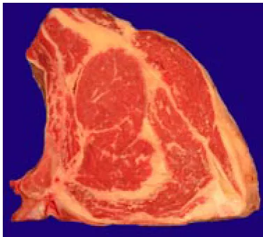
Meat samples were obtained directly in a meat factory where the cross slices of the loin were cut off from the hind quarter of ca�le carcass (Figure 1). The thickness of cross slice was nearly 20 mm. The dissection between the fore quarter and the hind one was carried out between the 8th and 9th rib and the 8th and 9th vertebra. The samples were transferred into a laboratory and stored at 4°C there. Before measurements the meat samples were cleaned from sawdust with knife and blo�ed with filter paper. A�er the image was shot with digital camera (see below) the slices were carefully divided into three parts: musculus longissimus lumborum et thoracis, bones (vertebra and a part of rib) and the rest of the loin (containing small muscles and
[image:2.595.320.509.548.718.2]muscular adipose tissue). Both the loin and the rest of the loin were weighed, separately homogenised using a laboratory blender, packed under vacuum into plastic bags and stored under freezing until analysed. For the analysis, 79 samples were selected to cover a sufficiently wide range of intramuscular fat content, e.g. samples of bulls and cows of dif-ferent breeds (predominantly Simmental) and live weights (381–927 kg).
Fat content determination using extraction method
5 g of each meat sample was dried with sand at the temperature of 103 ± 2°C. Then it was quan-titatively transferred into an extraction cartridge, and petroleum ether extraction was run for 5 h in the Soxhlet extractor (ISO 1444).
Image shooting
Most images were taken using a digital camera Nikon coolpix 800 (sensitivity “FINE”, 2.1 mega-pixels, without flash). The samples of meat (cross slices of loin) were lighted by two fluorescent tubes (11 W, 2700 K). The images in the form of *.jpg files were stored on the Compact Flash 64 MB and then transferred into a computer.
In a separate investigation we tested other ways of image shooting. Besides the above-mentioned digital camera Nikon coolpix 800 the camcorder Panasonic NV-DS 38EG was used. In the camcorder the “0 lux mode” was also used, when the infrared light that was reflected from the meat surface was captured by the camcorder. We tested different combinations of daylight and ultraviolet light (produced by the special tube Tesla RTU 125 W). Green and polarisation filters were also used in this experiment.
Video image analysis
The so�ware LUCIA 3.52b (Laboratory Imaging, Prague) was used to evaluate the images. White areas (fat, adipose tissue) and red (black) areas were distinguished in the muscle tissue. First, the background was removed from the image by means of image arithmetic and then the image of the loin cross-section was divided into an area belonging to
the muscle MLLT and to the remaining area of the loin cross-section.
The points of the “white” tissue were thresholded in the area of MLLT and the ratio of their area to the total area of muscle was measured. It is sup-posed that this area is created mostly by fat, so it was correlated with the fat content determined in this muscle by extraction.
The remaining part of the loin was processed in a similar way, this part, however, contains several small muscles, adipose tissue, connective tissue and bones. The bone area was removed by means of a binary editor. A�er calibration the measured areas in pixels were converted into areas in cm2.
RESULTS AND DISCUSSION
The fat content determined by Soxhlet extraction method appears to be in good accordance with the results obtained by video image analysis. Meat image shooting and subsequent discrimination of the adipose tissue from other tissues (connective, bones) and avoiding of distractive gloss effects are limiting factors.
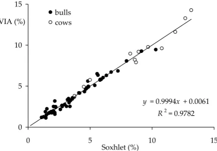
[image:3.595.307.529.543.697.2]A very good agreement between these two meth-ods of determination of intramuscular fat content was achieved in the musculus longissimus lumborum et thoracis (Figure 2). The digital camera takes only surface images of the cross-section and solely the fat particles that occur in the cross-section or in tightly adjoined layers could be measured while for fat de-termination by the extraction method a significantly thicker layer (approximately 10 mm) is used. But
Figure 2. The comparison of video image analysis with classical Soxhlet extraction – fat content in musculus longissimus lumborum et thoracis (cross-section behind the 8th rib)
y= 0.9994x+ 0.0061
R2 = 0.9782
0 5 10 15
0 5 10 15
Soxhlet (%)
we suppose that the distribution of intramuscular fat along the longitudinal axis is so homogeneous that this difference does not influence the results of measurement.
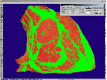
The problem is that shades from the surrounding musculature penetrate into intramuscular fat, so the fat particles are difficult to threshold. While the com-pact intermuscular adipose tissue can be separated quite clearly, this approach is more difficult during thresholding the thin particles in the muscle tissue. On the contrary, the fat particles closely under the surface stream in the red fragment of the image and it is also difficult to threshold and distinguish them from the muscle tissue (Figure 3).
In spite of all the above-mentioned problems the good accordance (r = 0.99) was a�ained between these two methods. The relation of both methods is linear; the rate is practically equal to 1.
To improve the contrast between the adipose tissue and surroundings different combinations of illumination and image shooting were examined. No satisfactory results are available for the time being; it is rather a question of the first qualitative results or experiences (see below).
On the other hand, the accordance between the results of extraction method and the results of video image analysis in the evaluation of whole
[image:4.595.128.473.86.344.2]slices of loin (including the adipose tissue and other muscles) is influenced by the fact that white areas in digital image are also created by the connective tissue, which however differs in the fat content (Figure 4).
The measured data can be interpreted in two ways. Although the simple linear correlation be-tween both methods leads to a high correlation coefficient, the limit value for image analysis at
y= 0.8408x + 13.441
R2 = 0.9118
y = 1.2169x R2 = 0.6883
0 20 40 60 80
0 10 20 30 40 50 60 70
Soxhlet (%) VIA (%)
[image:4.595.306.529.549.707.2]Figure 4. The comparison of video image analysis with classical Soxhlet extraction – fat content in the whole loin (cross-section behind the 8th rib)
zero fat content (results of extraction method) is a miscount. If we set a condition that the regression line will intersect the zero of y axis, the correlation coefficient is lower.
Then the fat content determined by the video image analysis has slightly higher values than in measurements by the extraction method. The ex-planation consists in the fact that the white objects in the image are both bone and connective tissue, which are then evaluated by the computer as “fat”. Hence it is necessary to remove these objects from the image. In the case of bone that has a regular shape it is very easily possible by the use of binary editor and image arithmetic. The greater problem is caused by the connective tissue. Here we consider suitable illumination (UV) or the use of filters. Though it is possible to use histological staining (Albrecht et al., 1996 – see above), the advantage of fast and non-destructive method is lost.
In spite of the expectations the recording of in-frared emission with digital camcorder in the “0 lux mode” was not successful. The thresholding was very difficult because the image is weakly contrasting and in addition it is illuminated ir-regularly as the camcorder also uses its own point infrared source. No improvement was achieved by the covering of this point source and by illumination from another source (visible light, UV). Besides it is monochrome, so binary image, where it is not pos-sible to use colour hue in thresholding. The images were weakly contrasting compared to images shot in the standard mode.
Conversely, the illumination of an object by ultra-violet light appears very suitable. A high contrast between the adipose and muscle tissue was reached by the combination of visible and ultraviolet light. So it was possible to threshold well the objects extra-neous to intramuscular fat and thus to reach good accordance with the results of extraction method. Since only a limited number of samples was used for this observation, we continue in observation and specify it. We also consider the use of another colour of light and/or its polarization. It is possible to take into account histological staining in the case of the use of some food colorants.
CONCLUSIONS
The video image analysis seems to be suitable for rapid estimation of intramuscular fat with the
accuracy necessary for the industrial conditions. The comparison of this method with the classi-cal Soxhlet extraction showed a good correlation between fat contents obtained by both methods, especially in the case of the musculus longissimus lumborum et thoracis.
The inaccuracies in fat determination by the video image analysis are the consequence of interfering influence of connective tissue and bones. The im-provement is supposed by modification of illumi-nation, the use of UV lights and filtration of some light components during shooting.
REFERENCES
Arneth W. (1998): Über die Bestimmung des intra-muskulären Fe�es. Fleischwirtscha�, 78, 218–220. Albrecht E., Wegner J., Ender K. (1996): A new technique
for objective evaluation of marbling in beef. Fleischwirt-scha�, 76, 1145–1147.
Bohuslávek Z. (2002): Estimation of beef carcasses con-formation carried out at a high-performance aba�oir line, based on an impedance method. Czech J. Anim. Sci., 47, 155–159.
Bohuslávek Z., Pipek P., Malý J. (2002): Use of BIA method for the estimation of beef carcass composition – weight of longissimus lumborum muscle, ratio of muscle tissue and fat on the cross-section of sirloin. Czech J. Anim. Sci., 47, 387–394.
Branscheid W., Dobrowolski A., Hoereth R. (1996): Video image analysis. A method for the on-line recording of the cut value of pig carcasses. Fleischwirtscha�, 76, 721–724.
Branscheid W., Dobrowolski A., Spindler M., Augustini Ch. (1999): Application of video image analysis in grad-ing of ca�le. Fleischwirtsch. Int., 4, 12–15.
Grau R. (1960): Grundlagen und Fortschri�e der Lebens-mi�eluntersuchung. Band 7, Fleisch und Fleischwaren. 1. ed. Berlin.
Gwartney B.L., Gao X., Tan J., Gerrard D.E. (1996): De-termining fat content in ground beef via colour image processing. J. Muscle Food., 7, 441–451.
Kuchida K., Yamaki K., Yamagishi T., Mizuma Y. (1992): Evaluation of meat quality in Japanese beef ca�le by computer image analysis. Anim. Sci. Technol., 63, 121–127.
ABSTRAKT
Využití analýzy obrazu pro odhad obsahu tuku
Složení vybraných částí hovězího masa bylo určováno v souvislosti s hledáním nových metod pro klasifikaci jatečně upravených těl skotu. Zvláštní pozornost byla věnována podílu tukové tkáně a obsahu intramuskulárního tuku v příčném řezu roštěncem. Celkem bylo odebráno 79 vzorků v širokém rozmezí složení. Byly zjišťovány korelace mezi výsledky extrakční metody podle Soxhleta s vyhodnocením podílu ploch odpovídajících svalové, resp. tukové tkáně pomocí analýzy obrazu. Byl zjišťován podíl svalu musculus longissimus lumborumet thoracis v příčném řezu roštěncem a jeho velikost i tvar. Ukazuje se dobrá korelace (r = 0,99, P < 0,05) intramuskulárního tuku stanoveného metodou VIA a metodou podle Soxhleta. Korelace v celém řezu roštěncem je ovlivněna skutečností, že zde i pojivová tkáň vytváří na digitálním obraze bílé plochy, které se však liší obsahem tuku.
Klíčová slova: jatečně upravené tělo; klasifikace; maso; intramuskulární tuk; analýza obrazu; bioelektrická impe-dance
Corresponding Author
Doc. Ing. Petr Pipek, CSc., Vysoká škola chemicko-technologická, Ústav konzervace potravin a technologie masa, Technická 3, 166 28 Praha 6, Česká republika