Effect of various co-culture systems on embryo
development in ovine
B. Heidari
1, A. Shirazi
1,2, M.-M. Naderi
1, M.-M. Akhondi
1, H. Hassanpour
2,
A. Sarvari
1, S. Borjian
11Reproductive Biotechnology Research Center, Avicenna Research Institute, ACECR, Tehran, Iran
2Department of Gametes and Cloning, Research Institute of Animal Embryo Technology, Shahrekord University, Shahrekord, Iran
ABSTRACT:Considering the advent of mesenchymal stem cells (MSCs) as a new source of somatic cells in embryo co-culture system, the current study was aimed to compare in vitro embryo development using embry-onic MSCs monolayer with embryembry-onic fibroblast cells (EFCs), oviductal epithelial cells (OECs), and cell-free culture system. The IVM/IVF presumptive sheep zygotes were randomly cultured in different culture condi-tions as follows: (1) SOFaaBSA medium for the whole culture period (SOF, n = 371), (2) SOFaaBSA medium for the first 3 days followed by co-culturing with MSCs for the next 5 days (SOF-MSCs, n = 120), (3) co-culturing with MSCs for the first 3 days followed by culture in SOFaaBSA medium for the next 5 days (MSCs-SOF, n = 133), (4) co-culturing with MSCs for the whole culture period (MSCs, n = 212), (5) SOFaaBSA medium for the first 3 days followed by co-culturing with EFCs for the next 5 days (SOF-EFCs, n = 132), (6) co-culturing with EFCs for the first 3 days followed by culture in SOFaaBSA medium for the next 5 days (EFCs-SOF, n = 165), (7) co-culturing with EFCs for the whole culture period (EFCs, n = 236), and (8) co-culturing with OECs for the whole culture period (OECs, n = 255). One-Way ANOVA by multiple pairwise comparisons using Tukey’s test was performed. Co-culturing in MSCs group had no superiority over EFCs and OECs groups. Though, when co-culturing with MSCs and EFCs was limited to the first 3 days of culture, the embryo development indices were improved compared to the other co-cultured groups. Considering both the hatching rate and total cell number, the application of MSCs for the first 3 days of culture (MSCs-SOF) was superior to the other co-culture and SOF groups.
Keywords: mesenchymal stem cell; fibroblast; oviduct; in vitro production; zygote
In an attempt to more closely mimic the in vivo
conditions, feeder cell lines (co-culture) were de-veloped. The application of somatic cells to support mammalian pre-implantation embryo development
in vitro was first applied to murine embryo culture in
mouse oviduct organ cultures (Biggers et al., 1962). Since then, embryo somatic co-culture has been ap-plied to a broad spectrum of animal species (Orsi and Reischl, 2007; Nematollahi-Mahani et al., 2009) and of course, humans (Teklenburg and Macklon, 2009).
Although the mechanisms by which somatic cells improve early embryo development remain elusive, the mode of action of co-culture systems
has been explained largely by two putative mecha-nisms. One of the modes is medium detoxification and the second mode is provision of required metabolites and specific growth stimulators. On the other hand, co-cultured cells during the course of their own proliferation can potentially provide bioactive factors and “cross-talk” which is absent in IVC media alone. This approach has been ef-fective in overcoming developmental blocks in most laboratory and domestic animals (Orsi and Reischl, 2007).
such as: higher and faster cleavage (Bongso et al., 1989), improved morphological appearance/ grade (Wiemer et al., 1989), increase of the aver-age number of blastomeres (Smith et al., 1992), improved post-thaw blastomere survival of cryo-preserved co-cultured embryos (Tucker et al., 1995), reduced apoptosis (Xu et al., 2000), higher blastocyst rate (Joo et al., 2001), facilitated hatch-ing (Ellhatch-ington et al., 1990), lower fragmentation rates, improved pregnancy rates (Wiemer et al., 1989), higher implantation ratio (Wetzels et al., 1998), and live births (Marcus and Brinsden, 1996). These effects are most pronounced with increasing the duration of co-culture (Wiemer et al., 1989), especially during the early cleavage stages which may be mediated by the expression of growth fac-tors (Yeung et al., 1992).
Despite the apparent benefits of co-culture on pre-implantation development, there are some reports indicating no significant improvement in early embryogenesis (Tucker et al., 1995; Hu et al., 1998) or the subsequent clinical pregnancy rates (Hu et al., 1998). There are even some reports indicating the adverse effects of co-culture (Ber-nardi et al., 1996) on embryo quality such as scant inter-cellular contacts between trophectoderm (TE) and inner cell mass (ICM), poorly developed trophoblast apical microvilli, cytoplasmic vacuola-tion, hood mitochondria, wide inter-cellular spaces and numerous cytoplasmic vesicles, phagosomes and lipid droplets (Shamsuddin and Rodriguez-Martinez, 1994).
Among different cell types employed in co-cul-ture systems, the embryotrophic properties of oviductal epithelial cells (OECs) and embryonic fibroblast cells (EFCs) have been well defined in
in vitro production of embryo in human
(Kervan-cioglu et al., 1997) and a broad spectrum of animal species (Orsi and Reischl, 2007). Besides their multipotent potential, adult mesenchymal stem cells (MSCs) can secrete a variety of cytokines and growth factors, such as MCP-1, VEGF-A, EGF, FGF-2, IL-6, LIF, or TGF-ß (Park et al., 2010; Tian et al., 2011). Some of these secreted bioac-tive materials could improve meiotic maturation
in vitro and the subsequent embryo development
(Ling et al., 2008).
Considering the controversial reports on ad-vantages of co-culture systems compared with chemically defined medium in in vitro production of mammalian embryos and the advent of MSCs as a new source of somatic cells in co-culture
system, this study was designed to compare the embryotrophic effects of MSCs, EFCs, and OECs with cell free culture system on in vitro produc-tion of embryo using sheep as the animal model.
MATERIAL AND METHODS
Except where otherwise indicated, all chemicals were obtained from Sigma-Aldrich (St. Louis, USA).
Preparation of OECs monolayer
The ewe oviducts were removed immediately after slaughter of the animal, placed in phosphate-buffered saline (PBS) containing penicillin/strep-tomycin, and transported to the laboratory on ice within 3 h. The oviducts were trimmed from the additionaltissue, washed with PBS containing anti- biotics (3 times), and their surface was disinfected with 70% ethanol. They were placed in 60-mm Petri dish (Falcon 3004;Becton & Dickinson, Frank-lin Lakes, USA) and 1 cm of the upper and lower portions of oviducts was separated. Trypsin-EDTA (0.5% trypsin and 0.25% EDTA in PBS) was injected into the lumen from infundibulum end and after 2–3 min, the lumen was squeezed with tissue forceps and its content was transferred into a conical tube. More trypsin-EDTA was added to the suspension for 5 min and the cellular clumps were dispersed through up and down by insulin syringe. After trypsin neutralization with TCM + 10% fetal calf serum (FCS), the suspension was centrifuged at 500 g for 5 min.The precipitate was washed twice by centrifugation and the pellet was resuspended in TCM supplemented with 10% FCS and penicil-lin/streptomycin. The cells were cultured in 50 µl droplets of TCM + 10% FCS at 39°C and 7% CO2. At 60–70% confluency and 2 h prior to embryo culture, the medium was changed for SOFaaBSA (synthetic oviductal fluid + essential and non-essential amino acids + 5 mg/ml BSA) medium.
Preparation of EFCs monolayer
peni-cillin/streptomycin on ice. Fetuses were rinsed in DPBS 3 times, the head, extremities, and internal organs were removed, and remaining tissues were finely chopped into small pieces with a scalpel blade and washed in DPBS with antibiotics. The fibroblasts were separated from the tissue pieces by a standard trypsinization procedure described elsewhere (Freshney, 1994) using trypsin-EDTA for 30–45 min at 37°C with occasional stirring. The cells were seeded into 60 mm tissue culture plates (Falcon; Becton & Dickinson, Franklin Lakes, USA) in Dulbecco’s Modified Eagle’s Medium (DMEM) supplemented with 10% FCS, 2mM glutamine, 0.1mM β-mercaptoethanol, and penicillin/strep-tomycin. The cells were sub-cultured after being reached >90% confluency and in passage 2–5 were used as a feeder layer at 40% confluency in 50 µl droplets at 37°C and 7% CO2. The medium of droplets was changed 2 h prior to embryo culture for SOFaaBSA medium.
Preparation of MSCs monolayer
Ovine 30–35-day fetuses were obtained from the slaughterhouse and transported to the laboratory as previously described. Bone marrow was collected by flushing femurs and tibias with DMEM. Mono-nuclear cells were harvested by Ficoll separation of marrow cells. After separation of cloudy corona and dilution with PBS, the suspension was centri-fuged in PBS (4–5 ml) at 600 g for 20 min (three times). The cells were then incubated in complete medium composed of DMEM, 10% FCS, non-essential amino acid (NEaa), NaHCO3 (3.7 mg/ml), l-glutamine, and penicillin/streptomycin at a cell density of 5 × 10⁶ cells/ml. After stemness verifica-tion, the cells were cultured in 50 µl droplets at 37°C and 7% CO2 until achieving 40% confluency. The medium of droplets was changed 2 h prior to embryo culture for SOFaaBSA medium.
Verification of MSCs
In addition to identification of MSCs based on their morphologic or phenotypic characteristics, their multilineage differentiation capacity into the bone, fat, and cartilage were evaluated. Moreover, the stemness property of MSCs and the expression of at least one related gene to each cell lineage were confirmed by molecular approach.
Multilineage differentiation capacity of MSCs
The osteogenic, adipogenic, and chondrogenic differentiation capacity of MSCs were examined using standard induction methods for each cell lineage which further confirmed using cytochemi-cal staining. The presence of osteogenic foci, in-tracellular accumulated lipid-rich vacuoles, and glycosaminoglycans within the extracellular matrix were evaluated using Alizarin-red, Oil red O, and Toluidine blue staining for the above cell lineages, respectively.
Molecular verification of MSCs
The RT-PCR analysis was performed to assess an expression of osteocyte, adipocyte, and chon-drocyte related genes in differentiated cell line-ages (one gene for each cell lineage) as well as two genes related to the stemness status of MSCs. Total RNA was extracted using RNA Extraction Kit (Rima zol; CinnaGen, Tehran, Iran) according to the manufacturer’s instructions.
Before RT, the extracted RNA samples were treat-ed by RNase-free DNaseI (EN0521; MBI Fermentas GmbH, St. Leon-Rot, Germany) to ensure that the extracted RNA for synthesis of cDNA was free of DNA contamination. The extracted RNA was re-verse-transcribed to cDNA using 1 mg of extracted RNA, random hexamer primers for ovine genes, and M-MuLV Reverse Transcriptase RNase H- (Vivantis Technologies Sdn. Bhd., Selangor D.E., Malaysia). The PCR reactions were performed using an Eppendorf Mastercycler (Eppendorf, Hamburg, Germany) using primer sequences listed in Table 1. The GAPDH was considered as a housekeeping gene. PCR products were analyzed in 1% agarose gel, stained with ethidium bromide and visualized by Uvitec gel documentation system. Primer se-quences, annealing temperature, the approximate sizes of the amplified fragments, and the GenBank Accession Nos. are shown in Table 1.
In vitro embryo production
fresh saline (37°C) and all visible follicles with a diameter of 2–6 mm were aspirated using gentle vacuum (30 mm Hg) via a 21 gauge short beveled needle. The follicular content was released in pre-incubated hepes-TCM, supplemented with penicillin (100 IU/ml), streptomycin (100 µg/ml), and 50 IU/ml heparin.
The cumulus-oocyte complexes (COCs) with at least 3 layers of cumulus cells, oocytes with a uniform granulated cytoplasm, homogenous dis-tribution of lipid droplets in the cytoplasm were selected for the experiments. The selected COCs were in vitro matured in TCM199 supplemented with 10% FCS and 0.1 IU/ml FSH. 10–15 COCs were transferred in 50 µl of the maturation me-dium in a 60 mm Petri dish, layered with sterile mineral oil, and cultured in 5% CO2 in air at 39°C for 24 h. The matured oocytes were exposed to motile spermatozoa obtained by centrifugation of frozen-thawed semen on a discontinuous Percoll density gradient (1 ml 40% Percoll over 1 ml 90% Percoll) at 700 g for 10 min at a concentration of 1× 10⁶ spermatozoa/ml. Fertilization was carried out in TALP medium supplemented with 5 mg/ml BSA, 10 µg/ml heparin, and 0.3mM sodium pyru-vate at 39°C for 22–24 h in maximum humidified air atmosphere with 7% CO2.
After fertilization (Day 0), the presumptive zy-gotes were mechanically denuded of their cumulus cells and randomly allocated into different culture systems. The composition of IVC medium was synthetic oviductal fluid with minor modification containing SOF supplemented with 2% (v/v)
BME-essential amino acids, 1% (v/v) MEM-nonBME-essential amino acids, 1mM glutamine, and 8 mg/ml fatty acid free BSA (SOFaaBSA). During IVC in SOFaaBSA medium, the embryos (6 embryos/30 µl drop) were cultured at 39°C in 7% CO2, 5% O2, and 88% N2 atmosphere with maximum humidity. The culture medium was refreshed on day 3 with SOFaaBSA supplemented with 10% Charcoal strip FCS. In co-culture system the embryos (10 embryos/50 µl drop) were cultured at 39°C under mineral oil in maximum humidified air atmosphere with 7% CO2. The culture medium in co-culture systems was SOFaaBSA without serum supplementation.
Experimental design
For evaluation of the effect of different culture systems in production of ovine embryos in vitro, the presumptive zygotes were randomly allocated into different culture systems as follows: (1) SOFaaBSA medium for the whole culture period (SOF, n = 371), (2) SOFaaBSA medium for the first 3 days followed by co-culturing with MSCs for the next 5 days (SOF-MSCs, n = 120), (3) co-culturing with MSCs for the first 3 days followed by culture in SOFaaBSA medium for the next 5 days (MSCs-SOF,
n = 133), (4) co-culturing with MSCs for the whole culture period (MSCs, n = 212), (5) SOFaaBSA me-dium for the first 3 days of culture followed by co-culturing with EFCs for the next 5 days (SOF-EFCs,
[image:4.595.65.533.104.301.2]n = 132), (6) co-culturing with EFCs for the first 3 days followed by culture in SOFaaBSA medium
Table 1. Details of primers used for RT-PCR
Gene Sequence (sense/antisense) Annealing temperature (°C) × cycle number Fragment size (bp) Accession No.GenBank
O.PPARα F: 5'- AGAACAAGGAAGCGGAAGTC-3' R: 5'- ATCCCGTCTTTGTTCATCAC-3' 58 × 28 199 FJ200440
O.Collagen1 F: 5'-CCCAGAACATCACCTACCAC-3'R: 5'-GGAGGGAGTTTACAGGAAGC-3' 55 × 38 317 FJ200442
O.Aggrecan F: 5'- TTGGACTTTGGCAGAATACC-3'R: 5’- CTTCCACCAATGTCGTATCC-3' 55 × 40 196 FJ200438
O.Oct4 F: 5'-CAATTTGCCAAGCTCCTAAA-3'R: 5'-TTGCCTCTCACTTGGTTCTC-3' 50 × 40 290 AY490804
O.Sox2 F: 5'-TGATACGGTAGGAGCTTTGC-3'R: 5'-GGTCTCTAAAGGGGCAAAAG-3' 50 × 41 362 X96997
for the next 5 days (EFCs-SOF, n = 165), (7) co-culturing with EFCs for the whole culture period (EFCs, n = 236), and (8) co-culturing with OECs for the whole culture period (OECs, n = 255).
For all experimental groups, embryonic develop-ment was evaluated on days 3, 5, 6, 7, and 8. Cleav-age rate was recorded on day 3 and the embryos were assessed for morphological development to blastocyst and hatched blastocyst until day 8.
The efficiency of different culture systems on supporting the embryo developmental potential were assessed by comparing the rates of cleav-age, blastocyst, expanded blastocyst, and hatched blastocyst. For further quality assessment the total cell number as well as ICM/total cells ratio were determined by a differential staining technique at day 7 blastocyst stage.
Staining
For differential staining of ICM and TE cells, blastocysts were washed in PBS supplemented with 0.1% PVP and then incubated in Triton X-100, prepared in the base medium (H-SOF containing 5 mg/ml BSA), for 20 s. The blastocysts were then stained in the base medium containing 30 µg/ml propidium iodide (PI) for 1 min. After two washes in base medium, the blastocysts were transferred in ice-cold ethanol containing 10 µg/ml Hoechst 33342
for 15 min. The blastocysts were then directly mounted into the small droplet of glycerol on glass slide and examined under an epifluorescent microscope Olympus IX71 (Olympus, Tokyo, Japan). ICM nuclei appeared blue, stained by DNA labeling with the membrane permeable Hoechst 33342, and TE cells appeared red due to staining of nuclear DNA with the membrane impermeable PI.
Statistical analysis
Data were collected over at least four replicates. All proportional data were subjected to an arc-sine transformation, and the transformed values were analyzed using One-Way ANOVA followed by Tukey’s post-hoc test using Jandel SigmaStat software (Verion 3.5, 2007). Differences with P < 0.01 were considered statistically significant. Data were expressed as mean ± SEM.
RESULTS
[image:5.595.64.532.491.709.2]No significant differences were observed in rates of cleavage and day 5 morula stage embryos among experimental groups with the maximum and minimum rates in MSCs-SOF and OECs groups, respectively. The blastocyst formation
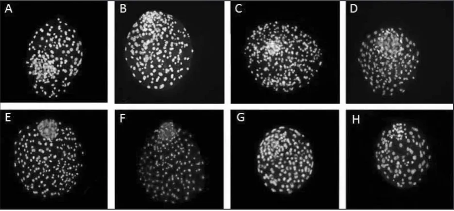
Figure 1. Double staining of blastocysts produced in different culture systems. The blue and pink stained cells (grayish in the photo) are representatives of Inner cell mass and Trophectoderm cells
Ta
ble 2. Eff
ec
t of diff
er en t c ult ur e s yst
ems on ov
ine embr yo de ve lopmen t in v itro C ult ur e condition O oc yt es No. C le av age n
(% ± S
EM)
D
ay 5
n
(% ± S
EM)
D
ay 6
n
(% ± S
EM)
D
ay 7
n
(% ± S
EM)
D
ay 8
n
(% ± S
EM) mor ul a* (c om pac te d) bl ast oc yst* bl ast oc yst (t ot al) bl ast oc yst† (e xp ande d) bl ast oc yst † (ha tc he d) bl ast oc yst bl ast oc yst (ha tc he d) bl ast oc yst bl ast oc yst (ha tc he d) SOF 371 325
(87.8 ± 2.0)
141
(36.8 ± 5.7)
25
(5.9 ± 1.7)
a
152
(40.1 ± 1.8)
a
86
(50.3 ± 7.0)
a,b
10
(4.5 ± 2.3)
190
(49.9 ± 3.0)
a
59
(25.8 ± 5.6)
a
197
(51.9 ± 3.2)
a
117
(56.3 ± 4.5)
a SOF-M SCs 120 109
(90.2 ± 2.9)
40
(32.7 ± 7.3)
0
(0.0 ± 0.0)
b
18
(16.1 ± 6.0)
b
6
(30.5 ± 16.3)
a,b
1
(2.8 ± 2.8)
25
(21.7 ± 5.1)
b
4
(22.2 ± 10.2)
a,b,c
27
(23.6 ± 5.7)
b
10
(32.9 ± 11.0)
a,c M SCs -S OF 133 121
(91.9 ± 2.8)
62
(51.3 ± 8.3)
4
(3.3 ± 1.6)
a,b
38
(28.7 ± 5.2)
c
17
(52.9 ± 9.7)
a
0
(0.0 ± 0.0)
49
(37.2 ± 5.7)
c
18
(39.4 ± 5.6)
a
52
(40.0 ± 7.0)
a,d
34
(69.0 ± 4.7)
b M SCs 212 184
(87.9 ± 3.0)
83
(40.6 ± 4.7)
0
(0.0 ± 0.0)
b
15
(6.8 ± 2.7)
b
4
(28 ± 2.8)
b
0
(0.0 ± 0.0)
23
(10.0 ± 2.6)
b
0
(0.0 ± 0.0)
b
24
(10.5 ± 2.9)
c
6
(17.6 ± 8.5)
c SOF-E FCs 132 119
(88.7 ± 3.2)
46
(35.7 ± 6.5)
0
(0.0 ± 0.0)
b
20
(16.3 ± 5.3)
b
10
(37.2 ± 12.5)
a,b
1
(4.2 ± 4.2)
30
(23.3 ± 6.7)
b,d
7
(22.7 ± 10.2)
a,b,c
33
(26.3 ± 6.6)
b
10
(33.2 ± 10.9)
a,c EFCs -S OF 165 146
(89.0 ± 3.2)
68
(44.6 ± 7.5)
6
(3.8 ± 1.6)
a,b
43
(27.3 ± 4.9)
c
24
(57.8 ± 11.6)
a
2
(4.1 ± 2.7)
49
(31.3 ± 5.0)
c,d
20
(36.7 ± 7.0)
a
56
(35.1 ± 4.3)
b,d
33
(55.4 ± 8.2)
a
EFCs
236
197
(84.1 ± 3.4)
97
(41.9 ± 5.3)
0
(0.0 ± 0.0)
b
21
(9.3 ± 2.1)
b
9
(30.1 ± 10.9)
b
2
(6.4 ± 4.4)
38
(16.5 ± 1.9)
b
9
(24.1 ± 8.5)
a,b,c
40
(17.2 ± 2.1)
c
19
(46.9 ± 8.9)
a,c OE Cs 255 214
(81.9 ± 4.3)
67
(28.8 ± 6.3)
18
(6.3 ± 2.4)
a
44
(16.0 ± 3.1)
b
28
(58.8 ± 9.5)
a
0
(0.0 ± 0.0)
64
(23.1 ± 4.7)
b,d
14
(17.0 ± 5.9)
c
67
(24.2 ± 4.9)
b,c
24
(32.4 ± 9.0)
c a-dnumb ers w ith diff er en t sup ers cr ipt le tt
ers in t
he same c
olumn diff er sig nific an tly ; P < 0.01 *pr op or
tions of mor
ul
a and bl
ast oc yst wer e e xpr ess ed b as
ed on t
he n
umb
er of o
oc yt es †pr op or
tions of e
xp
ande
d and ha
tc he d bl ast oc yst s wer e e xpr ess ed b as
ed on t
he n
umb
er of bl
ast
oc
yst
on day 5 was exclusively observed in SOF, OECs, and the co-cultured groups in which somatic cell co-culture was limited to the first 3 days of IVC (early co-cultured groups). On day 6 of culture the highest blastocyst rate was achieved in SOF group and the corresponding rates in early co-cultured groups were significantly higher than those in groups in which the embryos were first cultured in SOFaaBSA and then co-cultured with somatic cells monolayer for the next 5 days of IVC (late co-cultured groups). The lowest day 6 blastocyst and expanded blastocyst rates were observed in groups in which the embryos were cultured for the whole period of IVC in EFCs and MSCs monolayers. No significant difference was observed in hatching rate of day 6 blastocysts among groups. On day 7, the highest blastocyst rate was achieved in SOF and there was a tendency to the higher blastocyst rate in early co-cultured groups compared to the other co-culture groups. Day 7 hatched blastocyst rate in early co-cultured groups was higher than in OECs. On day 8, the blastocyst rate in SOF group was higher than in other groups except for the MSCs-SOF. Moreover, the total blastocyst rate in MSCs-SOF was higher than corresponding rates in other co-culture groups except for EFCs-SOF. The highest hatched blastocyst rate was achieved in MSCs-SOF group (Table 2).
Among groups, the highest total blastocyst cells number was observed in MSCs-SOF being sig-nificantly higher than corresponding numbers in SOF and OECs groups (Figure 1). Similarly, the highest and the lowest trophectoderm (TE) and inner cell mass (ICM) numbers were observed in MSCs-SOF and OECs groups, respectively. No
significant difference was observed in ICM/total cell ratio between groups (Table 3).
DISCUSSION
Despite the intrinsic quality of the oocyte as a key factor determining the proportion of oocytes developing to the blastocyst stage and the amazing plasticity and tolerance of mammalian embryos to the environment in which they are cultured, there are considerable volumes of evidence in the literature suggesting that the period of post fertilization embryo culture is the most critical period affecting blastocyst quality.
[image:7.595.65.532.102.256.2]In the current study, despite the insignificant difference in rates of cleavage and day 5 morula stage embryos between groups, the day 5 blasto-cysts were exclusively developed in SOF, OECs, and early co-cultured groups (MSCs-SOF and EFCs-SOF). Indeed, in term of cleavage and speed of embryo development, co-culture system had not only superiority over cell free culture system (SOF) but also, except for OECs, embryo devel-opment was deteriorated by culturing in MSCs, EFCs, and late co-cultured groups (SOF-MSCs and SOF-EFCs) (Table 1). This finding was contrasted to the majority of reports indicating the faster cleavage and higher blastocyst rate in co-culture systems (Bongso et al., 1989; Wiemer et al., 1989; Ellington et al., 1990; Smith et al., 1992; Wetzels et al., 1998; Joo et al., 2001). Though, there are some reports indicating no significant improvement in early embryogenesis (Tucker et al., 1995; Hu et al., 1998) and even reports indicating the adverse
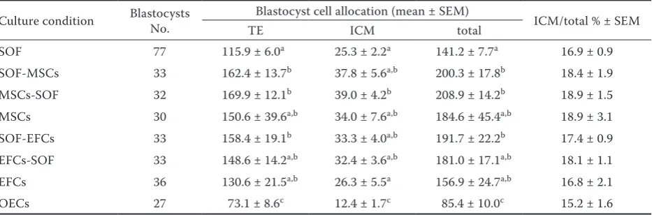
Table 3. Cell allocation of blastocysts derived from different culture systems
Culture condition Blastocysts No. Blastocyst cell allocation (mean ± SEM) ICM/total % ± SEM
TE ICM total
SOF 77 115.9 ± 6.0a 25.3 ± 2.2a 141.2 ± 7.7a 16.9 ± 0.9
SOF-MSCs 33 162.4 ± 13.7b 37.8 ± 5.6a,b 200.3 ± 17.8b 18.4 ± 1.9
MSCs-SOF 32 169.9 ± 12.1b 39.0 ± 4.2b 208.9 ± 14.2b 18.9 ± 1.5
MSCs 30 150.6 ± 39.6a,b 34.0 ± 7.6a,b 184.6 ± 45.4a,b 18.9 ± 3.1
SOF-EFCs 33 158.4 ± 19.1b 33.3 ± 4.0a,b 191.7 ± 22.2b 17.4 ± 0.9
EFCs-SOF 33 148.6 ± 14.2a,b 32.4 ± 3.6a,b 181.0 ± 17.1a,b 18.1 ± 1.1
EFCs 36 130.6 ± 21.5a,b 26.3 ± 5.5a 156.9 ± 24.7a,b 16.8 ± 2.1
OECs 27 73.1 ± 8.6c 12.4 ± 1.7c 85.4 ± 10.0c 15.2 ± 1.6
TE = trophectoderm, ICM = inner cell mass
effects of co-culture on pre-implantation embryo development (Bernardi et al., 1996).
From day 6 onward the difference in embryo de-velopmental indices between SOF and co-cultured groups became more pronounced in favour of cell free culture system (SOF) as such the day 6 blastocyst rate in SOF group was significantly (P < 0.01) higher than in partially (SOF-MSCs, MSCs-SOF, SOF-EFCs, and EFCs-SOF) and totally (MSCs, EFCs, and OECs) co-cultured groups. The presumptive zygotes, which had been totally cultured in MSCs and EFCs, had the least devel-opmental potential in terms of blastocyst rate on different days of culture (days 5–8). This finding was contrasting to the reports indicating the ex-tension of co-culturing time with somatic cells could improve the embryo development through reduction of environmental stress (Wiemer et al., 1989; Orsi and Reischl, 2007).
Among totally co-cultured groups, embryo de-velopment was better supported, though insig-nificant, by the OECs compared to the MSCs and EFCs. Interestingly, in partially co-cultured groups, when the co-culture period with MSCs and EFCs was limited to the first 3 days of culture (early co-cultured groups), the embryo developmental indices were improved.
The higher blastocyst rates on days 6, 7, and 8 in SOF compared to co-cultured groups, except for the day 8 MSCs-SOF, indicated that the cell-free culture system in terms of blastocyst rate was su-perior to the partially or totally co-cultured groups. Since the culture medium (SOFaaBSA) in SOF and co-cultured groups was identical, one explanation for the higher blastocyst rate in SOF group might be related to the addition of FCS after day 3 to the culture medium. Similarly, serum supplementation after day 3 in early co-cultured groups might be the reason for the higher blastocyst rate in these groups compared to the late co-cultured groups.
In terms of embryo quality, the hatching rates be-tween SOF and co-cultured groups on days 6 and 7, except for the day 7 MSCs group, were insignificant, though the corresponding rate on day 8 in MSCs-SOF was higher than in SOF group (P < 0.01).
In differential staining of day 7 blastocysts no significant difference was observed in ICM/TCN (total cell number) ratios between SOF and co-cultured groups. Though, the higher TCN in par-tially co-cultured groups compared to the SOF indicated that at least in term of embryo quality, co-culturing of ovine embryos with MSCs and EFCs
as a part of IVC could improve embryo quality. In contrast, the embryo quality was deteriorated in OECs compared to SOF and other somatic cell co-cultured groups.
The positive effect of co-culture system on viabil-ity markers, such as ICM/TE cell ratios and hatch-ability, has been previously confirmed (Bernardi et al., 1996). As shown (Table 1) among 3 different sources of somatic cells when they were employed for the whole culture period, the OECs could better support, though insignificantly, the development of pre-implantation embryos up to the blastocyst stage on days 5,6, and 7 while the embryo quality (TCN) in this group was significantly worse than in EFCs and MSCs groups. This finding was in contrast to what generally accepted indicating that among different cell types, oviduct epithelial cells are best suited to mimic the physiological milieu of early post-fertilization development (Gandolfi and Moor, 1987). This controversy may be explained by the fact that the oviductal cell cultures are mixed populations of several cell types that may change predominance during culture that in turn could affect the oviductal secretions in support or impairment of embryo development.
The embryotrophic effects of somatic cells, in co-culture systems, may be affected by various parameters, including: somatic cell origin (Galli et al., 2003), base medium composition (Ellington et al., 1990), timing of co-culture, presence of se-rum, microdrop/open culture, gas/oxygen tension, temperature, somatic cell substrate, maternal and paternal influences, inclusion of supplements, heavy metal ion chelators, and embryo develop-mental stage (Desai and Goldfarb, 1998; Rief et al., 2002; Orsi and Reischl 2007).
In the current study embryo development was adversely affected by the application of somatic cells for the whole culture period compared to the SOF group. Though, this adverse effect was decreased when co-culturing with MSCs and EFCs was limited to the first 3 days of culture. This finding was confirmed by the study in which more positive effect of somatic cells was achieved when somatic cells co-culturing was applied during the early cleavage stages (Yeung et al., 1992). Although, there are reports indicating the improvement of embryo quality by increasing the duration of embryo co-culture with somatic cells (Wiemer et al., 1989).
medium (SOFaaBSA) to address different require-ments of both somatic cells and pre-implantation embryos throughout the whole culture period has not been a good strategy. Under this condition, the somatic cells and embryos will compete for nutrient resources and the somatic cells cannot retain their proper morphological and functional properties (Rodriguez-Boulan and Nelson, 1989; Rief et al., 2002). This hypothesis was confirmed by previous reports indicating an incompatibility in the nutritional requirements of somatic cells and early embryos, which require nutritionally complex and dilute media, respectively (Leese, 1988; Bavister, 1992).
Concerning the effect of three types of somatic cells on embryo quality, the positive effect of MSCs was more evident. The MSCs can release several trophic factors including cytokines and growth factors (Orsi and Reischl, 2007; Park et al., 2010). The trophic effects of these bioactive factors in supporting follicular growth and in vitro
maturation of mouse oocytes have been shown (Ling et al., 2008). Moreover, there is difference between various types of stem cells in support of embryo development. It has been shown that bovine embryo development was better supported by amniotic epithelial stem cells compared to the bone marrow derived stem cells (Cremonesi et al., 2008).
Apart from all putative embryotrophic properties of MSCs, one possibility for the higher TCN in MSCs-SOF derived blastocysts might be related to the anti-apoptotic effect of MSCs through ex-pression of higher levels of anti-apoptotic signal molecules (e.g. X-linked inhibitor of apoptosis protein, Bcl-xL, Bcl-2, and heat shock protein-32) (Nematollahi-Mahani et al., 2009). The positive effect of MSCs on the quality of in vitro produced ovine embryos was further confirmed by the report indicating the improvement of poor quality hu-man embryos by culturing in MSCs conditioned medium (Mohamed et al., 2011).
CONCLUSION
In our study condition, the embryo development was better supported by the cell-free culture system compared to the co-culture system. Though, the application of the co-culture system, especially MSCs, for the first 3 days of culture could bet-ter improve the quality of resulting blastocysts
compared to other groups including the cell-free culture system.
Acknowledgement
The authors would like to thank the Avicenna Research Institute, ACECR, Tehran, Iran for fi-nancial and technical support, and also the staff members of the Research Institute of Animal Em-bryo Technology for their kind cooperation.
REFERENCES
Bavister B.D. (1992): Co-culture for embryo development: Is it really necessary? Human Reproduction, 7, 1339–1341. Bernardi M.L., Flechon J.E., Delouis C. (1996): Influence of culture system and oxygen tension on the development of ovine zygotes matured and fertilized in vitro. Journal of Reproduction and Fertility, 106, 161–167.
Biggers J.D., Gwatkin R.B., Brinster R.L. (1962): Develop-ment of mouse embryos in organ cultures of fallopian tubes on a chemically defined medium. Nauchni Trudove na Visshiia Meditsinski Institut, Sofiia, 194, 747–749. Bongso A., Soon-Chye N., Sathananthan H., Lian N.P.,
Rauff M., Ratnam S. (1989): Improved quality of human embryos when co-cultured with human ampullary cells. Human Reproduction, 4, 706–713.
Cremonesi F., Maggio V., Lange-Consiglio A. (2008): Equine amniotic epithelial or bone marrow mesenchymal stem cells differently support in vitro embryo development in a bovine in vitro culture model. Reproduction, Fertility and Development, 21, 156.
Desai N.N., Goldfarb J.M. (1998): Co-cultured human em-bryos may be subjected to widely different microenviron-ments: pattern of growth factor/cytokine release by Vero cells during the co-culture interval. Human Reproduc-tion, 13, 1600–1605.
Ellington J.E., Carney E.W., Farrell P.B., Simkin M.E., Foote R.H. (1990): Bovine 1–2-cell embryo development using a simple medium in three oviduct epithelial cell coculture systems. Biology of Reproduction, 43, 97–104.
Freshney I.R. (1994): Culture of Animal Cells. 3rd Ed.
Wiley-Liss, New York, USA.
Galli C., Duchi R., Crotti G., Turini P., Ponderato N., Colle-oni S., Lagutina I., Lazzari G. (2003): Bovine embryo technologies. Theriogenology, 59, 599–616.
Hu Y., Maxson W., Hoffman D., Ory S., Eager S., Dupre J., Worrilow K. (1998): Co-culture with assisted hatching of human embryos using Buffalo rat liver cells. Human Reproduction, 13, 165–168.
Joo B.S., Kim M.K., Na Y.J., Moon H.S., Lee K.S., Kim H.D.K. (2001): The mechanism of action of coculture on embryo development in the mouse model: direct embryo-to-cell contact and the removal of deleterious components. Fertility and Sterility, 75, 193–199.
Kervancioglu M.E., Saridogan E., Atasü T., Camlibel T., Demir-can A., Sarikamis B., Djahanbakhch O. (1997): Human Fal-lopian tube epithelial cell co-culture increases fertilization rates in male factor infertility but not in tubal or unexplained infertility. Human Reproduction, 12, 1253–1258.
Leese H.J. (1988): The formation and function of oviduct fluid. Journal of Reproduction and Fertility, 82, 843–856. Ling B., Feng D., Zhou Y., Gao T., Wei H., Tian Z. (2008):
Effect of conditioned medium of mesenchymal stem cells on the in vitro maturation and subsequent development of mouse oocyte. Brazilian Journal of Medical and Biological Research, 41, 978–985.
Marcus S.F., Brinsden P.R. (1996): In-vitro fertilization and embryo transfer in women aged 40 years and over. Human Reproduction, 2, 459–468.
Mohamed E.S., Osama M.A., Dina-Sabry A.F., Tamer F. (2011): Stem cell supernatant fluid as a co-culture me-dium may improve poor embryo quality. Evidence Based Women’s Health Journal, 1, 8–10.
Nematollahi-Mahani S.N., Pahang H., Moshkdanian G., Nematollahi-Mahani A.M. (2009): Effect of embryonic fibroblast cell co-culture on development of mouse em-bryos following exposure to visible light. Journal of As-sisted Reproduction and Genetics, 26,129–135. Orsi N.M., Reischl J.B. (2007): Mammalian embryo
co-cul-ture: trials and tribulations of a misunderstood method. Theriogenology, 67,441–458.
Park K.-S., Kim Y.-S., Kim J.-H., Choi B., Kim S.-H., Tan A.H.-K., Lee M.-S., Lee M.-K., Kwon Ch.-H., Joh J.-W., Kim S.-J., Kim K.-W. (2010): Trophic molecules derived from human mesenchymal stem cells enhance survival, function, and angiogenesis of isolated islets after trans-plantation. Transplantation, 15, 509–517.
Rief S., Sinowatz F., Stojkovic M., Einspanier R., Wolf E., Prelle K. (2002): Effects of a novel co-culture system on development, metabolism and gene expression of bovine embryos produced in vitro. Reproduction, 124, 543–556. Rodriguez-Boulan E., Nelson W.J. (1989): Morphogenesis
of the polarized epithelial cell phenotype. Science, 245, 718–725.
Smith S., Schmidt M., Purwantara B., Greve T. (1992): Ovi-duct epithelial cell co-culture of early porcine embryos. Acta Veterinaria Scandinavica, 33, 349–355.
Shamsuddin M., Rodriguez-Martinez H. (1994): Fine struc-ture of bovine blastocysts developed either in serum-free medium or in conventional co-culture with oviduct epithe-lial cells. Journal of Veterinary Medicine, A, 41, 307–316. Teklenburg G., Macklon N.S. (2009): Review: In vitro mod-els for the study of early human embryo-endometrium interactions. Reproductive Sciences, 16, 811–818. Tian L.L., Yue W., Zhu F., Li S., Li W. (2011): Human
mesenchy-mal stem cells play a dual role on tumor cell growth in vitro
and invivo. Journal of Cellular Physiology, 226,1860–1867. Tucker M.J., Kort H.I., Toledo A.A., Morton P.C., Wrigh G., Ingargiola P.E., Sweitzer C.L. (1995): Effect of coculture on subsequent survival and implantation of cryopre-served human embryos. Journal of Assisted Reproduction and Genetics, 12, 689–692.
Wetzels A.M.M., Bastiaans B.A., Hendriks J.C.M., Goverde H.J.M., van der Zalm A.P.E.M.P., Verbeet J.G.M., Braat D.D.M. (1998): The effects of co-culture with human fibroblasts on human embryo development in vitro and implantation. Human Reproduction, 13, 1325–1330. Wiemer K.E., Cohen J., Wiker S.R., Malter H.E., Wright G.,
Godke R.A. (1989): Coculture of human zygotes on fetal bovine uterine fibroblasts: embryonic morphology and implantation. Fertility and Sterility, 52, 503–508. Xu J., Cheung T.M., Chan S.T., Ho P.C., Yeung W.S. (2000):
Human oviductal cells reduce the incidence of apoptosis in cocultured mouse embryos. Fertility and Sterility, 74, 1215–1219.
Yeung W.S., Ho P.C., Lau E.Y., Chan S.T. (1992): Improved de-velopment of human embryos in vitro by a human oviductal cell co-culture system. Human Reproduction, 7, 1144–1149.
Received: 2012–09–22 Accepted after corrections: 2013–04–29
Corresponding Author
Associate Prof. Abolfazl Shirazi DVM, Ph.D., Avicenna Research Institute, ACECR, Reproductive Biotechnology Research Center, P.O. Box 19615-1177, Tehran, Iran