Evaluation of the Stability of Aneurysms after
Embolization Using Detachable Coils:
Correlation between Stability of Aneurysms and
Embolized Volume of Aneurysms
Shinichi Tamatani, Yasushi Ito, Hiroshi Abe, Tetsuo Koike, Shigekazu Takeuchi, and Ryuichi Tanaka
BACKGROUND AND PURPOSE:The long-term outcome of aneurysmal coil embolization has not been determined. We retrospectively analyzed the results of our cases treated with detach-able coils and evaluated the long-term stability of embolized aneurysms.
METHODS: This study involved 100 aneurysms in 93 patients who underwent follow-up angiography >3 months after initial treatment between December 1993 and December 1999.
The percentage of the coil volume occupying the aneurysm lumen (embolized volume) was used as an index to evaluate the stability of embolized aneurysms. The reliability of the embolized volume was also evaluated by comparing angiographic percentage occlusion.
RESULTS:Follow-up angiographic assessment was conducted 12ⴞ8.5 months after initial treatment. Angiographic evaluation of percentage occlusion at initial treatment did not always predict long-term stability of embolized aneurysms. Of 49 aneurysms judged as being totally occluded at initial treatment, 44 remained unchanged and five showed recanalization. The embolized volume of unchanged aneurysms was 30.8 ⴞ 10.2%, and that of recanalized aneu-rysms was 19.9ⴞ10.6%. There was a significant difference between these two groups (Pⴝ.03). Of 29 subtotally occluded aneurysms, nine had further thrombosis (embolized volumeⴝ31.8ⴞ 12.7%), nine remained unchanged (embolized volume ⴝ 23.2ⴞ 10.3%), and 11 had recanali-zation (embolized volume ⴝ 14.1 ⴞ 6.1%). The mean embolized volume of 11 recanalized aneurysms was significantly lower than in the thrombosed group and the unchanged group (Pⴝ .002 and P < .001, respectively). Large aneurysms tended to have recanalization more fre-quently (59%) than did small aneurysms (15%).
CONCLUSION:There is a significant correlation between embolized volume and stability of embolized aneurysms. Embolized volume is a more objective index than is subjectively angio-graphic percentage occlusion. In addition to angioangio-graphic assessment, measurement of embo-lized volume could be useful to predict angiographic changes of aneurysms.
During the last decade, endovascular coil emboliza-tion has become accepted as an effective method for the treatment of intracranial aneurysms (1–11). This technique was initially introduced as a means of treat-ment for patients at high surgical risk: those with aneurysms located in the posterior circulation or paraclinoid region, those with poor clinical status due to subarachnoid hemorrhage, those with a coexisting
medical condition, or those of extremely old age. As the efficacy and safety of this treatment have become known (1, 2, 4, 5, 8–11), indication for aneurysmal coil embolization has become widespread, and this technique is performed as a primary therapy at some institutes. However, several questions remain regard-ing advances in endovascular treatment usregard-ing detach-able coils. The long-term outcome of aneurysmal coil embolization is one of the big questions and is not well defined. Several articles report aneurysm recan-alization, coil compaction, or subsequent rebleeding during early short-term follow-up (1, 4, 5, 9, 12–16). It is true that some of the embolized aneurysms with detachable platinum coils cannot keep the shape of embolized coils for a long term. However, it is not well known how much percentage occlusion can
pre-Received June 14, 2001; accepted after revision January 2, 2002. From the Department of Neurosurgery, Brain Research Insti-tute, Niigata University, Niigata, Japan.
Address reprint requests to Shinichi Tamatani, MD, Department of Neurosurgery, Brain Research Institute, Niigata University, 1–757, Asahimachi, Niigata, 951-8585 Japan.
©American Society of Neuroradiology
vent recanalization of the embolized aneurysm. In this study, we evaluated the long-term angiographic stability of embolized aneurysms using embolized vol-ume (17), which is the objective index to show the percentage of coil volume occupying the aneurysm lumen.
Methods Patient Population
One hundred eighty-seven patients with 201 cerebral aneu-rysms were treated with detachable platinum coils between December 1993 and December 1999. This study involved 100 aneurysms in 93 patients who underwent follow-up angiogra-phyⱖ3 months after initial treatment. One hundred one an-eurysms in 94 patients were excluded from this study because of the following reasons. Twelve aneurysms in 12 patients, all of which were unruptured aneurysms, could not be embolized because of their wide necks. Three of the 12 aneurysms were treated by surgical clipping, and the other nine were followed without any treatment. Eighteen patients with 18 aneurysms died before undergoing follow-up angiography: six as a result of rebleeding, four as a result of initial brain damage, two as a result of vasospasm, two as a result of cardiac failure, and two as a result of other nonrelated diseases. Sixty-eight patients with 71 aneurysms had not yet undergone follow-up angiogra-phy at the time of this writing but were scheduled to do so. Twenty-five patients with 28 aneurysms refused to undergo follow-up angiography. Sixteen patients with 18 aneurysms had not been examined because of their medical conditions. Twen-ty-three patients with 25 aneurysms had moved to another place, and we were not able to contact them. Therefore, the remaining 100 aneurysms in 93 patients were evaluated in this study.
The study population was composed of 66 women and 27 men. The mean age of the patients at the time of presentation was 56 years (range, 23–80 years). Sixty-three of 100 aneurysms were unruptured, and the other 37 were ruptured. Sixty-three aneurysms in 60 patients were discovered incidentally by an-giography or MR anan-giography that was performed for unre-lated medical conditions. Thirty-three patients suffered from subarachnoid hemorrhage. Three of the 33 patients had mul-tiple aneurysms (two aneurysms in each of two patients and three aneurysms in one patient). These aneurysms were in-cluded in the ruptured aneurysms, because we could not de-termine which aneurysm caused the subarachnoid hemorrhage.
Indication for Aneurysm Treatment
Selection of patients for coil embolization rather than sur-gical clipping was conducted by consultation between vascular neurosurgeons and interventional neurosurgeons. The indica-tion for coil embolizaindica-tion of unruptured aneurysms included anticipated technical difficulty of clipping (32 aneurysms in 32 patients), refusal of surgery (30 aneurysms in 28 patients), and old age (one patient, 73 years old). The indication for coil embolization in cases of subarachnoid hemorrhage included anticipated technical difficulty of clipping (22 aneurysms in 21 patients), difficulty of detection of the responsible aneurysm (five aneurysms in two patients), poor medical condition (seven aneurysms in seven patients), attempted surgical exploration (two aneurysms in two patients), and old age (one patient, 80 years old).
Description of Aneurysm
Aneurysm Location.—Seventy-seven (77%) aneurysms were located in the anterior circulation and 23 in the posterior circulation. Of the 37 ruptured aneurysms, 21 (57%) were in the anterior circulation and 16 (43%) in the posterior
circula-tion. Of the 63 unruptured aneurysms, 56 (89%) were in the anterior circulation and seven (11%) in the posterior circula-tion (Table 1).
Size of the Aneurysms.—Seventy-one aneurysms (47 unrup-tured and 24 rupunrup-tured aneurysms) were small (⬍10 mm max-imal diameter), 27 (14 unruptured and 13 ruptured aneurysms) were large (10–24 mm), and two unruptured aneurysms were giant (⬎25 mm).
Size of the Neck.—A neck diameter⬍4 mm is defined as a
small neck. A neck size ⬎4 mm is defined as a wide neck. Fifty-seven (57%) aneurysms (36 unruptured and 21 ruptured aneurysms) had small necks, and 43 had wide necks. All large and giant aneurysms had necks⬎4 mm in diameter.
Embolization of Aneurysms
Interlocking detachable coils were used for the embolization of 16 aneurysms (nine unruptured and seven ruptured aneu-rysms) between December 1993 and February 1997. Guglielmi detachable coil (GDC) systems (18) have been used for aneu-rysmal embolization since March 1997. The embolization pro-cedure was performed with the patient under general anesthe-sia. Systemic heparinization (5000 U of bolus and then 1000 U/h) was used throughout the procedure and was reversed in all patients with ruptured aneurysms by the injection of protamine sulfate. In all patients with unruptured aneu-rysms, systemic heparinization was continued for 24 h after embolization.
[image:2.587.306.535.73.254.2]The size of the aneurysm in three planes (height, length, and width) was measured by digital subtraction angiography or by using a ruler, and the volume of the aneurysmal sac before embolization was calculated. Aneurysms were embolized with detachable coils by packing as densely as possible. The embo-lization was stopped when angiographically complete oblitera-tion was achieved, when the last coil could not be introduced inside the sac, or when occlusion of the normal branch next to the aneurysm might occur. Percentage occlusion at the end of the procedure was evaluated by embolized volume (17). Em-bolized volume was calculated by using the following algebraic equation: embolized volume ⫽ (volume of the embolized coil) / (volume of the aneurysm). The volume of the coil is calculated approximately on the supposition that the coil is a cylinder. The algebraic equation to calculate the volume of the coil is as follows: volume of coil⫽⫻(diameter of coil / 2)2 TABLE 1: Aneurysm location
Aneurysm Location
No. of Aneurysms (%)
Ruptured Unruptured Anterior circulation 21 (58) 56 (89)
ICA 11 42
Paraclinoid 1 25
PCoA 7 15
AchA 0 1
Bifurcation 1 1
ACA 8 7
MCA 2 7
Posterior circulation 16 (43) 7 (11)
BA tip 6 3
SCA 6 1
PICA 2 1
PCA 2 2
Total 37 63
⫻length of coil. The primary diameter of each type of coil is published by Boston Scientific, Target (Fremont, CA). The volume of the aneurysm is also approximately calculated based on the supposition that the aneurysm is ellipsoid: (volume of the aneurysm⫽4/ 3⫻(width / 2)2⫻(length / 2)2⫻(height / 2)2.
The rate of occlusion as measured by angiography was clas-sified as follows: total occlusion when the sac and neck were densely packed in any projection, subtotal occlusion when the sac was occluded but there was suspicion of a neck remnant or there was an obvious tiny neck remnant, and partial occlusion when there was loose packing and/or persistent opacification of the sac or neck remnant.
Angiographic Follow-up Studies
Each patient was scheduled for follow-up angiography at 3 to 6 months, 1 year, and 2 to 3 years after treatment. Recur-rence was defined as an increase in the amount of contrast material filling the aneurysm compared with the angiographic appearance of the aneurysm at the end of treatment, which was classified as an unstable aneurysm. An aneurysm was classified as stable at follow-up if the degree of occlusion was unchanged or had improved compared with the end-of-treatment study.
Statistical Analysis
All values are expressed as mean ⫾SD of the mean. Stu-dent’s t test was used for statistical analysis. P ⬍ .05 was considered significant.
Results
Immediate Angiographic Evaluation Total obliteration was achieved in 49 aneurysms (47 of 71 small aneurysms and two of 27 large rysms). Subtotal occlusion was obtained in 29 aneu-rysms (nine of 71 small aneuaneu-rysms, 18 of 27 large aneurysms, and two giant aneurysms), and partial occlusion was achieved in 22 aneurysms (15 of 71 small aneurysms and seven of 27 large aneurysms).
Relationship between Angiographic Percentage Occlusion and Embolized Volume
Seventy-eight aneurysms were shown to have total or subtotal occlusion on angiograms obtained at the end of the procedure. The mean embolized volume of 49 aneurysms that were totally obliterated on the angiograms was 29.6⫾ 10.7%. That of 29 subtotally occluded aneurysms was 22.4⫾12.1%. On the other hand, the mean embolized volume of 22 aneurysms showing partial occlusion was 17.2⫾7.2%. Statistical significance between the totally occluded group and the subtotally occluded group (P⫽.007) and between the totally occluded group and the partially occluded group (P⬍.001) was observed. No significant differ-ence existed between the subtotally occluded group and the partially occluded group (P⫽.07). However, there seemed to be a correlation between angio-graphic assessment and embolized volume. The mean embolized volume of small aneurysms showing per-centage occlusion of ⱖ90% on the angiograms was 30.3 ⫾ 10.4%. On the other hand, that of large and giant aneurysms was 16.7 ⫾ 8.2%. The embolized
volume of large and giant aneurysms was significantly lower, even if these aneurysms were shown to have satisfactory obliteration on angiograms.
Relationship between Frequency of
Recanalization and Embolized Volume (Table 2) Follow-up angiographic assessment of the degree of occlusion was made at 12⫾8.5 months after initial treatment. Twenty-eight (28%) of 100 aneurysms were observed to be unstable. Of the 57 small aneu-rysms with small necks, six (10.5%) were unstable. The embolized volume of the unstable aneurysms was 20.4 ⫾8.9%. On the other hand, the embolized vol-ume of the stable aneurysms was 30.5 ⫾ 8.9%. A significant difference between these two groups (P⫽ .03) was observed. Five (35.7%) of 14 small aneu-rysms with wide necks were shown to be unstable. The embolized volume of unstable small aneurysms with wide necks was 16.7 ⫾ 11.4% and that of stable aneurysms was 26.9 ⫾10.9%. Although unstable an-eurysms tend to have low embolized volume percent-ages, no statistical significance existed between these two groups because of their small number. Of 27 large aneurysms, 15 (55.6%) were unstable. The embolized volume of unstable large aneurysms was 12.9⫾5.7%. On the other hand, that of stable aneurysms was 20.2⫾8.2%. The embolized volume of unstable large aneurysms was significantly lower than that of stable ones (P ⫽ .012). Both of two giant aneurysms that were partially thrombosed showed recanalization af-ter embolization. The embolized volume percentages were 17.3% and 21.7%, respectively.
Comparison between Angiographic Evaluation and Embolized Volume during Long-Term
Follow-up
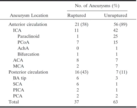
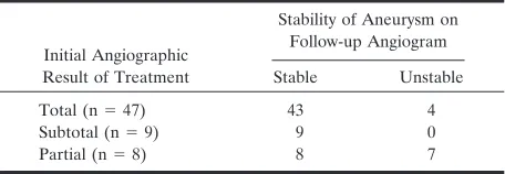
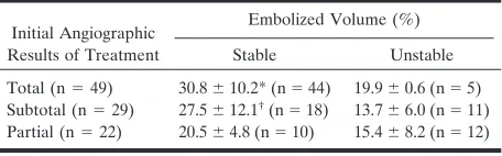
[image:3.587.305.533.73.153.2]Long-term angiographic evaluation is summarized in Tables 3, 4, and 5. No aneurysmal recanalization was observed in 44 (90.0%) of 49 totally occluded
TABLE 2: Embolized volume of aneurysms
Size of Aneurysm
Embolized Volume (%)
Stable Aneurysm Unstable Aneurysm Small (n⫽71)
Small neck (n⫽47) 32.5⫾9.8 19.3⫾7.3 Wide neck (n⫽13) 27.7⫾11.4 17.4⫾10.3 Large (n⫽27) 20.2⫾8.2 12.9⫾5.7
TABLE 3: Correlation between initial angiographic percentage occlusion and long-term stability in small aneurysms
Initial Angiographic Result of Treatment
Stability of Aneurysm on Follow-up Angiogram
[image:3.587.306.534.191.270.2]aneurysms (Table 5). The embolized volume of these 44 stable aneurysms was 30.8⫾10.2%. Five of 49 showed mild coil compaction, and the embolized volume of these five aneurysms was 19.9⫾10.6%. The embolized volume of the stable aneurysms was significantly higher than that of unstable aneurysms (P ⫽ .03). Of the 29 subtotally occluded aneurysms, nine (31%) showed fur-ther aneurysm thrombosis, nine (31%) remained angio-graphically unchanged, and 11 (38%) showed aneu-rysmal recanalization due to compaction of the coils. The mean embolized volume percentages of these three groups were 31.8⫾ 12.7%, 23.2⫾ 10.3%, and 14.1 ⫾ 6.1%, respectively. The mean embolized vol-ume of the recanalized aneurysms was significantly lower than those of the thrombosed and the un-changed aneurysms (P⫽ .0004 andP⫽ .02, respec-tively). No small aneurysm with subtotal occlusion showed recanalization regardless of embolized vol-ume (Table 3). Seven of nine small aneurysms and two of 18 large aneurysms with subtotal obliteration had further thrombosis (Tables 3 and 4). Of the 11 aneurysms showing recanalization, only one large an-eurysm received additional embolization. The other 10 aneurysms were followed conservatively because their recanalizations were minimal. Of the 22 partially occluded aneurysms, total obliteration was achieved in seven (32%) and three remained angiographically unchanged on the follow-up angiograms. The mean embolized volume of these stable aneurysms was 20.5⫾4.8%. On the other hand, the mean embolized volume of 12 unstable aneurysms was 15.4 ⫾ 8.2% (Table 5). Each of two cases underwent second em-bolization, and the other 10 aneurysms were treated conservatively. When the line of demarcation of em-bolized volume was drawn at 25%, 88% of the aneu-rysms with an embolized volume ⬎25% remained angiographically unchanged.
Discussion
The endovascular treatment of an intracranial an-eurysm using a GDC system was performed in 1990 (18), and its use has become widespread. According to the publication of data that provides efficacy and safety of endovascular embolization using detachable coils (1–11), indication for this technique has been extended to aneurysms that previously would have been treated by surgical clipping, although random-ized data have not been published.
On the other hand, coil embolization is a relatively new treatment modality, and several questions re-main. One of the big questions regards stability of the occlusion with detachable coils and efficacy in pro-viding protection against growth or regrowth of an-eurysms and consequent bleeding. In recent years, several authors reported long-term follow-up of an-eurysms treated with detachable coils (1, 2, 5, 8–12). According to their reports, aneurysmal recanalization and/or regrowth is apt to occur in cases of large aneurysms or in cases of aneurysms that were incom-pletely occluded at initial treatment. However, angio-graphic evaluation is very subjective, and it is very difficult to quantitatively assess percentage occlusion of the aneurysms. Our data show that angiographic assessment at initial treatment was not enough to evaluate actual and objective percentage occlusion of embolized aneurysms. Forty-nine aneurysms were judged to be totally occluded at initial treatment in this study; however, five of them showed recanaliza-tion on follow-up angiograms. The embolized volume of the five unstable aneurysms (19.9 ⫾ 10.6%) was significantly lower than that of the stable aneurysms (30.8⫾10.2%). For the 29 subtotally occluded rysms, the embolized volume of the unstable aneu-rysms was significantly lower than that of the stable aneurysms (P⫽.001). These results show that recan-alization can occur in embolized aneurysms with lower embolized volumes, even when angiographic evaluation at initial treatment is good. Comparing small aneurysms and large/giant aneurysms, the em-bolized volume of large/giant aneurysms was signifi-cantly lower, even if these aneurysms showed satis-factory obliteration on angiograms. Considering these results, embolized volume is a useful and objective index that may predict the outcome of an embolized aneurysm.
[image:4.587.54.280.84.163.2]When the line of demarcation of embolized volume was drawn at 25%, the incidence of recanalization was significantly lower in aneurysms with embolized volume percentages ⬎25%. This may indicate that the mark of embolized volume to obtain long-term stability of embolized aneurysms is 25%. Therefore, it is important to select adequate coils for achieving this embolized volume percentage before or during em-bolization. However, there were few large aneurysms that could achieve this embolized volume percentage. This may be the main reason that recanalization oc-curred frequently in large or giant aneurysms. Other reasons for low embolized volume percentages in large and giant aneurysms are considered.
TABLE 4: Correlation between initial angiographic percentage occlusion and long-term stability in large and giant aneurysms
Initial Angiographic Result of Treatment
Stability of Aneurysm on Follow-up Angiogram
Stable Unstable Total (n⫽2) 1 1 Subtotal (n⫽20) 9 11 Partial (n⫽7) 2 5
TABLE 5: Comparison between angiographic evaluation and embolized volume during long-term follow-up
Initial Angiographic Results of Treatment
Embolized Volume (%)
Stable Unstable Total (n⫽49) 30.8⫾10.2* (n⫽44) 19.9⫾0.6 (n⫽5) Subtotal (n⫽29) 27.5⫾12.1†(n⫽18) 13.7⫾6.0 (n⫽11) Partial (n⫽22) 20.5⫾4.8 (n⫽10) 15.4⫾8.2 (n⫽12)
[image:4.587.54.282.201.272.2]One of the reasons may be that the materials of embolic agents are solid. As the solid coil is inserted into the aneurysmal lumen, it piles up and parts of the coil cross each other, causing dead space that cannot be filled by the coils. The more coils are used, the more dead space is increased in the aneurysm lumen. Although many coils are used, isolated spaces may be created by the coils themselves. These spaces lead to a decrease in the embolization rate. Satoh et al (17) examined embolization rates using aneurysm models made of glass tubes. They showed that the maximum embolized volume was 32.0% to 33.3%, even though the aneurysms were packed as tightly as possible with platinum coils.
The limitation in the size of the helix of a coil may also cause a decrease of embolized volume. GDCs are manufactured with four primary wire diameters (GDC 10, GDC 10 soft, GDC 18, and GDC 18 soft). Because the maximum diameter of the helix of a GDC 10 is 10 mm, however, an aneurysm ⬎10 mm needs to be treated using a GDC 18 system or an interlocking detachable coil system, which has helical diametersⱕ20 mm. A GDC 18 coil or an interlocking detachable coil is thicker and stiffer than a GDC 10 coil, which may not allow the coil to better adapt to the aneurysmal shape or to the remaining space within a partially coiled aneurysm. These may also lead to more “dead space,” which may decrease em-bolized volume.
The size of the aneurysm neck influences emboli-zed volume. In our study, all large and giant aneu-rysms had wide neck diameters (ⱖ4 mm). This may be the third reason for the decrease of embolized volume. Zubillaga et al (19) showed complete aneu-rysm occlusion in 85% of small neck aneuaneu-rysms and in 15% of wide neck aneurysms. Vin˜uela et al (11) reported that complete obliteration could be ob-tained in only 31.2% of small aneurysms with wide necks. Debrun et al (12) also showed that absolute neck diameter significantly affects morphologic per-centage occlusion. They reported that aneurysms with neck diametersⱖ5 mm had a lower rate of complete occlusion and a higher rate of neck regrowth. The aneurysms with wider necks allowed prolapse of the coil into the parent artery that led to the prevention of complete occlusion.
Several ideas are considered to make up these weak points of embolization for large and/or wide neck aneurysms. Graves et al (20) described the im-portance of packing the inflow zone of an aneurysm for the durability of aneurysm obliteration. When coils are well placed in the inflow zone of an aneu-rysm, there is a greater chance that the immediate post-treatment percentage occlusion will persist or even increase than if the inflow zone is poorly coiled. New devices that may be useful for embolizing large aneurysms and/or those with wide necks are being developed. 3D GDC is one of the newly developed materials. Soft coils and intracranial vascular stents could be helpful in attaining better outcomes (21, 22). The “remodeling technique” that was initially pro-posed by Moret et al (23) is also useful for improving
outcomes in cases of embolized aneurysms. This tech-nique consists of remodeling the arterial wall by tem-porarily inflating a nondetachable balloon in front of the neck of the aneurysm during each coil placement. Moret et al (23) and Cognard et al (3–5) summarized their results. They did not measure the size of neck but evaluated the ratio between the sack and neck diameter. In their article, the frequency of total oc-clusion did not differ according to the sack-to-neck ratio when they used the remodeling technique (5). On the other hand, there are some problems with this technique, including the necessity to use various cath-eters and devices, making this procedure very com-plicated and increasing the chance of thromboem-bolic events. When the balloon is inflated near the perforating arteries, cerebral ischemia may occur. Aneurysmal rupture may be caused by the increased pressure in the aneurysm. The available diameter of the balloon is then limited to 4.0 mm, and the balloon would need to be overinflated to transiently occlude the internal carotid artery at the level of the aneurysm neck. Overinflation increases the risk of balloon fail-ure, either by rupture of the balloon or by loss of the wire’s ability to occlude the distal lumen (12). Expe-rience and skill of the physician are needed. Because of these problems, the use of this technique should be carefully considered.
It is very interesting that some of the subtotally embolized aneurysms at the end of initial treatment showed total obliteration on the follow-up angio-grams. This may be because of further aneurysmal thrombosis. However, this phenomenon was mainly observed in the small aneurysms, especially in small aneurysms with small necks. On the other hand, fur-ther aneurysmal thrombosis could not be expected for large or giant aneurysms because of their lower em-bolized volume. Hayakawa et al (8) reported similar results, that postembolization angiography revealed progressive thrombosis in 50% of small aneurysms with small necks and in 25% of small aneurysms with wide necks. They also showed that further thrombosis was not observed in large and giant aneurysms. Therefore, careful follow-up with angiography or MR angiography should be performed for large aneu-rysms after coil embolization. When recanalization occurs, additional treatment, such as re-embolization, surgical clipping, or parent artery occlusion, should be performed if necessary.
Conclusion
measurement of embolized volume could be useful to predict angiographic changes of aneurysms.
References
1. Bavinzski G, Killer M, Gruber A, Reinprecht A, Gross CE, Rich-ling B.Treatment of basilar artery bifurcation aneurysms by using Guglielmi detachable coils: a 6-year experience.J Neurosurg1999; 90:843–852
2. Byrne J, Sohn MJ, Molyneux AJ.Five-year experience in using coil embolization for ruptured intracranial aneurysms: outcomes and incidence of late rebleeding.J Neurosurg1999;90:656–663 3. Cognard C, Pierot L, Boulin A, et al. Intracranial aneurysms:
endovascular treatment with mechanical detachable spirals in 60 aneurysms.Radiology1997;202:783–792
4. Cognard C, Weill A, Castaings L, Moret J. Intracranial berry aneurysms: angiographic and clinical results after endovascular treatment.Radiology1998;206:499–510
5. Cognard C, Weill A, Spelle L, et al.Long-term angiographic fol-low-up of 169 intracranial berry aneurysms occluded with detach-able coils.Radiology1999;212:348–356
6. Graves VB, Strother CM, Duf TA, Perl J. Early treatment of ruptured aneurysms with Guglielmi detachable coils: effect on subsequent bleeding.Neurosurgery1995;37:640–648
7. Guglielmi G, Vin˜uela F, Duckwiler G, et al.Endovascular treat-ment of posterior circulation aneurysms by electrothrombosis us-ing electrically detachable coils.J Neurosurg1992;77:515–524 8. Hayakawa M, Murayama Y, Duckwiler GR, Gobin YP, Guglielmi
G, Vin˜uela F.Natural history of the neck remnant of a cerebral aneurysm treated with the Guglielmi detachable coil system.J Neu-rosurg2000;93:561–568
9. Malisch TW, Guglielmi G, Vin˜uela F, et al.Intracranial aneurysms treated with the Guglielmi detachable coil: midterm clinical results in a consecutive series of 100 patients.J Neurosurg1997;87:176–183 10. McDougall CG, Halbach VV, Dowd CF, Higashida RT, Larsen DW, Hieshima GB.Endovascular treatment of basilar tip aneu-rysms using electrolytically detachable coils.J Neurosurg1996;84: 393–399
11. Vin˜uela F, Duckwiler G, Mawad M. Guglielmi detachable coil embolization of acute intracranial aneurysm: perioperative ana-tomical and clinical outcome in 403 patients.J Neurosurg1997;86: 475–482
12. Debrun GM, Aletich VA, Kehrli P, Misra M, Ausman JI, Charbel
F.Selection of cerebral aneurysms for treatment using Guglielmi detachable coils: the preliminary university of Illinois at Chicago Experience.Neurosurgery1998;43:1281–1297
13. Hodgson TJ, Carroll T, Jellinek DA.Subarachnoid hemorrhage due to late recurrence of a previously unruptured aneurysm after complete endovascular occlusion.AJNR Am J Neuroradiol1998;19: 1939–1941
14. Makoui AS, Smith DA, Evans AJ, Cahill DW. Early aneurysm recurrence after technically satisfactory Guglielmi detachable coil therapy: is early surveillance needed? case report. J Neurosurg
2000;92:355–358
15. Manabe H, Fujita S, Hatayama T, Suzuki S, Yagihashi S.Rerupture of coil-embolized aneurysm during long-term observation: case report.J Neurosurg1998;88:1096–1098
16. Mericle RA, Wakhloo AK, Lopes DK, Lanzino G, Guterman LR, Hopkins LN.Delayed aneurysm regrowth and recanalization after Guglielmi detachable coil treatment: case report.J Neurosurg1998; 89:142–145
17. Satoh K, Matsubara S, Hondoh H, Nagahiro S.Intracranial aneu-rysm embolization using Interlocking detachable coils. Intervent Neuroradiol1997;3[suppl 2]:125–128
18. Guglielmi G, Vin˜uela F, Dion J, Duckwiler G.Electrothrombosis of saccular aneurysms via endovascular approach: part 2: prelim-inary clinical experience.J Neurosurg1991;75:8–14
19. Fernandez Zubillaga A, Guglielmi G, Vin˜uela F, Duckwiler GR. Endovascular occlusion of intracranial aneurysms with electrically detachable coils: correlation of aneurysm neck size and treatment results.AJNR Am J Neuroradiol1994;15:815–820
20. Graves VB, Strother CM, Partington CR, Rappe A.Flow dynamics of lateral carotid artery aneurysms and their effects on coils and balloons: an experimental study in dogs.AJNR Am J Neuroradiol
1992;13:189–196
21. Higashida RT, Smith W, Gress D, et al.Intravasculcar stent and endovascular coil placement for a ruptured fusiform aneurysm of the basilar artery: case report and review of the literature.J Neu-rosurg1997;87:944–949
22. Sekhon LH, Morgan MK, Sorby W, Grinnell V.Combined endo-vascular stent implantation and endoendo-vascular coil placement for the treatment of a wide-necked vertebral aneurysm: technical case report.Neurosurgery1998;43:380–384