The Accuracy of CT and MR Evaluation of the Sella Turcica for
Detection of Adrenocorticotropic Hormone-Secreting Adenomas in
Cushing Disease
Michael Buchfelder, Raymond Nistor, Rudolf Fahlbusch, and Walter J. Huk
PURPOSE: To document the accuracy of CT and MR of the sella turcica for detecting adrenocor-ticotropic hormone-secreting adenomas in Cushing disease. METHODS: The radiologic findings of the sella turcica prior to transsphenoidal surgery are reviewed in 141 patients who had biochemical evidence of pituitary-dependent Cushing disease. Axial thin-collimation CT scans with sagittal and coronal reformations before and after contrast enhancement were obtained in 125 patients. Seventy-eight patients had MR examinations with a 1.5-T superconducting magnet. In 11 of the patients gadolinium-enhanced MR scans were also obtained. The preoperative interpre-tation of the imaging studies was correlated with the surgical findings and patient follow-up.
RESOL TS: The sella turcica was enlarged in 43 cases (30% ). In 125 patients reformatted or direct coronal thin-collimation CT scans were available. Seventy-eight of the patients had MR. In the 12 patients with pituitary macroadenomas, the accuracy of CT (n = 1 0) and MR (n = 1 0) in respect to detection of the lesion was 100%. Of the 98 microadenomas assessed by CT, 47 (48%) were directly depicted as distinct hypodense lesions. In only 31 of 73 cases (42%), however, could CT predict the precise anatomic location and extent of the lesions. Only patients in whom the hypercortisolism was corrected by later surgery were considered for the correlation analysis. Of the 52 microadenomas assessed by MR, 28 (53%) were directly depicted as distinct lesions of reduced signal intensity on T1-weighted images, and in only 21 of 41 cases (52%) did MR show good correlation to the surgical findings. Some degree of partially empty sella was found in 22%
of the patients. CONCLUSIONS: Although both the sensitivity and the diagnostic accuracy of imaging methods of the sella turcica have been considerably improved in comparison with previous reports, they still provide only a minor contribution to the diagnosis and differential diagnosis of Cushing syndrome.
Index terms: Sella turcica, magnetic resonance; Sella turcica, computed tomography; Adenoma; Cushing disease
AJNR 14:1183-1190, Sep/Oct 1993
Adrenocorticotropic hormone (A CTH)-secret-ing pituitary adenomas causCTH)-secret-ing CushCTH)-secret-ing disease were originally described to be minute tumors (1).
Neurosurgical experience gained from numerous
sella explorations has confirmed that they rarely
attain a space-occupying character (2-5). The
problems associated with neuroradiologic
evalu-Received January 24, 1992; revision requested April 9; final revision received October 6 and accepted October 14.
All authors: Neurochirurgische Klinik mit Poliklinik der Universitat Erlangen-Niirnberg, 91054 Erlangen, Germany.
Address reprint requests to Dr. Michael Buchfelder, Neurochirurgische Klinik der Universitat Erlangen-Ni.irnberg, Schwabachanlage 6, D-8520 Erlangen, Germany.
AJNR14:1183-1190,Sep/Oct19930195-6108f0195-6108/93/1405-1183 © American Society of Neuroradiology
ation of the sella turcica in this rare kind of
pituitary hypersecretion syndrome are mainly
at-tributable to the small size of these lesions and
the fact that they frequently escape direct
radio-logic depiction, although surgical interventions at
the pituitary level frequently reveal an adenoma,
the resection of which usually leads to a remission
of the disease. The relatively low frequency of
ACTH-dependent Cushing syndrome was an
ob-stacle for compiling pertinent data on the
diag-nostic value of radiographs, thin-collimation
com-puted tomography (CT), and magnetic resonance
imaging (MR). To date, no such comparative
study allowing statistical evaluation has been
per-formed, to our knowledge. We therefore have
1184 BUCHFELDER
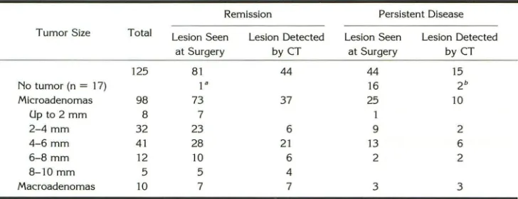
Fig. 1. Examples of different types of sella region CT findings in Cushing
dis-ease.
A, No evidence of abnormal
struc-tures.
8, Direct depiction of intrasellar
mi-croadenoma.
AJNR: 14, September/October 1993
A
8
TABLE 1: Tumor size and CT findings (n
=
125) in relation to treatment results after surgeryRemission Persistent Disease
Tumor Size Total Lesion Seen
at Surgery
125 81
No tumor (n = 17) 1"
Microadenomas 98 73
Up to 2 mm 8 7
2-4 mm 32 23
4-6 mm 41 28
6-8 mm 12 10
8-10 mm 5 5
Macroadenomas 10 7
• Hemihypophysectomy. b False-positive imaging.
consecutive patients who underwent transsphe-noidal surgery for Cushing disease in our depart-ment within an 8.5-year period up to March 31,
1991.
Patients and Methods
A consecutive series of surgically treated patients with ACTH-dependent Cushing disease were analyzed with re-spect to the radiologic findings of the sella turcica prior to transsphenoidal pituitary surgery. They underwent opera-tions between December 1, 1982 and March 31, 1991. The patients were shown to have Cushing disease by dynamic endocrine testing as described previously (2). Routine en-docrine investigation included low- and high-dose dexa -methasone testing, corticotropin-releasing hormone stim-ulation tests, and assessment of anterior pituitary function after administration of luteinizing hormone-releasing hor-mone and thyrotropin-releasing hormone with determina-tions of all pituitary hormones. The initial preoperative interpretations of the imaging studies were correlated with the surgical findings and patient follow-up. The interpreters were blinded to the diagnosis and surgical findings. Two readers were used to interpret each study.
Lesion Detected Lesion Seen Lesion Detected
by CT at Surgery by CT
44 44 15
16 2b
37 25 10
1
6 9 2
21 13 6
6 2 2
4
7 3 3
This series of patients included men and women, ranging from 23 to 66 years of age. They were divided into two groups: those with (n = 96) and those without (n = 45) remission from hypercortisolism after pituitary surgery.
One hundred twenty-five patients were studied by thin-collimation CT. The CT examinations were performed on Siemens (Erlangen, Germany) Somatom DR and DRH high -resolution scanners, respectively. Thin-collim<,~tion (2-mm) axial CT scans with sagittal and coronal computed refor-mations or direct ·Coronal views, befpre and immediately after bolus injection of contrast medium, were obtained in all 125 cases.
[image:2.612.211.556.77.259.2] [image:2.612.127.491.286.426.2]AJNR: 14, September/October 1993
A B
D
CT AND MR IN CUSHING DISEASE 1185
c
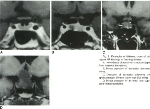
Fig. 2. Examples of different types of sella region MR findings in Cushing disease.
A, No evidence of abnormal structures (apart from cisternal herniation).
B, Direct depiction of intrasellar microade -noma.
C, Depiction of intrasellar adenoma with approximately 1 0-mm tumor size (full sella).
D, Direct depiction of an intra- and supra-sellar macroadenoma.
TABLE 2: Tumor size and MR findings (n
=
78) in relation to treatment results after surgeryRemission Persistent Disease
Tumor size Total Lesion Seen Lesion Detected Lesion Seen Lesion Detected at Surgery
78 54
No tumor (n = 16) 4"
Microadenomas 52 41
Up to 2 mm 3 3
2-4 mm 25 20
4-6 mm 19 14
6-8 mm 3 3
8-10 mm 2 1
Macroadenomas 10 9
• Hemihypophysectomy.
pituitary stalk shifting. Gadolinium-enhanced scans were additionally obtained in 11 of the 78 patients investigated. Enhanced scans were obtained immediately after gadolin-ium administration.
In microadenomas the CT and MR films were primarily evaluated with respect to the direct depiction of a typical intrasellar low density (CT) or low signal intensity region on the T1-weighted images (MR). Secondarily, indirect signs of an intrasellar tumor were searched for, namely an
by MR at Surgery by MR
33 24 5
12
24 11 4
7 5 1
13 5 2
3 1 9
upward bulging of the gland, a lateral displacement of the pituitary stalk, and an asymmetric sellar floor. Additionally, in the patients assessed by MR an eccentric displacement of the hyperintense signal of the posterior pituitary and the
disappearance of the hyperintense signal of blood flow in
the vein of the carotid sulcus or in the medial cavernous
sinus veins on gadolinium-enhanced scans were considered
[image:3.614.54.555.75.440.2] [image:3.614.124.491.469.610.2]1186 BUCHFELDER
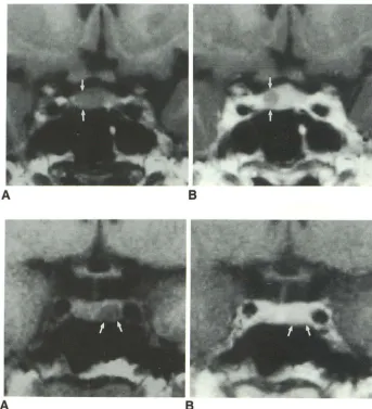
Fig. 3. Influence of paramagnetic
contrast medium (gadolinium-DTPA) on the direct depiction of intrasellar mi-croadenomas in Cushing disease. An
isointense, invisible microadenoma (A)
becomes evident after administration of
gadolinium DTPA (B).
Fig. 4. Influence of paramagnetic
contrast medium (gadolinium-DTPA) on the direct depiction of intrasellar
mi-croadenomas in Cushing disease. A clearly visible microadenoma (A) disap-pears after administration of gadolinium-DTPA (B).
A
as normal. Furthermore, the scans were reviewed for the
presence or absence of intrasellar herniation of the
sub-arachnoid space (partially empty sella). Of the total of 141
patients, only 62 were assessed by both CT and MR.
The radiologic findings were related to the intraoperative
findings with respect to the total size of the tumor resected
and its location. The outcome of surgery was classified
according to the result of low-dose dexamethasone testing
3 months after operation. The finding of cortisol levels
suppressible to less than 2 ,ug/dl was regarded as
biochem-ical proof of remission from the disease. The determination
of the specificity of the respective neuroradiologic
investi-gations was related to the group of 96 patients who were
found to have a remission from hypercortisolism after surgery. For the correlation of neuroradiologic and surgical findings, diameter, extension of the adenoma, and tumor
configuration were quoted. Identical or very similar findings were rated as "good." If the tumor was detected and located
only in part by neuroimaging methods, the correlation was
specified as "fair." A rating of "poor" denoted a false location
of the tumor by neuroimaging or the inability of the
neuroradiologist to correctly locate at least a part of the
tumor.
AJNR: 14, September /October 1993
B
B
Selective adenomectomy was performed whenever a discrete pituitary adenoma was identified during pituitary
microsurgery.
Results
Thin Collimation CT
[image:4.617.220.564.79.456.2]AJNR: 14, September/October 1993
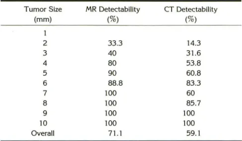
TABLE 3: Delectability by CT and MR imaging of surgically verified pituitary microadenomas in Cushing disease, in relation to tumor size as estimated during surgery
Tumor Size MR Delectability CT Delectability
(mm) (%) (%)
1
2 33.3 14.3
3 40 31.6
4 80 53.8
5 90 60.8
6 88.8 83.3
7 100 60
8 100 85.7
9 100 100
10 100 100
Overall 71.1 59.1
a low-density region in two cases. However, the
other 15 scans were reported to be normal. One
of the patients had a remission from
hypercorti-solism following a hemihypophysectomy
accord-ing to the ACTH gradient obtained duraccord-ing bilateral simultaneous inferior petrosal sinus blood
sam-pling. Of the 98 microadenomas found by the
surgeon, only 47 were directly depicted by a
clearly circumscribed low-density region. The re-lation between tumor diameter as found during pituitary surgery and the detection rate of indirect
and direct signs of an intrasellar adenoma is
summarized in Table 1.
MR
Of 78 patients assessed by MR, 52 had
mi-croadenomas, and 1 0 had macroadenomas. In 16
patients, no tumors were found during pituitary
microsurgery. Again, four different types of MR
presentation were distinguished (Fig. 2). All 10 macroadenomas were directly depicted. Invasion of structures surrounding the sella turcica was encountered in three patients during surgery but was preoperatively recognized by the MR findings in only two cases. There was no case of a false-positive demonstration of a discrete hypointense intrasellar lesion among the 16 cases in whom sella exploration did not reveal a pituitary micro-adenoma. Three of these patients had a remission from hypercortisolism following a hemihypophy-sectomy according to the ACTH gradient ob-tained during bilateral simultaneous inferior pe-trosal sinus sampling. The relation of tumor di-ameter as found during pituitary surgery and the detection rate of indirect and direct signs of an
intrasellar adenoma are summarized in Table 2.
CT AND MR IN CUSHING DISEASE 1187
In addition to the unenhanced MR scans, gad
-olinium-DTP A was used as a paramagnetic con-trast medium in 11 patients. The paramagnetic agent improved the quality of preoperative scans in only two of eight patients later shown during
surgery to harbor a pituitary microdenoma (Fig.
3). The microadenomas measured 5 to 6 mm and
8 to 9 mm, respectively. In both cases the
ade-nomas were isointense on nonenhanced MR scans and became visible as a nonenhancing intrasellar zone on gadolinium-enhanced scans.
However, in both of these cases obvious indirect
signs signaled the presence of an intrasellar ade
-noma even on nonenhanced scans. In two other
cases of microadenomas with diameters of 4 and
5 mm, respectively, the tumor was clearly de
-tectable as an intrasellar hypointense region on nonenhanced scans. After administration of gad-olinium the tumors enhanced in a way similar to the adjacent normal pituitary gland and thus
became less visible (Fig. 4). In the remaining four
adenomas the adenomas were clearly visible on
both nonenhanced and enhanced scans.
CT versus MR
A total of 62 patients had both CT and MR
prior to surgery. Of these, 41 had
microadeno-mas; eight had macroadenomas. In 13 of these
cases, no tumor was found during microsurgical
sella exploration. In the 8 cases with macroad-enomas, CT and MR were equally effective in depicting the size and extension of the tumors. However, in the three tumors with parasellar development, MR provided a better delineation of the adenomas and the connection of intra- and
parasellar tumor parts.
Of the 41 cases with microadenomas, 13 were
directly depicted by CT, and 20 were directly
depicted by MR. In only one instance was a
6-mm microadenoma directly depicted by CT but
not shown by MR. On the other hand, in six cases
MR directly depicted an intrasellar adenoma, al-though the CT scans had revealed only indirect
signs of an intrasellar lesion. In an additional five
cases there was neither direct nor indirect
detec-tion of a tumor on CT, but there was depiction
of a hypointense signal region corresponding to
a microadenoma on MR. Thus, in 11 out of 41
cases with microadenomas the MR findings were
clearly superior. In 29 cases the morphologic
information about the tumor provided by CT and
MR were equivalent. CT detected abnormalities
[image:5.612.54.298.117.259.2]1188 BUCHFELDER AJNR: 14, September /October 1993
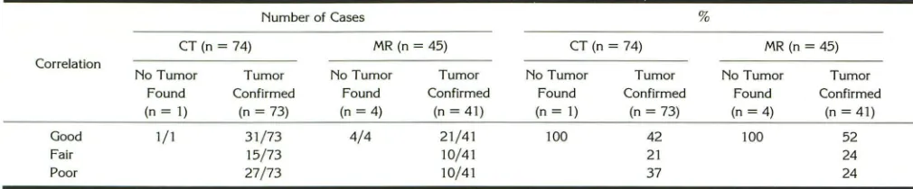
TABLE 4: Correlation of CT and MR images with the operative findings in surgically cured patients
Number of Cases %
CT (n = 74) MR (n = 45) CT(n=74) MR (n = 45)
Correlation
No Tumor Tumor No Tumor Tumor No Tumor Found
(n =I)
Tumor Confirmed
(n = 73)
No Tumor
Found (n = 4)
Tumor Confirmed
(n = 41)
Found Confirmed Found Confirmed
(n = 1) (n = 73) (n = 4) (n = 41)
Good Fair Poor
1/1 31/73 4/4 21/41 100 42
21 37
100 52
24 24 15/73
27/73
to harbor microadenomas; MR was considered
abnormal in 71. 1% of these lesions. The
detect-ability of microadenomas by CT and MR in cor-relation with the intraoperatively estimated size of the tumors are summarized in Table 3.
The results of pituitary imaging clearly were a prognostic factor for the outcome after pituitary surgery. When a distinct microadenoma was
de-picted by CT, 37 of 47 patients (78.7%) had
remission after surgery. However, if the scan was negative, only 28 of 58 patients (51. 7%) remitted. A hypointense, well-demarcated intrasellar lesion
on MR similarly was a favorable prognostic
indi-cator, because in 24 of 28 patients (85.7%) with
this finding the hypercortisolism was corrected
by pituitary surgery. Correspondingly, 17 of 31
patients with negative MR scans had persistent
disease after the operation. In 19 of the 141
patients no adenoma was found. Four out of eight patients in whom no discrete microadenoma was found during sella exploration, and who had a significant ACTH gradient when inferior petrosal sinus sampling was performed, had a remission
following respective hemihypophysectomy.
l'feuroradjofogjc vs Surgjcal Topography
In 16 cases in which no tumors were found,
the. MR findings were never false positive. In
contrast, the CT scan falsely suggested adenomas
that could not be confirmed by both surgeon and pathologist in two of the 17 cases. The correlation between CT and MR findings, respectively, and
the surgical topography is summarized in Table
4. There was an excellent correlation in the de-tection of intrasellar cisternal herniation, which was found in 31 out of the total 141 cases.
Discussion
Because hypercortisolism may be attributable
to a variety of different causes, and because it may be difficult even after extensive dynamic
10/41 10/41
functional testing to locate precisely the source of cortisol hypersecretion (6, 7), the neuroradiol-ogist is frequently expected to contribute to the
differential diagnosis of Cushing syndrome.
How-ever, the vast majority of pituitary adenomas
associated with ACTH-dependent Cushing syn-drome (Cushing disease) are small lesions. With
the development of thin-collimation CT and MR
neuroradiology of pituitary lesions, sella
tomog-raphy is no longer used and is today considered
to have only historical relevance.
The sensitivity of thin-collimation CT scans to detect microadenomas of the pituitary gland in Cushing disease directly was reported to range from 17% (8) to 58.3% (9). Indirect signs of an adenoma, such as upward bulging of the gland's upper surface or pituitary stalk deviation, were
more frequently found (8-1 0). There was some
debate as to whether direct coronal scanning with bolus contrast application could improve the di-agnostic accuracy (11). By using a specialized software system, and by carefully controlling the CT procedures, paying attention to the individual patient's requirements, Bonneville et al (12) have obtained results comparable to MR findings even when applied to patients with pituitary microad-enomas. However, Marcovitz et al (9), who have used direct coronal scans, report microadenomas found during pituitary microsurgery in 10 out of
15 patients with negative scans. Furthermore, the
detection of radiologic abnormalities does not
necessarily imply that the images depicted
rep-resent the morphologic reality. Teasdale et al (13)
have correlated the findings of reformatted
thin-collimation CT images with the operative findings
in 14 patients who underwent transsphenoidal
surgery for Cushing disease. In 5 of the cases CT gave false indications of the anatomic locations
of the ACTH-secreting adenomas. Only one of
three patients in whom no distinct microadenoma
was found during surgery had a small pituitary
gland without radiologic abnormalities on CT. In
[image:6.612.54.559.95.200.2]AJNR: 14, September/October 1993
visualized by reformatted CT images in a patient whose hypercortisolism was corrected by resec-tion of the tumor had an estimated diameter of 3 mm. Bone-hardening artifacts within the sella may be mistaken for pituitary microadenomas in coronal or sagittal reformatted images. In these, the streak artifacts may appear as rounded hy-podense lesions. These hyhy-podense regions may not be recognized as artifacts in reformatted im-ages, which is their major disadvantage. Although fat deposition in the cavernous sinus (14) is more frequently noted in Cushing disease than in other pituitary hypersecretion syndromes, it is not a pathognomonic sign.
The sensitivity of MR in directly detecting ACTH-secreting microadenomas was reported to
range from 44.9% (15) to 71% (16). Indirect
evidence of a microadenoma by some kind of pituitary stalk deviation was found in 59% of the patients (17). Although the use of gadolinium was believed by some authors to increase the sensi-tivity of direct detection (15, 18), we have seen two cases in which microadenomas became
in-visible after contrast enhancement. Generally, the capability of detecting small microadenomas seems to be slightly increased by MR if compared with CT. The smallest microadenoma directly depicted by MR in a patient who had a correction of hypercortisolism after pituitary microsurgery had an estimated diameter of 3 mm.
Although the association of pituitary microad-enomas with Cushing disease and intrasellar cis-ternal herniation has occasionally been reported
(17, 19), little attention has been paid to it in the literature. We have found some kind of intrasellar arachnocele in 22% of our patients. Among these, there was only one patient in whom en-largement of the sella turcica could not be
radi-ologically attributed to a distinct microadenoma.
It is obvious that the chance of detecting and
correctly localizing the adenoma intraoperatively
increases dramatically with its size.
In cases with the biochemical diagnosis of pituitary-dependent Cushing disease in which
there is no microadenoma visualized by
sophisti-cated imaging techniques, there is a smaller chance that a microadenoma is found during pituitary microsurgery. In addition, there is a smaller chance of remission following pituitary
microsurgery. We therefore would recommend
that this subgroup of patients should be
submit-ted for simultaneous petrosal sinus sampling to
confirm the diagnosis and to increase the chance of surgical cure by providing a basis for partial
CT AND MR IN CUSHING DISEASE 1189
hypophysectomy according to the ACTH
gra-dient (20, 21).
Our study was performed on a highly selected
group of patients, who were found to have the typical biochemical characteristics of Cushing disease during dynamic endocrine testing (2, 7). Therefore, it does not allow us to draw
conclu-sions on radiologic findings of patients who suffer
from hypercortisolism and who have neither hy-pothalamic nor pituitary disease. Nevertheless,
the differential diagnosis of the syndrome is still
predominantly based on the findings of classical
biochemical testing. Neuroradiologic assessment
of the sellar content provides a visualization of a
distinct microadenoma in only 40% to 50% of the patients found on the basis of dynamic
en-docrine testing to have Cushing disease, in whom
hypercortisolism was corrected by resection of a pituitary adenoma even when recent imaging
technology is used.
References
1. Cushing H. The basophil adenomas of the pituitary body and their clinical manifestations (pituitary basophilism). Bull Johns Hopkins
Hosp 1932;50:137-195
2. Fahlbusch R, Buchfelder M. Muller OA. Transsphenoidal surgery for Cushing's disease. J R Soc Med 1986;79:262-269
3. Laws ER. Cushing's disease: neurosurgical viewpoints, In: van H eer-den JA, ed. Common problems in endocrine surgery. Chicago: Year Book Medical, 1989:18-22
4. Mampalam T J, Tyrrell JB, Wilson CB. Transsphenoidal microsurgery for Cushing's disease: a report of 216 cases. Ann Intern Med
1988; 109:487-493
5. Nakane T, Kuwayama A, Watanabe M, et al. Long term results of transsphenoidal adenomectomy in patients with Cushing's disease.
Neurosurgery 1987;21 :218-222
6. Trainer PJ, Grossman A. The diagnosis and differential diagnosis of Cushing's syndrome. Clin Endocrinol (Oxf) 1991 ;34:317-330
7. Kaye TB, Crapo L. The Cushing syndrome: an update of diagnostic
tests. Ann Intern Med 1990; 112:434-444
8. Saris SC, Patronas NJ, Doppman JL, et al. Cushing syndrome: pituitary CT scanning. Radiology 1987;162:775-777
9. Marcovitz S, Wee R, Chan J, Hardy J. The diagnostic accuracy of preoperative CT scanning in the evaluation of pituitary ACTH-secret-ing adenomas. AJNR: Am J Neuroradiol 1987;8:641-644
10. Pojunas KW, Daniels DL, Williams AL, Thorsen MK, Haughton VM. Pituitary and adrenal CT of Cushing syndrome. AJNR: Am J Neuro-radio/ 1986;7:271-274
11. Bonneville JF, Cattin F, Moussa-Bacha K, Portha C. Dynamic com-puted tomography of the pituitary gland. The 'Tuft Sign." Radiology
1983;149:145-148
12. Bonneville JF, Cattin F, Dietemann JL. ACTH-secreting pituitary adenomas. In: Computed tomography of the pituitary gland. Berlin:
Springer, 1986:137-144
13. Teasdale E, Teasdale G, Mohsen F, Macpherson P. High-resolution computed tomography in pituitary microadenoma: Is seeing believ-ing? Clin Radio/ 1986;37:227-232
1190 BUCHFELDER
15. Dwyer AJ, Frank JA, Doppman JL, et al. Pituitary adenomas in patients with Cushing's disease: initial experience with Gd-DTPA
enhanced MR imaging. Radiology 1987;163:421-426
16. Peck WW, Dillon WP, Norman D, Newton TH, Wilson CB. High-Resolution MR imaging of microadenomas at 1.5 T: experience with Cushing disease. AJNR: Am J Neuroradio/1988;9:1085-1091 17. Johnson ZS, Phillips J, Devlin J, O'Donnell.). lntrasellar subarachnoid
space in Cushing's disease. lr Med J 1983;76:358
18. Doppman JL, Frank JA, Dwyer AJ, et al. Gadolinium DTPA enhanced
MR imaging of ACTH-secreting microadenomas of the pituitary gland. J Camp Assist Tomogr 1988;12:728-735
AJNR: 14, September/October 1993
19. Ganguly A, Stanchfield JB, Roberts TS, West CD, Tyler FH. Cushing's syndrome in a patient with an empty sella turcica and a
microade-noma of the adenohypophysis. Am J Med 1976;60:306-309
20. Miller DL, Doppman JL, Nieman LN, et al. Petrosal sinus sampling: discordant lateralization of ACTH-secreting pituitary microadenomas before and after stimulation with corticotropin-releasing-hormone. Radiology 1990; 176:429-431