With 13 figures Printed in Great Britain
THE FORCES EXERTED ON THE SUBSTRATE BY
WALKING AND STATIONARY CRICKETS
BY JACK HARRIS AND HELEN GHIRADELLA Biology Department, The University at Albany,
Albany, New York, 12222, U.S.A. (Received 26 June 1979)
SUMMARY
1. The gait and the protraction/retraction ratios (P/R ratios) for the cricket are described. They are essentially the same as for the cockroach and the grasshopper.
2. The vertical forces exerted on the substrate by all six legs of walking and stationary crickets are measured. On the basis of the ' forceprints' obtained and differences in P/R ratios among the legs of different thoracic segments, it is pointed out that all segments are not functionally identical. Specifically, the greater irregularity of the forceprints of the prothoracic legs, and the lower magnitude of peak force exerted on the substrate by the prothoracic legs suggest that the prothoracic legs are more involved in balancing or searching than in propulsion.
3. The metathoracic legs exert an increased vertical force on the sub-strate just before the initiation of protraction. This increase correlates with an extension of the leg apparently through extension of the femoral-tibial joint.
4. A slight decrease in the force exerted on the substrate by the meso-thoracic legs occurs when the leg is at right angles to the body.
5. Placing or lifting one mesothoracic leg does not affect the force exerted by the contralateral mesothoracic leg in a regular way. This argues against mechanical interactions between the legs and in favour of theories invoking central generation of pattern.
6. At a stepping frequency of below 2 steps s"1 the shapes of the force-prints of all legs are no longer repetitive. Also, below 2 steps s- 1 there is an increase in the variability of the peak force exerted on the substrate. It is possible that the animal switches to a more sensory sensitive mode below a step frequency of 2 s-1.
7. During stationary periods the forces exerted on the substrate continue to show oscillations which may be metachronal. This suggests a mechanism whereby a central oscillatory mechanism can account for the behaviour of an animal starting to walk following such a stationary period.
INTRODUCTION
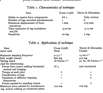
move-Table 1. Characteristics of technique
Item
Ability to resolve force components
Cruse (1976) All 3 Number of legs recorded simultaneously 1 Maximum displacement of force
transducer
Time resolution of leg touchdown and lift
Sensitivity
1 mm + 20 ms
20 mg
Table 2. Applications of technique
Item Insect used
Maximum stepping frequency Body weight (mean)
Filming speed Used for determining
Forces from insect walking horizontal, vertical and hanging
Torque at each joint Coordination of legs
Variations at different stepping frequencies
Forces exerted by standing insects Maximum force exerted by metathoracic
leg, animal walking on horizontal plane
Cruse (1976) Stick 2-3 s-» 820 mg 18 frames s"1 all 3 + — — — 252 mg
Harris & Ghiradella Only vertical
6
0-05 mm ± 17 ms 5 mg
Harris & Ghiradella Cricket
15-20 s"1 435 mg
32, 60, 80 frames s"1 only horizontal — + + + 220 mg
ments (Manton, 1973; Hughes & Mill, 1974). The regularities of these movements and their constancy in unrelated species has led to attempts to model and describe the gaits in terms of cybernetic rules of control (Graham, 1977a, b; Wilson, 1966). The regularity, simplicity, and elicitability of the gaits has led neurobiologists to utilize insect walking as a tool to explore the neural basis of behaviour (for reviews see Hoyle, 1976; Pearson & Duysens, 1976). The gait studies have described the mechanical movements of all six legs and the neurobiological studies have recorded the activities of a few muscles and nerves.
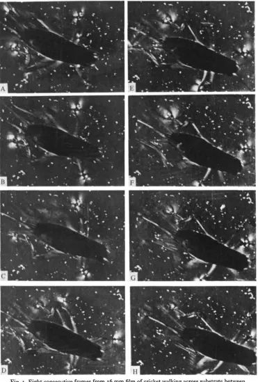
[image:2.451.57.397.48.355.2]Journal of Experimental Biology, Vol. 85
Fig. 1
Fig. 1. Eight consecutive frames from 16 mm film of cricket walking across substrate between crossed polars. The white patches at each tarsus are the photoelastic signals; the area of each patch corresponds to the force exerted by that leg. Note the tripod stance in B and F. Note also the surge in the signal from the right metathoracic leg in E, just before protraction (F). The fine parallel lines (B, F and G) are film-processing artifacts. The fine white dots littering the surface are caused by depolarizing dust on the surface, most of which was deposited by the cricket on a previous passage. Note (by reference to these dots) that the printing frame was shifted after C. This passage was made at a stepping frequency of 5-8 steps s"1. Time interval between frames is 16 ms.
To dissect the contributions of central tapes and sensory feedback to the co-ordination of walking, we need to know the normal leg movements of unrestrained insects and we also need to know how the animal maintains balance and produces propulsive forces. The leg movements have been characterized (Wilson, 1966) and Cruse (1976) measured the forces produced by the legs of a free-walking stick insect. By using strain-gauges he was able to measure the components of forces produced in the x, y and z directions from insects walking horizontally, vertically and hanging from a beam. The strain-gauge technology permitted recording from one leg at a time. This present study reports quantitative measurements of forces exerted on the substrate (in the vertical direction only) by all six legs of unrestrained walking and stationary crickets. For comparisons of this work and Cruse's see Tables 1 and 2.
MATERIALS AND METHODS
The recent work on insect walking has utilized large insects: the stick insect (Wendler, 1966; Cruse, 1976; Graham, 1972), the cockroach (Hughes, 1967; Wilson, 1966; Delcomyn, 1971a, b\ Pearson & lies, 1970, 1973) and orthopterans (Hoyle, 1964, Runion & Usherwood, 1968). For this work, adult female common house-crickets, Acheta domesticus, were used. Although the cricket can jump, it is not so specialized for jumping as other orthopterans, and field and laboratory observations suggest that the cricket prefers to walk' rather than to jump. (In the laboratory the younger, smaller instars are more likely to jump than the older, larger instars.)
The technique used to measure the forces exerted by the crickets is photoelastic substrate transduction. A complete description of the technique is available elsewhere (Harris, 1978). Briefly, the passing cricket tarsus induces temporary strain bire-fringence in a polymeric (gelatin based) substrate. This birebire-fringence is visible when the substrate is viewed in polarized light (Fig. 1). The area of the birefringence is quantitatively related to the stress (force per area) exerted on the substrate by the cricket tarsus (Harris, 1978). Dividing the stress by the area of contact gives the force exerted by the tarsus. It is important to emphasize that the animal does not noticeably indent or deform the substrate; the optical pattern results from induced strain.
Fig. 2. Diagram of film sequence in Fig. i. In this diagram, load-bearing legs are represented by solid lines, protracting legs are represented by dashed lines, and the photoelastic signal is represented by stipple.
distortion or misrepresentation of the vertical component of the force. If at each angle of setting of the micromanipulator, a compensatory weight is added which serves to keep the vertical component of the force constant while constantly increasing the horizontal component of the force, it is found that the area of birefringence remains constant.
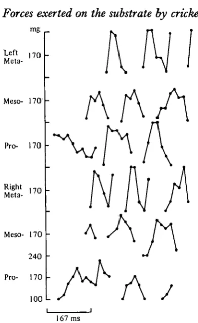
mg
Left 1 7 O f.
MetaMeso 170
-Pro- 170
Right ) 7 0 Meta- "U
Meso- 170
240
Pro- 170
100 L
[image:6.451.122.318.34.353.2]167 ms
Fig. 3. Graphical representation of forces exerted on the substrate over 0-5 s by a cricket (including the sequence shown in Fig. 1). The forces are calculated from the area of bi-refringence measured for each foot in consecutive frames of the film. A break in the line represents protraction.
21-i °C. A heat filter was placed between the light source and the stage to prevent heating of the substrate.
Since the photoelastic substrate technique measures stress (force per unit area) it is impossible to state the ultimate sensitivity of the technique for resolving forces (Harris, 1978). For the stresses applied by walking crickets, we could routinely measure differences in force of less than 2 mg. However, some error is to be expected from the photographic and analysis procedures and we therefore chose 5 mg force as a conservative estimate of sensitivity. Accuracy and repeatability of the inter-pretation were assured by calibrating each film as described below.
The pattern of birefringence was filmed on 35 mm Tri-X film at 48 frames s~x with a Bell & Ho well type A-7 camera and at 60 and 80 frames s"1 on 16 mm Tri-X film with an Arriflex camera. The films were processed in Diafine developer and the area of birefringence for each foot in each frame was measured on an Inter-national Imaging Systems false colour densitometer. The system was calibrated on each film by applying known stresses by a needle with an end of known area applied at varying angles as described above.
convention introduced by Wilson (1966) is reversed in that retractions are in black and protractions are represented by breaks in the line. In this way vertical deflexion can be used to indicate the magnitude of the vertical force exerted on the substrate.
RESULTS
(A) Description of gait
The gait of the cricket is essentially the same as reported for the cockroach (Delcomyn, 1971a), independent of speed over most of its range. Fig. 4 shows the angles swept by the legs. These are slightly more anterior than in the grasshopper (Burns, 1973). Figs. 5-7 illustrate the changes in the gait characteristics with increasing stepping frequency and consequent velocity. As was found to be the case with the cockroach (Delcomyn & Usherwood, 1973) and the locust (Burns, 1973), the duration of protraction is approximately constant for all velocities above two hertz and the duration of retraction varies with step frequency. Step frequencies were determined as an average for each passage of the cricket through the filming area rather than for each step. This has the effect of averaging over several steps the rapid changes seen in instantaneous velocity of the animal and reflects the time averaged rate of progress of the animal for that particular stepping frequency.
(B) Forceprihts
Each passage of the cricket was plotted as in Fig. 3. Comparisons of these 'force-prints' for different animals and of the same animal walking at different speeds demonstrates that the shape of the forceprint is more consistent (i.e. the same shape occurs in all forceprints) for the mesothoracic and metathoracic legs than it is for the prothoracic legs for animals walking above 2 steps s- 1 (Fig. 8). For animals walking slower than this frequency the forceprints are non-repetitive for legs of all segments.
A small dip usually occurs in the forceprint of the mesothoracic legs (Figs. 3, 8). This dip occurs when the leg is at 93 + 50 to the central axis of the body. When the leg is bent, such as occurs when the animal is turning towards that side (i.e. not extended straight out from the body through all joints as is the case normally), the dip is not present.
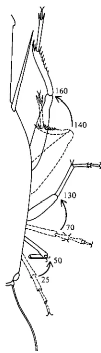
Fig. 4. Angles relative to body axis swept by legs during retraction. This data is taken from films made from above the animal, using polarizing optics, which does not permit resolution of the angles between segments of individual legs. Note that unlike the legs of other segments, the prothoracic flex legs during retraction.
(C) Walking behaviour
Fig. 10 is a histogram of observed walking speeds. The cricket walks between 2 and io steps s"1 on this substrate under experimental conditions. Speeds slower than 2 steps s"1 involve starts and stops and may correspond to the slow walking of cockroaches that Delcomyn & Usherwood (1973) termed 'ambling'. At speeds faster than 10 steps s- 1 the cricket resorts to small hops interspersed with normal steps. From Fig. 10 it can be seen that adult female crickets walking on the substrate show a preference for walking about 4-5 steps s-1.
The mean peak force (and standard deviation) exerted on the substrate as a function of stepping frequency is shown in Fig. 7. There is a small, but significant, change in peak forces at different speeds.
250
~ 2 0 0
I 150
Q
100
50
Retraction
Protraction
1 2 3 4 5 6 7 8 - 9
Stepss"1
Fig. 5. Duration of retraction and protraction at various stepping frequencies. Data points not showing error bars had standard deviations too small to indicate.
0-9
0-8
0-7
0-6
0-5
0-4
0-3
0-2
0 1
I I I
1 2 3 4 5 6 7 8 9
Stepss"1
271
240
200
160
120
80
40
-l i i
I
]/
I 1
AX
T
I M 1
}
T
1 8
Stepss"1
Fig. 7. Peak forces exerted on the substrate at various stepping frequencies. The dots represent the mean and the error bars indicate the standard deviation of the vertical component of the force. This figure represents the average highest force recorded for each leg at each stepping frequency without reference to when that peak force occurs in the retraction. For typical forceprints, see Figs. 3 and 8. • , Metathoracic legs; x , mesothoracic legs; O, prothoracic legs.
Pro
Meso-200 mg
Meta-167 ms
Fig. 9. Tracing of metathoracic leg position from one frame before protraction (solid line) and two frames before protraction (dashed line) from sequences showing a surge in the signal just before protraction. The tracings were superimposed to show that the leg has extended in that interval (16 ms). The arrow indicates how much the tracings had to be moved to superimpose them, which is how far the body had moved forward over the legs in that interval. These two examples represent the range of variability of leg extension and body movements of all examples examined.
35
30
1 25
0
S 20
D a =1
* 15 10 5
-1 2 3 4 5 6 7 8 9
Steps s"1
Fig. 10. Histogram of observed stepping frequencies for crickets walking on gelatin-based substrates. Each 'case' represents the average stepping frequency for one passage through the filming area consisting of at least three steps.
on one mesothoracic leg when the other is placed or lifted. Fig. n considers the effects on the weight borne by one mesothoracic leg when the other is placed or lifted. The effect can be seen to be small and irregular.
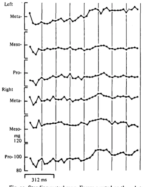
(D) Standing metachrony
273
Place
Lift
[image:12.451.40.396.40.172.2]\
Fig. 11. Changes in the force exerted on the substrate by one mesothoracic leg when the other mesothoracic leg is either placed on the substrate or lifted from the substrate. Each arrow represents three data points from three consecutive film frames. The end of the arrow represents the frame before the opposite mesothoracic leg is placed or lifted, the middle of the arrow (where the arrow touches the line) represents the frame when the contralateral leg is lifted or placed, and the pointer on the arrow represents the frame after the contralateral leg is lifted or placed. A downward deflexion of the arrow means the force on the substrate increased, an upward deflexion means the force on the substrate decreased. Brackets represent 80 mg.
Pro100
-80 L
Fig. 12. Standing metachrpny. Forces exerted on the substrate by a stationary cricket. Same conventions as Fig. 3.
[image:12.451.86.319.274.578.2]274
Left meso-iRight
pro-Amplitude X
Fig. 13. The amplitude of force for two legs from Fig. 12 (the forceprint for the two legs is given at the top of the figure, drawn to a different scale than in Fig. 12). For each frame of the film, one data point is generated. The amplitude of force exerted by one leg is plotted on the abscissa and the amplitude of force exerted by the other leg is plotted on the ordinate. The data points so generated are connected consecutively. The presence of loops is indicated by marking the connecting loops differently; the direction of the loops is indicated by arrows. The narrow line represents the linear regression. For these two legs, the correlation coefficient is 073.
exerted on the substrate during standing metachrony, phase diagrams (Fig. 13) were constructed and correlation coefficients between two legs were calculated. It is not strictly correct statistically to calculate correlation coefficients between two potentially independent parameters, but while more sophisticated tests are being made (Harris, in preparation), the results obtained (r = o-6o to 0-93) are at least suggestive.
DISCUSSION
The stepping behaviours of different segments are not identical. The metathoracic legs spend a higher percentage of the step cycle protracting than do legs of other segments (Fig. 6). The same was found for the cockroach (Delcomyn & Usherwood, 1973). Pearson & lies (1970) found that the bursts to the metathoracic coxal levators were indeed longer than the bursts to the mesothoracic coxal levators in the cock-roach. The longer protraction time may be necessary because the metathoracic legs are longer or because the mesothoracic and prothoracic legs may be more involved in balancing.
The greater irregularity of the forceprints of the prothoracic legs and the lower magnitude of peak force exerted on the substrate by the prothoracic legs (Fig. 7) suggest that the prothoracic legs are more involved in balancing or searching than in propulsion. The prothoracic legs protract and retract in the proper metachronal order (Fig. 3), but during each retraction there is greater variability in the pattern in the force exerted on the substrate than is seen in other segments. It is interesting to note that the prothoracic legs flex during retraction (Fig. 4) while the legs of the other segments extend.
Previous studies on the contributions of central and peripheral nervous elements to the generation of walking patterns have focused on the (larger) mesothoracic and metathoracic legs. It is possible that these studies are not generalizeable to the prothoracic legs and that the coordination patterns of the prothoracic legs are con-siderably more sensitive to sensory feedback than the legs of other segments. This was suggested also by Burns (1973), who found greater variability in protraction and retraction durations in the prothoracic legs than in the legs of other segments. At very slow stepping frequencies (less than 2 steps s-1) the forceprints of all legs show no repeating pattern, and resemble the forceprints of prothoracic legs. One possible explanation for this change from a systematic pattern of forceprints to a variable pattern is that at slower walking speeds the animal is now feeling her way more carefully; that is, that she is making use of sensory feedback to make fine adjustments in each retraction.
The differences in shapes of the forceprints of the legs from different segments could mean the legs behave differently and/or carry different loads. This means that a more careful definition of 'metachrony' needs to be made since it has been heretofore tacitly assumed that the legs of all segments are functionally identical. 'Metachrony' now should be seen to be simply the proper temporal sequence of stepping of the legs relative to each other, which is independent of detailed events during the protraction or retraction of any one leg.
legs are the highest and the prothoracic legs produce the lowest peak force. This serves as another illustration that the legs of the different segments are not functionally identical during normal walking. The difference in peak forces may be accounted for in part by the fact that the centre of gravity of the female cricket lies between the coxal articulations of the metathoracic legs, so the posterior legs must, presumably, carry a larger load. However, this is only a partial explanation, since what is being compared is the peak force exerted on the substrate, not the minimum force which would support the animal's weight; and in the case of the metathoracic legs, that peak force occurs early in the retraction.
A dip in the forceprint of the mesothoracic legs occurs when the leg is at right angles to the body and does not occur if the leg is bent at the femoral-tibial joint, when, for example, the insect is turning. These observations suggest that the dip is caused by a mechanical alignment of force angles such that the force vectors are momentarily reduced, rather than by a feature of the nervous-muscular system. Nonetheless, such a dip could serve as a sensory signal. Cruse (1976) found similar dips in some of the forceprints, and human dog forceprints also show a double peak in force exerted on the substrate (Alexander, 1977) but the causes are probably different.
The surge in force exerted on the substrate by the metathoracic legs just before protraction (Figs. i(e), 3, 8, 9) is apparently caused by sudden extension of the femoral-tibial joint, sometimes with the involvement of other joints as well. The push resulting from this extension may be necessary to shift the weight the leg was carrying to a more anterior leg, or may be the result of a hurriedly completed retraction mediated by a reinforcing reflex (Pearson & Duysens, 1976). Again, it is possible that the sensory feedback from such an extension may signal other segments of the imminent lifting of the leg.
Forceprint details like the mesothoracic dip and the metathoracic surge occur during single retractions and therefore independent of protraction and retraction cycles. They are seen at all stepping frequencies (Fig. 8). The metathoracic surge appears to be largely mediated by the flexor and extensor muscles in the leg (so-called 'intrinsic' muscles), compared to retraction and protraction, which are more strongly mediated by coxal levators and depressors (' extrinsic' muscles). Since both 'intrinsic' and 'extrinsic' muscles are normally employed in walking, it is plausible that nervous control of walking in insects consists of two superimposed systems: a central pattern generator which mediates metachrony through coxal levators and depressors, and a system mediating complex balancing which acts through intrinsic leg muscles. Presumably, the second system is more sensitive to sensory feedback. According to this hypothesis, a mechanical model for an insect leg would be a rotating gear operating the coxal-femoral joint and a spring representing the tibial-femoral joint.
On the other hand, Hoyle (1964), Ewing & Manning (1966) and Runion & Usherwood (1968) found that stimulation of the tibial flexors and extensors was continual and simultaneous to flexors and extensors. Specifically, phasic stimulation of tibial extensors and flexors through the fast excitatory nerve is involved in jumping. Tonic stimulation of tibial extensors and flexors through the slow excitatory nerve follows the pattern of force production from extension of the tibial-femoral joint (Runion & Usherwood, 1968). Further, Runion & Usherwood (1968) describe a reflex con-nection between the tarsal hairs and the tibial extensors, mediated through the slow excitatory nerve, which functions while the animal is stationary and which pre-sumably therefore is involved in maintaining balance. Tonic stimulation and sensory feedback are just what would be expected in a system undergoing small and rapid adjustments, as would be the case in this postulated balancing system.
Pearson & lies (1973) found that severing one of the two nerves carrying sensory information from the cockroach metathoracic leg did not abolish rhythmic leg movements nor prevent normal patterns of stepping. However, severing both nerves caused the animal to adopt an unusual posture and to walk with great difficulty, without being capable of normal adjustments, although using an apparently normal tripod gait. This lends additional support to the hypothesis that pattern generation and sensory feedback to muscles involved in balancing are anatomically separate, but neurologically superimposed systems.
To postulate superimposed pattern-generating and balancing systems offers a way out of a current dilemma. Sensory feedback has been shown to influence the cycle of protraction and retraction. Wong & Pearson (1976) found that trochanteral hair plates and cuticle stress receptors are normally involved in the timing of protractions. However, animals with ablated sense organs, and completely de-afferented preparations can cycle approximately normally (Wendler, 1966; Pearson & lies, 1973), so the sensory feedback is not required for central generation of stepping even though it may be normally utilized by intact animals. The finding in this study of varying forceprint details within normal metachronal steps suggests that central pattern-generation and sensory-feedback mechanisms are operating simul-taneously but with different principal targets. This hypothesis would predict that deafferentation would not affect protraction and retraction (metachronal cycles) very much but would profoundly affect balancing.
more fluctuation than an animal responding to the repetitive playing of the same tape. At very slow speeds (less than 2 steps s"1) there does appear to be a change in variability and this may signal the change to a more sensory-sensitive kind of walking. This is illustrated by the increased variability in peak force and in the nonrepetitive shapes of the forceprints. Delcomyn & Usherwood (1973) found a change in character in the burst patterns below 3-4 steps s- 1 in the cockroach and suggested that below that frequency the animal used a different gait termed 'ambling'.
Pearson (1972) found that the coxal depressor in the cockroach spiked spontaneously at a rate of 5-30 Hz (his diagrams show a spontaneous rate of 3-4 Hz. This compares with the rate of about 5 Hz of the standing metachrony and suggests that the standing metachrony may be caused by the activity of the coxal levators and depressors working at levels too low to cause movement, but applying sufficient isometric pressure to be picked up by the substrate transduction technique. The preferred rate of stepping of the cricket in our conditions is around 4-5 steps s"1 (Fig. 10), which suggests that this rate of stepping may be generated by the spontaneous rate of the de-pressor motor neurones. Delcomyn & Usherwood (1973) found that the slow dede-pressor motor neurone continued to spike for 15 min during a stationary period and only stopped spiking while the animal was crouched down with all its weight borne by the body instead of by the legs.
The standing metachrony is not the effect of a single leg oscillator because there is relative coordination with other legs. This suggests that oscillating programs persist during stationary periods. These oscillator programs may be either central oscillator programs or continuously active proprioceptive oscillator programs and may be linked individual segment oscillators (Graham, 1977).
Graham (1977) concluded that coordination of metachrony must be by sensory cues since a central pattern generating system would not know where the leg was positioned following a stationary period. However, the existence of standing meta-chrony suggests a mechanism whereby a central oscillatory mechanism may indeed provide the necessary information for starting up following a stationary period. To make an analogy, the cricket has its motor running and its clutch disengaged. This suggestion is consistent- with the evidence discussed above that pattern generation is a centrally mediated event.
REFERENCES
ALEXANDER, R. M C N . (1977). Terrestrial locomotion. In Mechanics and Energetics of Animal Locomotion (ed. R. McN. Alexander and G. Goldspink), p. 180. London: Chapman and Hall.
BASSLER, U. (1977). Sensory control of leg movement in the stick insect Carausius morosus. Biol. Cyber-netics 25, 61-72.
BURNS, M. D. (1973). The control of walking in orthoptera. I. Leg movements in normal walking. J. exp. Biol. 58, 45-58.
CRUSE, H. (1976). The function of the legs in the free walking stick insect, Carausius morosus. jf. Comp. Physiol. 112, 235-262.
DELCOMYN, F. (1971a). The locomotion of the cockroach, Periplaneta americana. J. exp. Biol. 54,
443-45*-DELCOMYN, F. (19716). The effect of limb amputation on locomotion in the cockroach, Periplaneta americana. J. exp. Biol. 54, 453-469.
DELCOMYN, F. & USHERWOOD, P. N. R. (1973). Motor activity during walking in the cockroach, Periplaneta americana. I. Free walking. J. exp. Biol. 59, 629-642.
EWING, A. W. & MANNING, A. (1966). Some aspects of the efferent control of walking in three cock-roach species. J. Insect Physiol. 12, 1115-1118.
GRAHAM, D. (1972). A behavioral analysis of the temporal organization of walking movements in the first instar and adult stick insect Carausius morosus. J. comp. Physiol. 81, 23—32.
GRAHAM, D. (1977a). The effect of amputation and leg restraint on the free walking coordination of the stick insect Carausius morosus. J. comp. Physiol. 116, 91-116.
GRAHAM, D. (19776). Simulation of a model for the coordination of leg movements in free walking insects. Biol. Cybernetics 26, 187-198.
HARRIS, J. (1978). A photoelastic substrate technique for dynamic measurements of forces exerted by moving organisms. J. Microscopy 114, pt. 2, 219-228.
VON HOLST, E. (1943). Uber relative Koordination bei Arthropoden Pflilgers Arch. ges. Physiol. 246, 847-865.
HOYLE, G. (1964). Exploration of neural mechanisms underlying insect behavior. In Neural Theory and Modeling (ed. R. F. Reiss). Stanford Press. Adv. Behav. Biol 18, 346—376.
HOYLE, G. (1976). Arthropod walking. In Neural Control of Locomotion (ed. Herman, Grillner, Stein and Stuart), pp. 137-179. New York: New York.
HUGHES, G. M. (1957). The coordination of insect movements. II. The effects of limb amputation and the cutting of commissures in the cockroach Blatta onentalis. J. exp. Biol. 34, 306-333. HUGHES, G. M. & MILL, P. J. (1974). Locomotion: terrestrial in The Physiology of Insecta, 2nd ed,
vol. Ill (ed. Rockstein), pp. 335-382. New York: Academic Press.
LAND, M. F. (1972). Stepping movements made by jumping spiders during turns mediated by the lateral eyes. J. exp. Biol. 57, 15-40.
MANTON, S. M. (1973). The evolution of arthropodan locomotory mechanisms. II. Habits, morphology, and evolution of the Uniramia and comparisons with the Arachnida. Zool. J. Linn. Soc. 53, 257-
375-PEARSON, K. G. (1972). Central programming and reflex control of walking in the cockroach. J. exp. Biol. 56,
173-193-PEARSON, K. G. & DUYSENS, J. (1976). Function of segmental reflexes in the control of stepping in cockroaches and cats. In Neural Control of Locomotion (ed. Herman, Grillner, Stein and Stuart), PP- 579-539- New York: Plenum.
PEARSON, K. G. & ILES, J. F. (1970). Discharge patterns of coxal levators and depressor motorneurons of the cockroach Periplaneta americana. J. exp. Biol. 52, 139-165.
PEARSON, K. G. & ILES, J. F. (1973). Nervous mechanisms underlying intersegmental coordination of leg movements during walking in the cockroach. J. exp. Biol. 58, 725-744.
RUNION, H. I. & USHERWOOD, P. N. R. (1968). Tarsal receptors and leg reflexes in the locust and grasshopper. J. exp. Biol. 49, 421-436.
USHERWOOD, P. N. R. & RUNION, H. I. (1970). Analysis of the mechanical responses of the meta-thoracic extensor tibiae muscles of free walking locusts. J. exp. Biol. 52, 39-58.
WENDLER, G. (1966). The coordination of walking movements in arthropods. In Nervous and Hormonal Mechanisms of Integration. Symp. Soc. exp. Biol. no. XX, 229-251.
WILSON, D. M. (1966). Insect walking. A. Rev. Entom. 11, 103-122.