Development of Posterior Fossa Dural Sinuses, Emissary Veins, and
Jugular Bulb: Morphological and Radiologic Study
Toshio Okudera, Yun Peng Huang, Tatsuhiko Ohta, Akira Yokota, Yasuhiro Nakamura, Fumiaki Maehara, Hidetsuna Utsunomiya, Kazuo Uemura, and Hitoshi Fukasawa
PURPOSE: To report the anatomic and radiologic development of the transverse, sigmoid, and
occipital sinuses, the emissary veins, and the jugular bulb formation from the jugular sinus in humans before and after birth. METHODS: Roentgenograms of 33 injected brains showing the cranial venous system in human fetuses from 3 to 7 months of gestational age and cerebral
angiograms of newborns and infants up to 6 years of age (23 clinical cases) were made and
analyzed in detail. Special attention was focused on the inner diameters of the transverse and
sigmoid sinuses and of the internal jugular veins, particularly at the sigmoid sinus-internal jugular vein junction. RESULTS: Marked increase in venous flow from the rapidly growing cerebral
hemispheres leads to ballooning of the transverse sinuses in the absence of an increase in the inner diameters of the sigmoid and jugular sinuses. The ballooning also results in formation of the occipital sinus, marginal sinus around the foramen magnum, and emissary veins. The formation of
the jugular bulbs from the jugular sinuses begins after birth when a shift from a fetal to a postnatal type of circulation (or from a lying-down position to an erect posture) takes place. CONCLUSION:
The morphological changes of the posterior fossa dural sinuses, emissary veins, and jugular bulb are closely related to the development of the brain, shift to postnatal type of circulation, and
postural hemodynamic changes.
Index terms: Fetus, growth and development; Dural sinuses; Posterior fossa; Veins, jugular; Veins, anatomy
AJNR Am J Neuroradio/15:1871-1883, Nov 1994
With recent advances in the care of ture infants, it is possible for extremely prema-ture infants (600 gm in weight, 6-month-old fetuses) to survive the critical postnatal period.
Received August 12, 1992; accepted after revision April 18, 1994. This work was supported by a grant (63-A) from the National Center of
Neurology and Psychiatry of the Ministry of Health and Welfare, Japan.
From the Departments of Radiology (T.O., K.U.) and Pathology (H.F.), Akita Research Institute of Brain and Blood Vessels, Akita, Japan; D epart-ment of Radiology, Mount Sinai Medical Center, New York, NY (Y.P.H.); Department of Neurosurgery, Fukuoka University School of Medicine,
Fukuoka, Japan (T.O.); Department of Neurosurgery, School of Medicine, University of Occupational and Environmental Health, Kitakyushu, Japan (A.Y.); Departments of Pathology (Y.N.) and Neuroradiology (H.U.), St
Mary's Hospital, Kurume, Japan; and Department of Radiology, Kumamoto University School of Medicine, Kumamoto, Japan (F.M.).
Address reprint requests to Toshio Okudera, MD, Department of Radi
-ology, Akita Research Institute of Brain and Blood Vessels, 6-10 Senshu-Kubota-Machi, Akita City, Akita 010, Japan.
AJNR 15:1871-1883, Nov 1994 0195-6108/94/1510-1871 © American Society of Neuroradiology
It is therefore important to understand the ana-tomic development of the dural sinuses in in-fants of this age.
We report gross morphological findings
based on: (a) dissection of injected fetal brains of 3 to 7 months of gestational age; (b) multi-directional stereoroentgenograms of injected specimens; and (c) cerebral angiograms of ne-onates and infants up to 6 years of age. Partic-ular attention has been paid to the development of the posterior fossa dural sinuses (the trans-verse, sigmoid, and occipital sinuses), the em-issary veins, and the (superior) jugular bulb. We describe the cause of (a) enlargement (or bal -looning) of the transverse sinuses, (b) marked but transient enlargement of the occipital and marginal sinuses, (c) development of the emis-sary veins, and (d) formation of the (superior) jugular bulb from the jugular sinus.
1872 OKUDERA
Materials and Methods
lryected Aborted Fetuses
Thirty-three brains of aborted fetuses ranging from 3 to 7 months of gestational age were obtained. Contrast ma-terial was injected into the venous system through a cath-eter introduced into the superior vena cava or into both internal jugular veins. Gestational ages of the aborted in-jected fetal brains were at latter half of third month (3 brains), at fourth month (8 brains), at fifth month (12
brains), at sixth month (7 brains) and at seventh month
(3 brains). The gestational ages of the aborted fetuses
were determined using Boving's chart ( 1), measuring the
crown-rump length, body weight, and foot length, and
expressed in months from the first day of the last
men-strual period. The contrast materials used were
water-suspension of fine-grain barium sulfate containing 1.5% gelatin or microfil silicone rubber. Injections were
per-formed either gently by hand or by using a 500-ml irri-gator with an average pressure of 34 mm of water. After injection of contrast material, stereoroentgenography was carried out in multiple directions using soft-tissue techniques. After opening of a part of the skull with scis-sors, we fixed the fetus in a 10% formalin solution for 3 months. This was followed by multidirectional
stereo-roentgenography of the dural sinuses and veins, dissec-tion, and further photography. In some brains serial fron-tal, horizonfron-tal, or sagittal sections were made.
Clinical Material
Cerebral angiograms (taken in stereoscopic pairs) of 23 children were analyzed: 8 less than 1 year of age; 2
be-tween age 1 and 2 years; 6 between age 2 and 3 years; 2
between age 3 and 4 years; 2 between age 4 and 5 years; 2 between age 5 and 6 years; and 1 more than 6 years of
age. The transverse and sigmoid sinuses, emissary veins, jugular sinuses (from which the jugular bulbs develop),
and internal jugular veins were systematically investigated
on both sides, especially as to course, caliber, and mode of
drainage.
Radiologic measurements of the inner diameters of
these veins and sinuses were made in the anteroposterior projection (Towne view). These include measurements of the inner diameters of the transverse sinuses, sigmoid sinuses, jugular sinuses (jugular bulbs over age 2), and internal jugular veins on both sides.
Results
Transverse Sinus
The terms transverse sinus and lateral sinus
are confusing and have been used interchange-ably in the past. For the sake of clarity, the term
lateral sinus is best used to include both the transverse and sigmoid sinuses.
At the fetal age of 3 months (11th and 12th weeks), the transverse sinus (on each side) is
AJNR: 15, November 1994
small (Fig 1). It can be said to begin at the junction with the straight sinus and run infero-laterally before turning inferomedially and pos-teriorly to become the sigmoid sinus. Its posi-tion, represented by the outer tentorial margin, continues to descend with growth of the cere-bral hemispheres. On each side, the superior sagittal, the transverse, and the sigmoid sinuses form a single narrow curved course that is con-tinuous inferiorly with the internal jugular vein. The superior sagittal sinus is divided posteriorly into right and left limbs. The straight sinus, lo-cated rather high at this age, drains mainly into the left limb.
Until the age of 4 V2 fetal months (17th and 18th weeks), the transverse sinus has a rei a-tively even caliber. At this age, it begins to en-large from its lateral border on each side (Figs 2 and 3). This enlargement (or ballooning) (4, 7) rapidly progresses medially and reaches the primitive torcular approximately 1 to 1 V2 months later (Fig 4). This ballooning may be most conspicuous at the age of the 5th month (19th and 20th weeks) and may further extend into the posterior portion of the superior sagittal and superior petrosal sinuses. With the increas
AJNR: 15, November 1994
A
c
Sigmoid Sinus and Jugular Bulb
The sigmoid sinus begins at the junction of the transverse sinus and the superior petrosal sinus. It runs inferiorly along the posterior por-tion of the petrous pyramid (retropyramidal segment). The sinus then runs anteroinferiorly along the inferior border of the petrous pyra-mid (infrapyrapyra-midal segment). At the fetal age of 3 months the sigmoid sinus is small in cali-ber. Because the cerebellar tentorium is lo-cated rather high in this stage, undulation in
DEVELOPMENT 1873
8
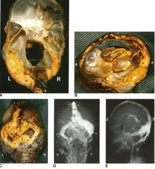
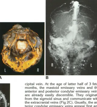
Fig 1. A 12-week-old fetus (65-mm crown-rump length).
A, Photograph of the brain showing injected veins and dural
sinuses. The left cerebral hemisphere has been removed except for the left thalamus (Th) so that the transverse sinus (7), the free
margin (row of arrowheads) of the tentorium, the internal cerebral vein ( 1), the great cerebral vein of Galen (2), and the straight sinus
(3) can be well demonstrated. The superior sagittal sinus (4), the
ventral diencephalic vein (5) (draining into the tentorial sinus [6]),
and the stem of the middle cerebral vein ( 7) are also labeled. The
relatively small transverse sinus ( 7) is somewhat obscured by the superimposed tentorial sinus (6) and other adjacent veins.
B, Towne view and C, lateral view roentgenograms. The tran
s-verse ( 1) and sigmoid (2) sinuses on each side form a continuous
narrow gentle curve that is continuous inferiorly with the jugular sinus (3) and the internal jugular vein (4). In the Towne projection,
the segment of the sigmoid sinus, which lies behind the petrous
pyramid (retropyramidal segment [X]) courses inferomedially in an arcuate fashion convex superomedially, whereas the infra
-pyramidal segment ( Y), which runs below the petrous pyramid
courses almost vertically. The mastoid and condylar emissary
veins (open white arrowheads, mastoid above, condylar below) originate from the sigmoid sinus on each side. The occipital si
-nuses (5) are plexiform in shape and are not dilated at this stage.
The primitive internal cerebral vein ( 6), great cerebral vein of Galen (7), straight sinus (8), superior petrosal sinus (9), superior
sagittal sinus ( 10) and internal vertebral venous plexus (asterisk), deep cervical vein (arrowhead), and inferior petrosal sinus ( 11)
are labeled.
[image:3.613.62.487.72.608.2]1874 OKUDERA AJNR: 15, November 1994
c
D
E
Fig 2. A 16-week-old fetus (115-mm crown-rump length). Photographs of the base of the skull (anterior, middle, and posteriorfossa) showing the contrast-filled dural venous sinuses.
[image:4.612.53.562.96.649.2]AJNR: 15, November 1994 DEVELOPMENT 1875
B
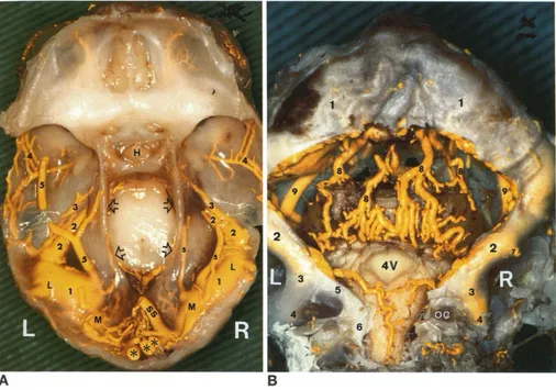
Fig 3. Photographs of the brain of a 17 -week-old fetus ( 130-mm crown-rump length) showing injected dural sinuses.
A, Base of the skull (view from above). The ballooned transverse sinuses ( 1) are uneven in size. The lateral parts (L) are larger than the medial parts (M). The superior petrosal sinus (2) are slightly dilated posteriorly because of anterior extension of ballooning of the transverse sinus into the posterior part of the superior petrosal sinus. The torcular is plexiform, showing many cut surfaces of the veins (asterisks). The straight sinus (SS) opens into the medial portion (M) of the transverse sinus on the right side. The prootic sinus (3),
middle meningeal vein (4), tentorial sinus (5), hypophysis (H), and free margins of the tentorium (open arrows) are labeled.
B, Posteriorfossa dural sinuses (same specimen, view from behind and below). The markedly dilated transverse sinuses (J) are seen. This ballooning continues medially to reach the torcular at the age of the fifth fetal month (Fig 4). The inner diameters of the sigmoid (2) and jugular sinuses (3) and internal jugular veins (4) remain unchanged since the 3rd fetal month (Fig 2C). The right anterior condylar vein (5) emerging from the jugular sinus (3) runs medially and inferiorly through the hypoglossal canal and joins the internal vertebral venous plexus (6). The mastoid emissary vein ( 7) emerging from the sigmoid sinus (2) can be identified on the right. The well-developed dorsal cerebellar veins (B) running upward on the dorsal aspect of the cerebellum, the lower part of the fourth ventricle (4\1), the tentorial sinus (9), and part of the occipital condyle (oc) are also labeled.
Fig 2, cont'd. C, View from behind. The occipital sinuses ( J) are markedly increased in number and size-five to seven large venous channels. They stream inferiorly (from the primitive torcular area as well as from the medial portions of both transverse sinuses [2]) toward the developed marginal sinus (3) around the foramen magnum. The marginal sinus then drains into the deep cervical veins (B) (not well opacified on this specimen) and/or the internal vertebral venous plexus. The sigmoid sinuses ( 4) are not dilated, and the jugular sinuses (5) are small. The dilated transverse sinuses with incomplete multiple septations (arrows), mastoid emissary veins (6), and the dilated posterior part of the superior sagittal sinus ( 7) are labeled. The medial part of the transverse sinus is defective on the left (asterisk).
[image:5.614.55.561.76.431.2]1876 OKUDERA
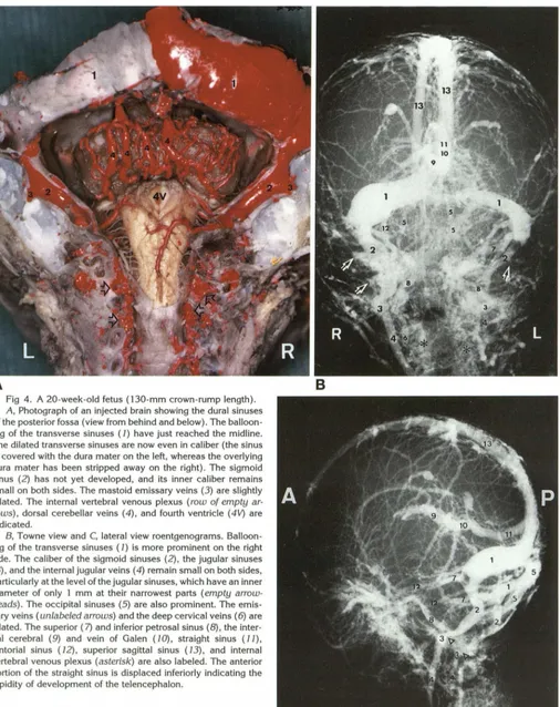
Fig 4. A 20-week-old fetus (130-mm crown-rump length). A, Photograph of an injected brain showing the dural sinuses of the posterior fossa (view from behind and below). The balloon-ing of the transverse sinuses ( 1) have just reached the midline. The dilated transverse sinuses are now even in caliber (the sinus is covered with the dura mater on the left, whereas the overlying dura mater has been stripped away on the right). The sigmoid
sinus (2) has not yet developed, and its inner caliber remains
small on both sides. The mastoid emissary veins (3) are slightly dilated. The internal vertebral venous plexus (row of empty a r-rows), dorsal cerebellar veins (4), and fourth ventricle (4V) are indicated.
B, Towne view and C, lateral view roentgenograms. Balloon-ing of the transverse sinuses ( J) is more prominent on the right
side. The caliber of the sigmoid sinuses (2), the jugular sinuses
(3), and the internal jugular veins ( 4) remain small on both sides, particularly at the level of the jugular sinuses, which have an inner
diameter of only 1 mm at their narrowest parts (empty
arrow-heads). The occipital sinuses (5) are also prominent. The
emis-sary veins (unlabeled arrows) and the deep cervical veins ( 6) are dilated. The superior (7) and inferior petrosal sinus (8), the inter-nal cerebral (9) and vein of Galen ( JO), straight sinus ( 7 7), tentorial sinus ( 72), superior sagittal sinus (13), and internal
vertebral venous plexus (asterisk) are also labeled. The anterior
portion of the straight sinus is displaced inferiorly indicating the
rapidity of development of the telencephalon.
[image:6.615.60.566.74.712.2]AJNR: 15, November 1994 DEVELOPMENT 1877
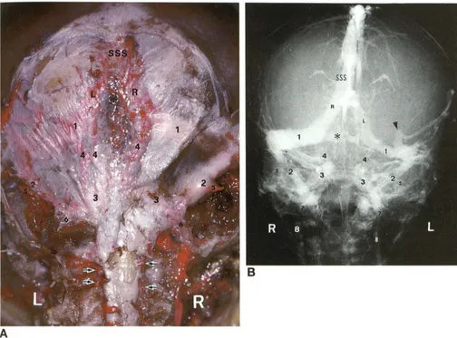
Fig 5. A 24-week-old fetus (215-mm crown-rump length).
A, Photograph of an injected brain showing the occipital sinuses. The posterior fossa dura is left intact. The occipital sinuses ( 4) show a marked regression in caliber and in number. The caliber of the sigmoid sinuses (2) remain narrow. The marginal sinus (3) around the foramen magnum also shows regression. The transverse sinus ( 1) is labeled. The superior sagittal sinus (SSS) is divided posteriorly into the right (R) and left (L) limb. The mastoid (5) and condylar ( 6) emissary veins, internal vertebral venous plexus (arrows), and plexiform torcular (black asterisk) are also labeled. Internal vertebral venous plexus are located in the wall or periphery of the spinal canal.
B, Roentgenogram (Towne view). Although the transverse sinuses ( 7) are asymmetric (the right one being larger), they are relatively even in caliber on each side. On the left, the transverse sinus has irregular dural pouches (arrowhead), which receive several superficial cortical veins. The sigmoid (2) and jugular sinuses ( 7) and the internal jugular vein ( 8) are still poorly developed and small calibers. Unlike in the younger fetus (3 to 4 months), the retropyramidal portion of the sigmoid sinus has a gentle curved course convex slightly inferolaterally (instead of superomedially); its course begins to resemble that of an adult, because of more rapid development of the flat bones of the posterior fossa relative to the growth of the petrous pyramids. The occipital sinuses ( 4) are less prominent than those of earlier stages. The marginal sinus (3) around the foramen magnum is also smaller and less conspicuous. Note the connecting (somewhat plexiform) dural sinus (black asterisk), which is located in the region of torcular bridging between the two posterior limbs of the superior sagittal sinus (R, L). Several small occipital sinuses originating from the connecting sinus run toward the marginal sinus. The mastoid emissary veins (5) and the superior sagittal sinus (SSS) are also labeled.
this stage takes a gentle inferomedial curve
convex superomedially and the infrapyramidal
segment courses almost vertically downward
(Figs 18 and 20). However, these changes
be-come less obvious with increasing age (fourth
and fifth month). Above the age of 6 months in
the fetus, the sigmoid sinuses take gentle
curves laterally and begin to resemble those in
adults. The inner diameters of the sigmoid and jugular sinuses remain extremely small during the intrauterine life, only 1 to 2 mm (Fig 7).
Emissary Veins
Extracranial drainage of the posterior fossa
[image:7.615.53.557.78.451.2]1878 OKUDERA
Fig 6. A 7 -month-old (25-week) fetus
(225-mm crown-rump length).
A, Photograph (view from above). The
transverse sinuses ( 7) maintain their large shapes. Their calibers are large and are rel-atively even on each side. The cut surface
(row of arrowheads) of the torcular (2),
straight sinus (3), tentorial sinuses (4), and the superior petrosal sinus (5) are labeled.
B, Roentgenogram (anteroposterior
view). The transverse sinuses ( 7) maintain their large size. They are of even inner di-ameters. The sigmoid sinuses (2), the jug-ular sinuses (3), and the internal jugular veins ( 4) are not yet developed. In compar-ison with those of earlier stages, each sig-moid sinus (2) at this stage takes a sharper
medial curved course convex inferolaterally
before reaching the jugular sinus. The oc-cipital sinuses ( 5) and the mastoid emis-sary veins ( 7) are well seen. The internal vertebral venous plexus (asterisk) and the deep cervical veins (arrowheads) are also well demonstrated. The superior sagittal si-nus (SSS), the marginal sinus (6), and the inferior petrosal sinus (8) are also labeled.
A
Fig 7. Roentgenogram of injected cranium of a 8-month-old
aborted fetus (lateral view). The relationships of the transverse (J), sigmoid (2), and jugular sinuses (3) and the internal jugular vein ( 4) are similar to those of the 7 -month-old fetus (Fig 6), and their calibers remain small. Similarly the internal cerebral vein (5) and the straight sinus ( 7) and the torcular (9) have the relation-ships similar to those of an adult. The vein of Galen (6), however, takes a more horizontal course because of the somewhat short straight sinus (normal variation). Note the position of the splenial
vein (row of arrows). The superior sagittal sinus (10) is also
labeled.
vein, are (a) via the anterior condylar vein or the marginal sinus into the internal vertebral venous plexus, (b) via the posterior condylar vein into the posterior external vertebral vein or posterior jugular vein, (c) via the mastoid emissary, or (d) via the occipital emissary vein into the
oc-AJNR: 15, November 1994
8
[image:8.614.78.561.81.627.2] [image:8.614.235.556.85.441.2]AJNR: 15, November 1994
sinus and/or the marginal (occipital) sinus with the vertebral, paravertebral, and/or deep cervi-cal veins. On the other hand, the mastoid em -issary veins connect the sigmoid sinus with the occipital (or posterior auricular) veins.
Occipital Sinus
At the age of 3 fetal months ( 11 to 12 weeks), fine plexiform vascular channels originate from the primitive torcular plexus (and the medial portion of the primitive transverse sinus) and course towards the distal portion of the sigmoid sinus on each side. The occipital sinuses rapidly increase in caliber and exhibit the greatest de-velopment at the age of 4 to 5 months. Five to seven venous channels, some of them very large, originate from the primitive torcular area as well as from the medial portion of both trans -verse sinuses. They stream toward the already markedly developed prominent marginal sinus around the foramen magnum (Fig 2C). Some channels may, however, be connected to the distal portion of the sigmoid sinus or sinuses. They decrease in caliber and in number by dim-inution and fusion (Fig 5). By the time the fetus reaches the sixth or seventh month, only a few prominent occipital sinuses can be recognized. They connect the medial portions of the trans-verse sinuses to the well-developed marginal sinus or to the distal ends of the sigmoid sinuses.
Most of the findings described above are identical or nearly identical in the fetuses examined.
After Birth
Alterations with age in the diameters of the transverse and jugular sinuses (the latter be -come the jugular bulbs) are well demonstrated in Figures 8 and 9. From birth to age 1 year, the diameters of the transverse sinuses decrease somewhat compared with late fetal life. At 1 year of age, the transverse sinuses have their adult appearance. They increase by only a few millimeters in diameter by age six years.
After birth, the diameters of the sigmoid s i-nuses increase rapidly to 5 to 7 mm; the rate of increase is similar to that of the jugular sinuses. The jugular sinuses grow rapidly and become approximately 9 to 1 0 mm in diameter at 1 year of age (Fig 10). Eventually, they acquire the same diameters as the transverse sinuses
(Fig 9).
DEVELOPMENT 1879
Fig 8. Roentgenogram of an injected full-term (40-week-old) neonate. The transverse sinuses ( 7) are relatively equal in caliber.
The union of the posterior limbs of the superior sagittal sinus (2)
is similar to that of an adult. The connecting sinus (3) and the posterior end of the straight sinus (SS) are labeled. The
retropy-ramidal and infrapyretropy-ramidal segments (X and Y) of the sigmoid
sinus (4) take a smooth inferomedial curved course convex
in-ferolaterally before reaching the jugular sinus (arrow). This
infero-medial curved course has, however, a less inferolateral convexity
than that of an adult, although it is more convex than that of an earlier fetus. Note the small calibers of the sigmoid sinus ( 4) and the internal jugular vein (5, 6), particularly at their junctions (the jugular sinus, arrow). The right and left innominate (or brachi o-cephalic) veins ( 7) unite to form the superior vena cava (8).
Because of unusual tortuosity of the internal jugular veins (5, 6) on
both sides and the shorter and more vertical course of the left
innominate vein, the relatively straight course formed by the jug
-ular sinus, internal jugular veins, innominate vein, and superior
vena cava on the right is not demonstrated, compared with the
more circuitous course made up by the corresponding veins on
the left, which eventually has to join the superior vena cava
located on the right side.
Discussion
Morphological Changes of the Transverse Sinuses
1880 OKUDERA AJNR: 15, November 1994
A
B
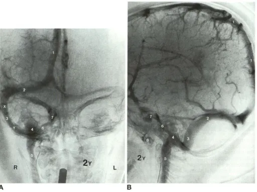
Fig 9. Carotid angiogram of a 2-year-old boy, venous phase.
A, Anteroposterior view and 8, lateral view. The superior sagittal sinus ( 1) drains mainly into the right transverse sinus (2). The diameter of the sigmoid sinus (3) is now equal to that of the transverse sinus (2). Note in the anteroposterior view the prominent jugular bulb (4), which projects upward. Because of a more rapid development of the occipital squama relative to that of the petrous pyramid, the retropyramidal segment (X) of the sigmoid sinus runs more vertically and is located more laterally. The infra pyramidal segment ( Y) of the sinus (3), on the other hand, takes a much more horizontal course in the anteroposterior view before reaching the jugular bulb, so that the sigmoid sinus takes a smooth, large, curved-course convex inferolaterally before turning upwards to form a jugular bulb. The sinus then courses downward to emerge as the internal jugular vein (5). Also labeled are the inferior petrosal sinus ( 6), cavernous sinus (7), and pterygoid venous plexus (8).
from the transverse sinus down to the internal jugular vein are of roughly even diameter and are easily discernible. Between the latter half of the third and fourth fetal months, both the trans-verse and sigmoid sinuses have the same diam-eter (3, 7). However, there is a segment of nar-rowing between the sigmoid sinus and the internal jugular vein, particularly at their junc -tion. We have designated this junction the
jug-ular sinus. The latter eventually becomes
di-lated to form the superior jugular bulb on each side. After the fourth month ( 15 to 16 weeks) of gestation, there is ballooning of the transverse sinuses. This ballooning, according to Masaki ( 4), begins at the fifth fetal month and lasts until
the seventh month. He states that the balloon-ing starts from the midportion of the transverse sinus, gradually subsides, and finally disap-pears at 8 to 10 intrauterine months. Our obser-vation, however, indicates that ballooning be-gins at 4 V2 fetal months in the lateral portion of the transverse sinus and proceeds medially on each side.
[image:10.614.54.566.73.454.2]AJNR: 15, November 1994
15 . - - - ,
~ 5
"' c: c:
• TS r;;J Jll
<16 week
n.s.
16 week< Birth Birth < 1 ycJr 1 year<
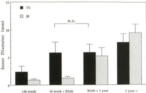
Fig 10. Diagram showing alterations with age in the inner diameters of the transverse sinus and the (superior) jugular sinus (or, later, the bulb). The maximum inner diameters of the trans -verse and the jugular sinuses were measured on the roentgen o-grams (in Towne view, taken in stereoscopic pairs) of injected fetal heads (3 to 7 months of age) and on cerebral angiograms (Towne view, taken in stereoscopic pairs) in clinical cases (new
-born to infants up to age 6 years). Ballooning of the transverse sinuses (TS) is noted in fetus of 16 to 26 weeks (4 to 7 months).
After 20 weeks, the inner diameter of the transverse sinus is gradually equalized. The diameter of the fetal jugular sinus (JS) (from which the jugular bulb [JB] develops postnatally) is approx -imately 1 to less than 2 mm with only 1 mm increase between the 11th and 26th weeks. From birth to age 1 year, the inner diameter of the transverse sinus decreases somewhat; however, after 1 year of age, the sinus has an adult appearance. It increases only slightly in diameter (a few millimeters) by 6 years of age. On the other hand, the inner diameter of the jugular sinus (JS) rapidly increases after birth, becoming approximately 9 to 10 mm at the age of 1 year, and becomes the jugular bulb at the age of 2 years.
The jugular bulb and sigmoid sinus eventually acquire the same diameter as that of the transverse sinus.
ballooning. On the other hand, the sigmoid
sinus-internal jugular vein junctional area (the jugular
sinus) is poorly developed; their inner diameters
usually remain at only 1 to 2 mm until the age of
7 fetal months in all fetuses examined (we have
investigated fetuses only up to 7 months old).
Beginning at 41/2 to 5 fetal months, the
superfi-cial cortical veins of the expanding cerebral
hemispheres rapidly increase in size and drain
into the transverse sinus on each side (6-8).
This corresponds to the period of ballooning of
the transverse sinuses and to the rapid increase
in the total blood draining into the transverse
sinuses. The narrow internal lumens of the
sig-moid sinuses and the internal jugular veins,
par-ticularly at their junctions (the jugular sinuses),
results in physiologic intraluminal venous
sta-sis, intraluminal venous hypertension,
second-ary ballooning of the transverse sinuses, and
enlargement and some formation of multiple
emissary veins for better extracranial drainage
(formation of physiologic collateral venous
DEVELOPMENT 1881
channels). Another outcome of the ballooning and followed by a relative decrease in the diam-eter of the transverse sinus is formation of dural
outpouches, into which a cortical vein or veins
may join. As the ballooned transverse sinuses
gradually return to their normal size, small dural
pouches (joining the transverse sinus on the side of the convexity dura or of the tentorial
dura) are frequently formed. This is the reason
that, in some cases, a cortical vein or veins
indirectly join the transverse sinus through a small tentorial sinus or a dural sinus over the
convexity. In addition, irregular ballooning and
shrinkage of the transverse sinus frequently re
-sults in formation of septa, fenestration, or even
defective or missing segment (particularity
me-dial portion) of the transverse sinus (3, 7). The
latter may also be caused by irregularity or de-fect in one of the posterior limbs of the superior sagittal sinus near the torcular. Also clinically those findings can be seen on the venous phase
of routine cerebral angiograms. Poor communi
-cation between the two transverse sinuses at the
level of the torcular is of paramount importance
when one considers using a combined
ap-proach to resect a tumor that involves both the
middle and posterior fossae and to ligate and sever the sinus surgically near its knee for a
better exposure.
If one postulates that the torcular is located at the junction of the superior sagittal sinus and
the straight sinus and that the superior sagittal
sinus has right and left posterior limbs, the tor
-cular may be said to be situated on one side
depending on where the straight sinus joins.
Usually, the straight sinus drains into the left limb, whereas the superior sagittal sinus more frequently joins the transverse sinus on the right side. The reason for the more frequent drainage of the superior sagittal sinus into the right transverse sinus may be related to the relatively linear courses of the transverse
si-nus, the sigmoid sinus, the internal jugular
vein, the superior vena cava, and the atrium
on the right side (10). On the left side, the in-ternal jugular vein has to take an extra-long inferomedial bend through the left brachio-cephalic vein before joining the superior vena cava located on the right. Thus drainage of cranial venous blood is more circuitous on the
left than on the right (Fig 11).
The asymmetry of the heights of the medial
portions of the transverse sinuses is clearly re
-lated to the asymmetric heights of the tentorial
[image:11.615.51.297.72.229.2]1882 OKUDERA
makes it difficult to evaluate the exact location of the torcular in fetus on external inspection. The difference between the relatively straight connection from the transverse sinus through the sigmoid sinus down to the internal jugular vein seen in the fetus and the more sinuous course of the same segments seen in adults is in most part caused by the development of the telencephalon and secondary posterior and in-ferior displacement of the transverse sinuses ( 14 ).
Morphological Changes of the Sigmoid, Occipital, and Marginal Sinuses with Emissary Veins and Jugular Bulb
Although a drainage mode from the primitive transverse sinuses through the primitive sig-moid sinuses to the primitive internal jugular veins is already detectable at the 30-mm stage
(9, 10), the sigmoid sinuses are easily
identifi-able after the 80-mm stage (9, 1 0). Our studies, however, indicate that the sinuses are poorly developed at this stage and maintain their tiny inner calibers until after birth. On the other hand, the change in the volume of the posterior fossa, influenced greatly by the development of the cerebellum, is most marked between the ages of 30 and 40 weeks in fetus (16). Alter-ations in the course of the sigmoid sinuses ap-pear to occur during this period.
Reports on the occipital and the marginal si-nuses along the foramen magnum have been scanty. Even a classical monograph by Padget
(8, 9) failed to include any description until the
fetus reaches the 80-mm stage. Our cases (65-mm stage, 3 months of gestation) and Masaki's cases (4) (4 months of gestation) show the plexiform occipital and marginal si-nuses extending from the region of the tentorial
attachment down to the foramen magnum. The
transient enlargement followed by diminution in number and size of the occipital sinuses simi-larly is related to the physiologic hemodynamic response to the increased venous drainage from the intracranial to the extracranial spaces.
Each internal jugular vein of the adult has an enlargement in two places ( 4). The superior jugular bulb is located in the jugular foramen and forms the origin of the internal jugular vein.
Inferiorly, there is dilatation in the course of the
vein located a short distance above the junction with the brachiocephalic vein. The latter corre-sponds to the site of the two-leaf valves. These
AJNR: 15, November 1994
~BcV
Fig 11. Schematic diagram showing (in adult) the relatively linear course of the superior jugular bulb, the internal jugular vein,
the inferior jugular bulb, and the brachiocephalic (also called innominate) vein on the right side before opening into the superior vena cava and the right atrium. Labeled are the pounding effects of ascending negative pulse waves (arrow), the inferior jugular bulb (IJB), internal jugular vein (IJV), left brachiocephalic vein (LBcV), lateral sinus (LS), which includes the transverse and sigmoid sinuses, left subclavian vein (LSciV), pulmonary artery (PA), right brachiocephalic vein (RBcV), right subclavian vein
(RSciV), superior jugular bulb (SJB), sigmoid sinus (SS), superior
sagittal sinus (SSS), superior vena cava (SVC), and transverse sinus ( TS). In the fetus, the superior sagittal sinus splits posteriorly into two posterior limbs. The posterior limbs of the superior sag-ittal sinus and the transverse, sigmoid, and jugular sinuses on the right therefore take a much less laterally curved (more linear) course (convex laterally). The fetal jugular sinus is also much less tortuous than the adult jugular bulb.
[image:12.612.316.560.75.345.2]AJNR: 15, November 1994
bulbs are usually seen only in children aged 2 years or older. This appears to correspond to
the period when a shift takes place from a
"fetal" to a "postnatal" circulatory type (or from a "lying down" position to an erect posture). For this reason, it appears more appropriate for the portion of the sinus that later becomes the
jug-ular bulb to be called the jugular sinus before it
becomes the jugular bulb at age 2 years.
Because the jugular sinus is surrounded by
cartilaginous and osseous structures, it is
diffi-cult to expand during fetal life. This needs the
pounding effects of ascending negative pulse
waves originating from the right atrium,
travers-ing upward to strike the jugular sinus at near
right angle at the base of the skull (jugular foramen). This takes place only after the infants assume erect posture. The hammering effects
of the negative pulse waves hitting the roof of
the jugular sinus eventually create the (supe-rior) jugular bulb and enlarge the surrounding osseous structure, creating the osseous jugular fossa (6) (Fig 11 ). The reason for the frequently larger transverse sinus and sigmoid sinus on the
right side is also explained. The dynamic
change of the inner caliber of the sigmoid si-nuses is similar to that of the superior jugular bulb. According to Hirakoh (2), the volume of
each (superior) jugular bulb in adults far
ex-ceeds the total combined blood volume of the
sigmoid and the inferior petrosal sinuses. In
ad-dition, there is a low pressure area (empty
space) within the (superior) jugular bulb that acts as a reservoir for venous blood. This works
as a suction pump, smoothly conveying the
in-tracranial venous blood through the internal jugular vein into the heart. Occasionally there
may be an excessive enlargement of the jugular
bulb causing an upward arching of the internal
auditory canal and the adjacent inner ear, which
might be a cause of tinnitus, dizziness, or mild
hearing impairment (ectasia of the jugular bulb) (3).
The present study focused mainly on the
morphological development of the posterior
fossa dural sinuses, emissary veins, and jugular
bulb. On the other hand, recent functional
ana-tomic studies, such as the higher hydrostatic pressure of the dural sinuses in children than in
adults ( 13), the lack of autoregulation of blood
DEVELOPMENT 1883
pressure in premature infants ( 11), and the
pre-dilection to hemorrhage in the germinal matrix
in premature neonates in cases of increased
intracranial venous pressure (5, 15) await
fu-ture deeper understanding of the correlation
between the morphological and functional
changes of the intracranial venous structures.
References
1. Boving BG. Anatomy of reproduction. In: Greenhill JP, ed.
Obstet-rics. Philadelphia: WB Saunders; 1965:3-101
2. Hirakoh G. On the fossa jugularis and outflow cranial venous blood through it [in Japanese]. J Kurume Med Ass 1962;25 :965-971
3. Huang YP, Okudera T, Ohta T, Robbins A. The torcular, the straight, sagittal, lateral, occipital and tentorial sinuses: the vari-ations and clinical significance. In: Kapp JP, Schmideck HH, eds.
The Cerebral Venous System and Its Disorders. Orlando, Fla:
Grune and Stratton; 1984:109-167
4. Masaki S. Studies on the development of the dural sinuses of the
human fetus [in Japanese]. Fukuoka Acta Medica
1959;50:2769-2788
5. Nakamura Y, Okudera T, Hashimoto T. Germinal matrix h
emor-rhage of venous origin in preterm neonate. Human Pathol 1990; 21:1059-1062
6. Noda R. Development of the cerebral vessels of the human fetus
[in Japanese]. Fukuoka Acta Medica 1958;1057-1072
7. Okudera T, Ohta T, Huang YP. Development of the torcular, adjacent dural sinuses and related draining veins in human fetus. In: Kapp JB, Schmideck HH, eds. The Cerebral Venous System
and Its Disorders. Orlando, Fla: Grune and Stratton; 1984:93-107
8. Okudera T, Ohta T, Huang YP, Yokota A. Development and ra-diological anatomy of the superficial cerebral convexity vessels in the human fetus. J f'ieuroradiol 1988; 15:205-224
9. Padget DH. Cranial venous system in man in reference to devel
-opment of adult configuration and relation to arteries. Am J A nat
1956;98:307-355
10. Padget DH. The development of the cranial venous system in
man: from the viewpoint of comparative anatomy. Conlrib
Em-bryol1957;247:81-138
11. Pape KE, Wigglesworth JS. Haemorrhage, ischemia and the peri-natal brain. Clinical Developmental Medicine. London: Spastics International Medical Publications, 1979:69/70:1-185
12. Shinohara M. Morphological study on the bulbus venae jugularis
internae of embryo [in Japanese]. Acta Anal 1957;32:41 13. Sobata E, Ebina K, Hikichi M, lwabuchi T. Distinctiveness of
children in terms of dural sinus pressure [in Japanese]. Nervous System in Children 1985:10:257-266
14. Streeter GL. The development of alterations in the vascular
sys-tem of the brain of the human embryo. Contrib Embryol1918;24:
7-38
15. Takeshige Y. Bedeutung des Venensystems fUr die Haemody-namik des Hirnstammes. Anal Anz 1967;125:166-192 16. Yamaguchi K, Goto N, Nara T. Development of the human fetus