MR Cisternography: Initial Experience in 41 Cases
Taher El Gamma! and Betty Sue Brooks
PURPOSE: To present our initial experience with MR cisternography, an application of fast spin-echo MR with fat suppression, and compare it with routine MR cranial studies in the evaluation of the subarachnoid cisterns and their contents. METHODS: MR cisternography is a heavily T2-weighted fast spin-echo technique with high spatial resolution; it uses fat suppression and video
reversal of the images. A small number of individual sections (two to four) are compressed into a composite image by a maximum-intensity projection algorithm, providing better depiction of anatomy in three dimensions. MR cisternography enhances the signal intensity of the cerebrospinal
fluid (CSF) with suppression and subtraction of the background. A total of 41 patients were examined during a period of 6 months. MR cisternography was performed as an additional one (n = 31) or two (n = 1 0) sequences after conventional MR study. RESULTS: Twenty-one cases of disease were examined by MR cisternography, including 8 neoplasms, 4 CSF fistulas, and 3 large intracranial aneurysms. MR cisternography provided information unavailable by conventional MR studies in 17 cases. These included visualization of fistulous tracks in patients with CSF rhinorrhea,
origin of a large suprasellar aneurysm, an additional loculus of a posterior fossa aneurysm and its relation to surrounding structures, and proper location of three tumors (intraaxial versus
ex-traaxial). Clear depiction of the pituitary gland separate from the cavernous sinus was noted in 60%
of the cases, and a new observation of a CSF sleeve around the third nerve in the posterior
cavernous sinus was made in 85% of the cases. CONCLUSION: MR cisternography is superior to conventional MR studies in depicting anatomic structures within the subarachnoid spaces. This technique is recommended in the evaluation of cranial CSF fistulas and suprasellar and posterior
fossa masses and in diagnosis of intraaxial versus extraaxial location of intracranial tumors.
Index terms: Cisternography; Magnetic resonance, comparative studies; Magnetic resonance,
technique; Subarachnoid cisterns
AJNR Am J Neuroradiol 15:1647-1656, Oct 1994
Positive-contrast cisternography, an invasive technique, was used in the past to depict normal and abnormal images of the subarachnoid spaces and their contents in the cranial cavity and at the craniovertebral junction. The images were enhanced with the use of polytomography (1) and magnification and subtraction
tech-niques (2-4). These studies were found useful to delineate tonsillar herniation ( 1), small tu-mors (3, 4), and large posterior fossa aneu-rysms (5).
Computed tomographic (CT) cisternography after intrathecal contrast injection has been used to evaluate cerebrospinal fluid (CSF) leaks from the cranial cavity (6) and in the evaluation of posterior fossa normal anatomy (7), but with magnetic resonance (MR) the need for these Received September 28, 1993; accepted after revision January 24,
1994.
From the Department of Radiology, University of Alabama at Birming -ham.
Address reprint requests to Taher El Gamma!, MD, Department of Radiology, University of Alabama at Birmingham, 619 S 19th St, Birming -ham, AL 35233-6830.
AJNR 15:1647-1656, Oct 1994 0195-6108/94/1509-1647 © American Society of Neuroradiology
invasive procedures is greatly diminished. Re-cently fast spin-echo MR sequences have pro-vided heavily T2-weighted images of the cranial cavity with excellent depiction of the subarach-noid spaces and their contents.
We report an MR technique, MR cisternogra-phy, which greatly enhances the details of the
1648 EL GAMMAL AJNR: 15, October 1994
anatomic structures within the subarachnoid spaces of the brain and the craniovertebral junction, and present our initial experience in 41 cases. The technique is essentially an extension of the application of MR myelography (8) (El Gamma! T, Brooks BS, Freedy M, Magnetic Resonance Myelography: Initial Experience in 80 Cases, presented at the 31st Annual Meet-ing of the American Society of Neuroradiol-ogy, Vancouver, Canada, May 1993) into the cranial cavity and and the craniovertebral junction.
Subjects and Methods
We examined 41 patients over a period of 6 months starting in February 1993. They were 24 male and 17 female patients ranging in age from 4 to 82 years with a mean age of 45.9 years. The patients were selected in a random fashion. Volunteers and patients with normal MR findings were included at the beginning of the study. Later, patients with suspected disease in the parasellar regions and in the posterior fossae were included. MR cisternog-raphy was not attempted on uncooperative patients.
All scans were performed on a 1.5-T Signa unit with 5 X
software using a standard head coil. In two cases a spine
MR cisternography findings in patients with disease (n = 21)
Cases
Cranial CSF fistula (n = 4) 1 Anterior fossa (Fig 1 )"
3 Petrous bone and posterior fossa•
Parasellar and orbit (n = 8) 2 Pituitary adenomas
Hemorrhagic Postoperative
2 Suprasellar aneurysms (Fig 5)" (both large)
2 Suprasellar tumors•
Craniopharyngioma•
Subfrontal glioma• 2 Optic nerve injury
With intraconal hemorrhage (trauma)" With scar from previous granuloma (Fig 2)•
(local anesthesia) Posterior fossa (n = 8)
2 Basilar aneurysms
Anterior inferior cerebellar artery (large) (Fig 7)•
Anterior inferior cerebellar artery origin (small fusiform)"
1 lntracanalicular acoustic neurinoma 2 Meningiomas
Tentorial"
Posterior•
2 Intrinsic lesions
Quadrigeminal plate glioma•
Cavernous angioma of fourth ventricle"
1 Mild Chiari II malformation Cerebral hemispheres (n = 1)
Renal cell carcinoma with two supratentorial masses (Fig 8)"
MR Cisternography Findings
Fistula track into right ethmoid sinus was only demonstrated by MR cisternography Fistula communication between left temporal horn parencephalic cyst and middle ear
was only seen by MR cisternography in one patient
Two cases of postoperative rhinorrhea, in one no fistula seen by MR cisternography, in the other a possible communication between middle fossa and petrous bone was not confirmed by surgery
Showed high-signal tumor
Chiasma! deformity not clearly seen on conventional MR
Aneurysm origin only seen on MR cisternography; other case, wide neck and dolichoectasia only seen on MR cisternography
Displaced chiasm only seen on MR cisternography
lntraaxial location seen on MR cisternography and unclear on conventional MR In both cases conventional MR failed to show normal optic nerves seen on MR
cisternography
Relation to brain stem and cranial nerves and depiction of a second loculus; these findings were not clear on conventional MR
Not seen on conventional MR and seen on MR cisternography only
Shown by both composite MR cisternography and enhanced conventional MR
MR cisternography showed normal tentorium; conventional MR could not rule out tumor invasion
Extraaxiallocation by MR cisternography, indeterminate on conventional MR
Shown by MR cisternography and conventional MR
Perilesional high signal seen only on MR cisternography, most likely caused by gliosis.
Shown by MR cisternography and conventional MR
Inferior mass shown to be extraaxial (meningioma}; MR cisternography showed CSF around tumor bed, and conventional MR showed hyperostosis; superior mass shown to be intraaxial by MR cisternography, and conventional MR was inconclusive
AJNR: 15, October 1994
l "
l0 'Ef
32kHz
:m ·e. esp/-C
l0
A
B
Fig 1. Posttraumatic recurrent CSF leak with meningitis.
MR CISTERNOGRAPHY 1649
c
A, Coronal and B, sagittal MR cisternography show the high signal of abnormal gyrus rectus and CSF herniating into the right anterior ethmoid sinus (arrows on right). In A, note the linear high signal in the left anterior ethmoid (left arrow). A preoperative diagnosis of fistula on the left was not made.
C, Coronal CT cisternogram shows a defect in the roof of the right anterior ethmoid sinus (arrow), opaque ethmoid sinuses, and no fistulous track. Only a very small amount of contrast media reached the anterior fossa probably because of posttraumatic adhesions.
phased-array coil was used to evaluate the craniovertebral junction. The MR cisternography was performed as one or two sequences (coronal and/or sagittal) after the conven-tional MR brain studies. The conventional MR studies in-cluded fast spin-echo axial sequences. In four patients coronal fast spin-echo sequences were performed as well. MR cisternography, a fast spin-echo sequence with fat suppression, used an echo train length of 16, a 16-cm field of view with no phase-wrap option, 3-mm-thick sections,
10 000/200/4 (repetition time [TR]/echo time/excita-tions), and a 512 X 192 matrix. A total of 16 images were
obtained in 8 minutes. Image video reversal was used in all the MR cisternography studies.
Twenty-seven cases had coronal studies; 4 had sagittal studies only, and 10 had both coronal and sagittal images. A small number of images were compressed into compos-ite images using a maximum intensity projection algo-rithm. The number of sections used was usually two to four with a maximum of eight. If more sections are used in the composite image this will result in a complex distorted image.
The image quality of the cisternograms was subjec-tively rated as poor, average, good, or very good. Com-parison of MR cisternography with conventional MR stud-ies was done by one unblinded observer.
Results
Thirteen cases were normal, and seven cases
showed only atrophy without other
abnormali-ties. Findings of disease were noted in 21 cases
(Table). As shown in the Table, MR
cisternog-raphy was superior to conventional MR
exami-nations, which were only axial fast spin-echo in
the majority of cases. MR cisternography
pro-vided valuable additional information in 1 7 of
21 cases with pathologic changes. In some of
these cases MR cisternography also proved su-perior to other neuroradiologic procedures.
Specifically, MR cisternography was better than
catheter angiography in locating the origin of
one suprasellar aneurysm and in depicting an additional aneurysm loculus of an anterior infe-rior cerebellar artery aneurysm. MR
cisternog-raphy was superior to CT cisternogcisternog-raphy in the
diagnosis of CSF fistulas (Fig 1 ). In 35 cases,
image quality of the MR cisternography studies
was rated as very good, in five cases as good,
and in one case as average.
Discussion
MR cisternography, a fast spin-echo heavily
weighted T2 sequence with fat suppression,
provides detailed anatomic information of the
subarachnoid cisterns, their contents, and
adja-cent structures by virtue of its ability to increase
the signal intensity of the CSF and to suppress
the background. MR cisternography depicts the
cranial nerves with anatomic detail unequaled
by any other imaging technique. Because the
outer walls of blood vessels are outlined, a rep
-resentative detailed "extraluminal" angiogram
[image:3.612.56.556.80.265.2]-1650 EL GAMMAL
Fig 2. Left optic nerve injury after local anesthesia to the left orbit in a patient who developed diminished visual activity.
A, Transaxial MR (6500/85) shows ap -parent enlargement of the left optic nerve (arrow).
B, Oblique sagittal (composite MR cis-ternography) image shows the normal left optic nerve in its entire course (arrows). In the orbit, the nerve is surrounded by a nar -row CSF sleeve compressed by a post -granuloma scar. Coronal MR cisternogra -phy failed to outline the optic nerve in the left orbit because of the compressing scar.
A
vasively reemphasized the importance of ex tra-parenchymal imaging.
In this study, a long TR of 10 000 millisec -onds was used in all 41 cases. We agree with Krudy (8) that an increase of the TR to more than 5000 milliseconds would not add to the scan quality. This long TR was used mainly to include 16 contiguous 3-mm-thick sections suf
-ficient to cover a large area such as the poste-rior fossa. For evaluation of a limited area such as the sellar region, a TR of 5000 milliseconds is now being used, and the exam is accomplished in only 4 minutes.
Unlike MR myelography, only a few individual sections need to be compressed to obtain the needed information in MR cisternography. A composite image of all individual sections will provide a complex and nonmeaningful image. The compressed or composite images provide useful information, although some loss of detail occurs, especially if more than three sections are used. Composite images are useful in de-tection of fistulous tracks in patients with CSF rhinorrhea, especially when the track is irregular and tortuous. Composite images are essential in the evaluation of the internal auditory recess. A defect seen on an individual section should be confirmed by a composite image. Apparent de-fects can occur in individual sections because of partial volume effect of adjacent bone and/or narrow internal auditory canals. Composite im-ages are also useful in the evaluation of the optic nerve along its entire angular course (Fig 2). Composite images were also useful in the evaluation of various mass lesions in the sub-arachnoid space.
Most of the conventional MR studies included gadolinium enhancement. Ideally, we should
AJNR: 15, October 1994
8
have compared MR cisternography with coronal fast spin-echo MR with high spatial resolution.
In this series only four patients had coronal fast spin-echo MR studies, but MR cisternography provided better anatomic information in all four cases.
We reviewed an earlier series (n = 34) of normal MR studies of the pituitary region using coronal fast spin-echo high-resolution scans (6500/108/4 and 512 X 256 matrix or 6500/ 108/2 and 512 X 384 matrix). We found that 23.5% of cases (n = 8) showed good definition of the pituitary glands, clearly separate from the surrounding structures. We also found that in 30% of cases (n = 10), the CSF sleeves of the third cranial nerves in the posterior cavernous sinuses were clearly seen. In the present series the normal pituitary regions were included in 20 MR cisternography scans. In 60% of these cases (n = 12), the pituitary glands were clearly de-lineated separate from the adjacent cavernous sinuses by linear high signal probably caused by venous plexus (Fig 3). We also found that 85% of cases (n = 17) depicted the third-nerve CSF sleeves in the posterior cavernous sinuses (Fig 4). The higher percentage of depiction of these anatomic findings by MR cisternography suggests its superiority over conventional fast spin-echo, because associated pulsation arti -facts degrade the anatomic details in the sellar regions on the conventional studies. These are greatly eliminated in MR cisternography by background suppression (Figs 3 and 4).
CSF Fistula
[image:4.615.230.560.77.256.2]AJNR: 15, October 1994 MR CISTERNOGRAPHY 1651
A
B
c
Fig 3. Normal pituitary gland is well depicted by MR cisternography but not by conventional MR.
A and B, MR cisternography shows clearly defined pituitary glands in two patients probably by periglandular venous plexus (arrows).
C, In another patient, fast spin-echo coronal (6500/1 08/4), 512 X 256 matrix, video reversal image. Note the distorted sellar anatomy
by pulsation artifacts.
patient demonstrated a CSF track from a left temporal horn communicating porencephalic cyst into the left middle ear cavity. At surgery the fistulous communication was confirmed. The other patient had herniation of a portion of porencephalic right gyrus rectus and CSF into the right anterior ethmoid sinus (Fig 1). A linear high signal was also noted in the left anterior ethmoid sinus but was not seen continuously with the cranial CSF. The right fistula was con-firmed at surgery; however, there was also an-other fistula extending into the left ethmoid sinus that was not diagnosed with MR cister-nography. Although MR cisternography had a false-negative result on the left, we maintain that the high signal extending outside the
era-A
B
nial cavity should be continuous with the re
-mainder of the CSF for a definitive diagnosis
to be made. Neither CT cisternography nor
conventional MR could reveal the fistula com
-munication in either case. MR cisternography
accurately depicted the fistulous tracks in these patients. It is noninvasive and cost
ef-fective, and we recommend it in the evalua-tion of patients suspected to have cranial CSF fistulas.
Parasellar Region and Orbit
The detailed anatomic information by MR cis
-ternography provided an important preopera
-tive finding in a patient with suprasellar crania
-Fig 4. Normal CSF sleeve around third
nerve in the posterior cavernous sinus.
A, Fast spin-echo (6500/1 08) in a case with cerebral hemorrhage. The third nerve
within the CSF sleeve is well seen on the
fast spin-echo image (arrow). Note, how -ever, the lack of detail in the pituitary
re-gion caused by pulsation artifacts.
[image:5.612.49.554.79.283.2] [image:5.612.50.382.554.733.2]1652 EL GAMMAL AJNR: 15, October 1994
A
B
Fig 5. Giant supersellar aneurysm.
A, Coronal MR cisternography shows a well-defined round aneurysm displacing and compressing the optic chiasm (upper arrow). The medial border of the aneurysm extends close to left terminal internal carotid artery (lower arrow).
8, More-anterior coronal MR cisternography shows the normal positions of proximal anterior cerebral arteries (upper curved arrow). The medial border of the aneurysmal sac is shown by the lower curved arrow. Origin of the aneurysm is seen below the right carotid bifurcation (upper straight arrow). The normal right middle cerebral artery (lower straight arrow) is seen in a wide subarachnoid space because of upward displacement of the right basal ganglia.
C, Axial fast spin-echo (6500/1 08) shows the giant supersellar aneurysm.
pharyngioma showing proper location of the displaced normal optic chiasm that was not ap-parent on the conventional study. MR cisternog-raphy was also helpful in the evaluation of two cases with suprasellar aneurysms. The origin of a giant right internal carotid aneurysm could not be determined by catheter angiography be-cause the very large sac covered all the seg-ments of the carotid artery in various projec-tions. MR cisternography determined the origin of the aneurysm below the carotid termination and above the cavernous sinus (Fig 5). In the
second case, MR cisternography showed a
moderate-size aneurysm arising by a wide neck from a diffusely ectatic distal carotid artery with dolichoectasia of both anterior and middle
ce-rebral arteries. It was concluded that the
aneu-rysm was inoperable, and catheter angiography
was not performed.
There were two patients with orbital trauma. Conventional MR in both cases could not eval-uate the suspected injuries of the optic nerves properly because of intraconal hemorrhage in one case and a granuloma scar in the other. MR cisternography depicted normal optic nerves through their entire courses in both cases (Fig 2) with the use of oblique sagittal composite MR cisternography images. Coronal MR cisternog-raphy, as well as conventional MR studies, failed to depict the normal optic nerves properly.
Ob-lique sagittal composite MR cisternography is the procedure of choice for proper depiction of disease in relation to the optic nerves in the orbit.
Posterior Fossa
The posterior fossa cranial nerves were well depicted by the MR cisternography studies in this series. The fifth cranial nerve was clearly shown in appropriate sections, and in 50% of
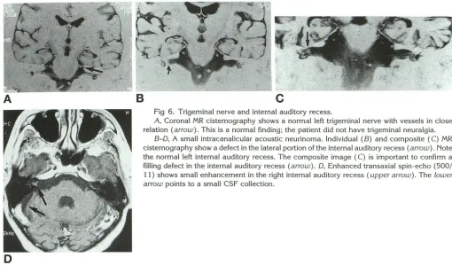
cases (n = 12 of 25) MR cisternography
de-picted vessels closely applied to the fifth nerves (Fig 6A). None of these patients had trigeminal neuralgia (9). Postoperative changes in the fifth nerve were revealed in two cases. In one case of trigeminal neuralgia, there was swelling of the fifth nerve after an isolation procedure. In the other case, postoperative MR cisternography showed residual acoustic neurinoma compress-ing and flattencompress-ing the right fifth nerve. This find-ing was not seen on conventional MR.
In 25 cases in which sections were made through the petrous bones, the internal auditory subarachnoid recesses were outlined in 100%
(n = 25). In only one case, an apparent defect
was noted in the lateral portion of the left
inter-nal auditory subarachnoid recess on an
individ-ual section. A compressed image of three
[image:6.612.57.561.83.276.2]AJNR: 15, October 1994 MR CISTERNOGRAPHY 1653
Fig 6. Trigeminal nerve and internal auditory recess.
A, Coronal MR cisternography shows a normal left trigeminal nerve with vessels in close
relation (arrow). This is a normal finding; the patient did not have trigeminal neuralgia.
8-0, A small intracanalicular acoustic neurinoma. Individual (B) and composite (C) MR
cisternography show a defect in the lateral portion of the internal auditory recess (arrow). Note the normal left internal auditory recess. The composite image (C) is important to confirm a filling defect in the internal auditory recess (arrow). 0, Enhanced transaxial spin-echo (500/
11) shows small enhancement in the right internal auditory recess (upper arrow). The lower arrow points to a small CSF collection.
D
Compressed-image MR cisternography is
es-sential in the evaluation of intracanalicular masses. In a case of small acoustic neurinoma examined by MR cisternography a small defect was noted on the individual section and was also seen on the composite MR cisternography
image. The corresponding conventional MR
study showed a mildly enhancing lesion (Fig 68-D). It is possible to use MR cisternography in the evaluation of intracanalicular lesions without the use of gadolinium enhancement, which is cost effective and also reduces scan-ning time. The internal jugular recess has a
variable appearance. It may appear oblong,
similar to the internal auditory recess, but also can have a globular or linear appearance. The lower cranial nerves were clearly depicted by
MR cisternography.
As in the suprasellar region, MR cisternogra-phy delineates the outer surface of the posterior fossa vessels, providing an extraluminal angio-gram. In one case of giant basilar artery aneu-rysm, MR cisternography provided valuable preoperative information (Fig 7). In addition to the basilar aneurysm, this series included two meningiomas presenting as posterior fossa
masses. In one case of falx meningioma,
con-ventional enhanced MR showed a thick
enhanc-ing right tentorial leaflet, a "dural tail," which could have been caused by either en plaque tumor or nonneoplastic reactive change ( 10,
11 ). MR cisternography showed the right
ten-torium to be of normal thickness. This was
use-ful preoperative information, consistent with
absence of tumor extension in the tentorium. Dural thickening seen on MR cisternography probably represents true pathologic change. One of our atrophy cases also had healed intra-cranial sarcoidosis, and MR cisternography de-picted a thickened tentorial leaflet caused by healed granuloma. In another case of posterior fossa meningioma, the extraaxiallocation of the tumor was shown on MR cisternography with a
layer of CSF separating the meningioma from
the cerebellum.
Cerebral Hemispheres and Ventricles
MR cisternography showed in great detail the
surface brain gyri and important sulci. Cortical
veins were easily depicted as they coursed in
the adjacent CSF on the coronal sections.
De-piction of normal cortical veins on conventional
MR sagittal views was reported to be a helpful
sign in the differentiation between focal wide
[image:7.612.49.559.77.376.2]1654 EL GAMMAL
A
Fig 7. Giant right anterior inferior
cerebellar artery aneurysm.
A, Catheter angiogram lateral projection
shows a small loculus in the upper part of
the aneurysmal sac (arrow).
B, Coronal gadolinium-enhanced
spin-echo (650/11) shows the aneurysmal sac compressing the brain stem with
enhance-ment in its upper wall and in the upper
portion of the sac. Note the pulsation
artifacts.
C, Coronal MR cisternography at the
same level as B. The aneurysm (lower ar-row) displaces and compresses the right
middle cerebellar peduncle (upper arrow).
0, Coronal MR cisternography shows significant compression of the pons and a
small infarct (not seen on the conventional
AJNR: 15, October 1994
MR study) (left arrow). The fifth nerve is seen lateral to the aneurysmal sac (right arrow).
£, Composite coronal image MR cisternography shows the neck of the aneurysm from the basilar artery close to right anterior inferior
cerebellar artery (small right arrow). The left anterior inferior cerebellar artery is shown by the left upper arrow. Left inner ear structures (semicircular canals, cochlea, and vestibule) are depicted by the left middle arrow, and the left vertebral artery is shown by the left lower arrow. The large right arrow points to the origin of the upper loculus of the aneurysm, which was better depicted on sagittal MR
cisternography (not shown).
accumulation (12). However, we had two cases of subdural hygroma in which straight cortical veins were apparent coursing through the fluid separating the brain from the inner table shown on conventional sagittal MR studies. We believe that coronal MR cisternography is more reliable in the diagnosis of "cortical vein sign," helping
to differentiate a wide subarachnoid space from subdural fluid accumulation.
MR cisternography with suppression of the brain parenchyma signal better depicts the
ven-tricles and their contours. MR cisternography should be able to show small changes in relation to the foramina of Monro. MR cisternography
more effectively depicts small areas of brain
disease having increased water content in close
proximity to the ventricles as was seen in three cases in this series (n = 3 of 41) and also near the brain surface (n = 1 of 41) (Fig 70). The aqueduct could not be clearly depicted by MR cisternography because of the increased CSF pulsatile flow within the aqueduct.
MR cisternography was valuable in the differ-entiation of intraaxial and extraaxial lesions in
three patients in this series. These included a patient with a suprasellar tumor blocking the
foramen of Monro and involving both frontal
intra-AJNR: 15, October 1994 MR CISTERNOGRAPHY 1655
A
B
Fig 8. MR cisternography determines intraaxial versus extraaxiallocation of masses
in a patient with renal cell carcinoma.
A and B, Gadolinium-enhanced coronal spin-echo (650/11) images; in A, a small
enhancing mass in the left posterior hemicranium associated with hyperostosis (arrow)
suggests the diagnosis of meningioma. B, Another mass noted in the right parasagittal parietal region (arrow). The location was indeterminate by conventional MR; another
meningioma could not be excluded.
C and 0, Coronal MR cisternography. The left meningioma (C) is seen surrounded by CSF separating it from the indented brain (arrow). The parasagittal lesion on the right (D) is seen as an intracerebral mass by MR cisternography (arrow). The high signal (reversed video) in the right hemisphere below the mass represents an old infarct.
D
cranial tumors with renal cell carcinoma (Fig 8),
the determination of the intraaxial location of a high parasagittal lesion as determined by MR cisternography averted unnecessary catheter angiography or surgical biopsy in the treatment of the patient. MR cisternography was also able to determine the correct extraaxial location of a posterior fossa meningioma.
In conclusion, the accurate and noninvasive
delineation of fistulous tracks by MR cisternog
-raphy now makes it the procedure of choice in
the evaluation of patients with suspected cranial
CSF fistulas. MR cisternography provides
im-portant additional preoperative and
postopera-tive information in patients with intracranial
masses projecting into the subarachnoid space
in the suprasellar region and in the posterior
fossa. Depiction of small intracanalicular
masses by MR cisternography in patients with
suspected acoustic neurinoma provides a cost
-effective alternative to conventional MR with ga
-dolinium enhancement. Composite sagittal
ob-lique MR cisternography images also proved
excellent in the delineation of the entire course
of the optic nerve and are recommended when
additional or more detailed evaluation is
indi-cated for patients with orbital disease.
References
1. Roberson J, Brismar J, Davis K, et al. Metrizamide cisternography with hypocycloidal tomography: preliminary results. AJR Am J
Roentgenol1976;127:965-967
2. El Gamma! T. Cervical myelography and posterior fossa exami
-nations with amipaque: use of magnification and subtraction.
Radiology 1980;136:219-222
3. El Gamma! T, Brooks BS. Serial biplane magnification and
sub-traction myelocisternography: normal and pathological findings.
AJNR Am J f'leuroradiol 1981 ;2:55-63
4. El Gamma! T, Brooks BS. Myelography in the evaluation of the craniovertebral junction. In: Taveras M, Ferrucci JT Jr, eds. Ra di-ology: Diagnosis/lmaging/lnlervention. Philadelphia: Lippincott,
[image:9.612.48.564.80.458.2]1656 EL GAMMAL
5. Massey CE, El Gamma! T, Brooks BS. Giant posterior inferior
cerebellar artery aneurysm with dysphagia. Surg Neuro/1984;22:
467-471
6. Drayer BP, Wilkins RH, Boehnke M, Horton JA, Rosenbaum AE.
Cerebrospinal fluid rhinorrhea demonstrated by metrizamide CT
cisternography. AJR Am J Roentgeno/1977;129:149-151
7. Maw ad M, Silver A, Hila! S, Ganti S. Computed tomography of the
brainstem with intrathecal metrizamide, 1: the normal brain stem.
AJNR Am J Neuroradiol1983;4:1-11
8. Krudy AG. MR myelography using heavily T2 weighted fast
spin-echo pulse sequences with fat presaturation. AJR Am J
Roent-genol 1992:159:1315-1320
9. Tash RR, Sze G, Leslie DR. Trigeminal neuralgia: MR imaging
features. Radiology 1989; 172:767-770
AJNR: 15, October 1994
10. Goldsher D, Litt AW, Pinto RS, Bannon KR, Kricheff II. Dural "tail" associated with meningiomas on Gd-DTPA-enhanced
MR images: characteristics, differential diagnostic value, and
possible implication for treatment. Radiology 1990; 176:
447-450
11. Tokumaru A, O'uchi T, Eguchi T, et al. Prominent meningeal
enhancements adjacent to meningioma on Gd-DTPA-enhanced
MR images: histopathologic correlation. Radiology 1990; 175:
431-433
12. McCluney KW, Yeakley JW, Fenstermacher JM, Baird SH,
Bonmati CM. Subdural hygroma versus atrophy on MR brain