Noctuid moths have auditory systems sensitive to ultrasound (Roeder and Treat, 1957). It is widely believed that these ears have evolved in response to predation pressure from echolocating bats which hunt using ultrasonic sonar (Roeder, 1967a; Fullard, 1987). Stimulation of these auditory units triggers escape behaviours which range from negative phonotactic flight at low sound intensities to erratic dives and turns at high sound intensities (Roeder, 1962, 1967b, 1975). For a noctuid moth to make a decision as to whether an echolocating bat is present in its environment, it must evaluate the information from only two sensory cells per ear, the more sensitive A1 cell and the less sensitive A2 cell. There is no ability to differentiate frequency. The tuning curves of the two sensory cells are matched, with the A2 cell being less sensitive than the A1 by about 20–25 dB (Roeder, 1964). No other sensory apparatus is known to modulate the escape behaviour. A major problem in relating the activity of the A1 cell to the initiation of the escape behaviour is that the precise levels of A1 cell activity required to trigger an escape response are unknown (Fullard, 1987; Surlykke, 1988). Roeder (1967b) suggested that the required level of activity is close to the physiological threshold of the A1 cell, though quantitative data are lacking. It appears that stimulus levels sufficient to excite
the less sensitive A2 cell are required to augment or inhibit the synaptic drive to the longitudinal flight muscles (Madsen and Miller, 1987). This is probably responsible for the complex diving manoeuvres observed (Roeder, 1962, 1975). Whether these two types of behaviour exist in isolation or whether there are intermediate forms of behaviour remains unknown (Surlykke, 1984). In common with many other sensory cells, the A cells exhibit two types of associated noise. The first is spontaneous discharge (Douglass et al. 1993), which may produce an incorrect decision that a bat is present. The second is inaccurate encoding of stimulus level (Zimmerman, 1978). This latter error would cause the decision-making process (which type of escape behaviour to use and when to initiate it) less reliable. This study examines the ability of the A1 cell to encode information at the peripheral level using biologically relevant stimulus types. Since information cannot be created once encoded by the peripheral system, examination of the way information is channelled and the reliability of the encryption puts an upper boundary on the reliability of the receiver. From this, the most important features of bat echolocation calls for distinguishing bat from non-bat stimuli can be established. This paper examines the response of the A1 cells to stimuli varying in intensity, duration and presentation rate over ranges Printed in Great Britain © The Company of Biologists Limited 1996
JEB0036
The ability of the noctuid A1 cell acoustic receptor to encode biologically relevant information from bat echolocation calls is examined. Short-duration stimuli (less than approximately 6 ms) reduce the dynamic resolution of the receptor, making intensity, and hence range, estimates of foraging bats unreliable. This low dynamic range is further reduced by inaccurate encoding of stimulus intensity, reducing the real dynamic range of the A1 cell to 1 bit at stimulus durations below 3.1 ms. Interspike interval is also an unreliable measure of stimulus intensity at low stimulus levels and/or for short-duration stimuli. The quantity of information encoded per stimulus is reduced as the presentation rate of stimuli is increased. The spontaneous generation of A1 cell action potentials may
reduce the ability of the moth to discriminate bat from non-bat signals. Even with a recognition criterion of three A1 cell spikes per call, the moth would regularly make wrong decisions about a bat being present in the immediate environment. Removing this noise would necessitate a considerable loss of information through filtering at the interneurone level. It is proposed that, for bats using short-duration calls, the moth would only be able to recognise an approaching bat from the repetitious nature of the incoming signal.
Key words: noctuid moth, audition, auditory sensitivity, Agrotis segetum, bat, echolocation.
Summary
THE PERIPHERAL AUDITORY CHARACTERISTICS OF NOCTUID MOTHS:
INFORMATION ENCODING AND ENDOGENOUS NOISE
DEAN A. WATERS*
School of Biological Sciences, University of Bristol, Woodland Road, Bristol BS8 1UG, UK
Accepted 5 January 1996
*Present address: Department of Biology, University of Leeds, Leeds LS2 9JT, UK.
similar to those likely to be encountered in the field from echolocating bats. The results are interpreted in relation to the noise levels of the A1 cell, the accuracy of encoding information and the need to separate bat from non-bat stimuli.
Materials and methods
The turnip moth Agrotis segetum (Denis & Schiffermuller) was selected for these experiments and was supplied from an existing culture. All experiments were performed in a 4 m34 m32.6 m room lined with 1 cm thick sound-absorbing foam. The temperature was maintained at 12 °C and 85 % relative humidity, similar environmental conditions to those experienced by moths in the field. All apparatus was covered in sound-attenuating foam and was free from unwanted ultrasonic noise above approximately 10 dB SPL (no difference in noise level recorded from the high-frequency output of an Ultra Sound Advice S-25 bat detector with the apparatus on or off; the S-25 has an estimated lower sensitivity of approximately 10 dB SPL, K. Maries, personal communication). Tympanic preparations were made, derived from the methods of Roeder (1966) and Agee (1967). Briefly, the moth was decapitated, its legs removed, and the moth was fastened dorsal side up in a shaped polystyrene block with the wings clamped spread open. A groove in the block allowed sound to reach the tympanic organ situated at the base of the hindwing. The notum was brushed free of scales and a circular incision made around it. On removal of the notum, the mesophragma was gripped with a pair of forceps and pulled free, the longitudinal flight muscles being severed at the anterior end. The resulting cavity was flooded with saline (Fielden, 1960). A bipolar silver hook electrode was scanned over the surface of the exposed dorsoventral muscle until the tympanic nerve IIIN1 was encountered, characterised by the regular firings of the non-auditory B cell. The nerve was then hooked over the tip of the electrode. Output was preamplified (CEP 8120) and bandpassed between 50 Hz and 10 kHz with a gain of 10003. Output was passed to an audio amplifier and Tektronix 5113 dual-beam storage oscilloscope. Once established, preparations remained stable for between 2 and 6 h, with no sign of lowered auditory sensitivity. All experiments were completed within 1 h of establishing the preparation.
Ultrasound stimuli were generated by a custom-made sine-wave generator and pulse shaper, amplified and broadcast through an Ultra Sound Advice amplifier and matched capacitance loudspeaker (total harmonic distortion <1 %). Stimulus level could be attenuated in 1 dB steps over the range 0–30 dB using a custom-made decibel attenuator (each step accurate to ±0.05 dB, ±0.23 dB overall). Where required, oscilloscope voltages were converted to sound pressure levels immediately after the experiments by a Brüel and Kjær 2204 sound pressure meter equipped with a 6.35 mm 4135 microphone (grid off) placed at the ear of the preparation (frequency response ±2 dB 0.01–120 kHz). The measurement of the sound field at the preparation controlled for the
reflection, refraction and diffraction effects caused by the polystyrene block and the proximity of the apparatus.
Stimulus intensity encoding
Constant-frequency trapezoidally shaped pulses with a rise and fall time set to 10 % of the plateau duration (total 20 %) were generated and broadcast at the preparations. The carrier frequency of the pulses was set to 50 kHz. Pulses were presented in two sets of halved durations over the range 50–12.5 ms and 6.25–1.5 ms. Three moths were used for each of the sets. For each set of presentations, a threshold level of one A1 cell action potential in four out of five stimulus presentations was determined for the longest stimulus in the set at the +4 dB level on the attenuator. The attenuator was then switched to the 0 dB level to make the stimulus subthreshold. The output of the preamplifier and stimuli were recorded onto two direct record (DR) channels of a Racal Store 4DS instrumentation recorder at 38 cm s21, giving a ±3 dB
bandwidth of 100 Hz to 75 kHz. Five stimuli were presented at each intensity level, increasing from 0 dB up to +30 dB. The order of presentation of the pulse durations was a Latin square design, the three durations being matched with three replicates for each duration set. Analysis was performed on a Kay DSP 5500 Sonagraph configured as a dual-channel storage oscilloscope. The numbers of stimulus-locked A1 action potentials were measured, as were the interspike intervals in the A1 cell spike train. At higher stimulus levels, the superimposition of A1 and A2 cell spikes was problematic. In some cases, it was possible to identify A2 cell spikes where they occurred between consecutive A1 cell spikes, though this was not reliable at higher stimulus levels when the A1 and A2 spikes became superimposed. This may mean that A2 spike number may be underestimated (Fig. 1).
Encoding of repetition rate
increased in intensity by 3 dB. This is because the auditory system is believed to be an energy detector with a reduction in sensitivity of 3 dB per halved stimulus duration (Adams, 1971; Surlykke et al. 1988). The stimulus protocol resulted in 60 types of stimuli per preparation. The experiment was performed on four moths, the order of stimulus presentation being alternated for each one.
Adaptation rates
Threshold values were determined using a criterion of 2–3 action potentials per 50 kHz 6.25 ms pulse at the 0 dB setting of the attenuator. The signal was then switched to produce a constant-amplitude output for 2 s and presented at 0, +10, +20 and +30 dB amplitude, 0 dB corresponding to approximately 50 dB SPL. The preparation was allowed to recover for 30 s between stimuli, the spontaneous discharge rate returning to normal within this time. The stimuli and neural output were recorded at 38 cm s21onto two channels of the Racal recorder.
The speaker was calibrated immediately afterwards to determine the absolute sound pressure level of the stimuli. The number of A1 action potentials were counted within the first 0–25, 25–50, 50–100 and 100–200 ms intervals and each
200 ms thereafter, following the method of Pérez and Coro (1985). At high stimulus levels, coincident A1 and A2 spikes made quantification difficult. Where coincidence was clear, i.e. double the normal spike amplitude or double spikes, these were counted as evidence for an A2 cell spike. Where adaptation resulted in lower spike rates, the distinction was less clear and the A1 cell data are hence less reliable.
Spontaneous generation of action potentials
Six preparations of A. segetum were established and allowed to run with no stimulus for approximately 30 min, the output from the preamplifier being recorded on the Racal Store 4DS at 38 cm s21. The tape length used was divided into ten equal
sections, and the 5 s at the start of each section was analysed, the time from the start of the section to each A1 cell action potential being recorded.
Results
Intensity information encoding
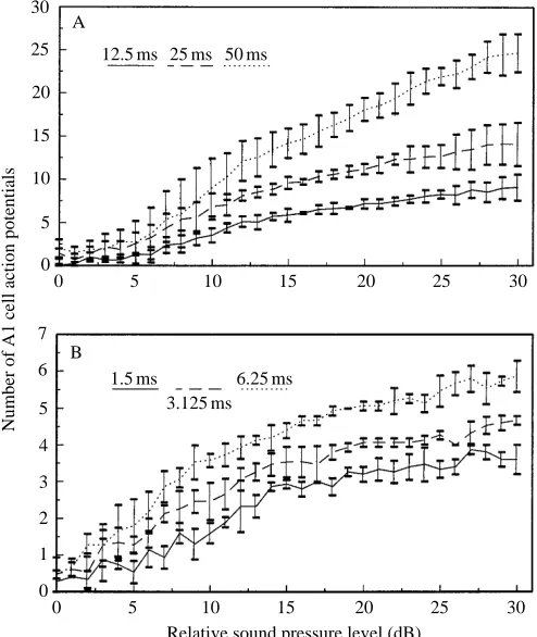
The effect of increased stimulus intensity with stimulus duration on the number of A1 cell action potentials is presented in Fig. 2A,B. Mean values from five stimulus presentations to three individuals are given for each point. The number of
B
C
D
*
A
[image:3.609.65.282.350.668.2]1 mV 5 ms
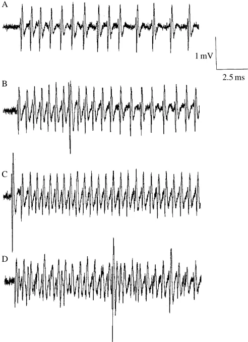
[image:3.609.319.566.374.667.2]Fig. 1. Response of the A1 and A2 cells to a 50 kHz 6.25 ms pulse with increasing stimulus level. (A) 54 dB SPL; (B) 60 dB SPL; (C) 66 dB SPL; (D) 72 dB SPL. Only A1 action potentials are shown in A–C. The position of closely spaced A1 and A2 action potentials is marked with an asterisk in D. The bottom trace shows the time course of the stimulus. The large spike is from the non-auditory B cell.
Fig. 2. The mean ± S.D. number of stimulus-locked A1 action potentials for three stimulus durations with increasing stimulus intensity (N=3 individuals, N=5 stimulus presentations per individual). (A) 50, 25 and 12.5 ms stimuli; (B) 6.25, 3.125 and 1.5 ms stimuli. Stimulus levels are relative to the SPL at 4 dB below the A1 cell threshold value in response to the longest-duration stimulus in the set.
A 30
25
20
15
10
5
0
Number of A1 cell action potentials
0 5 10 15 20 25 30
12.5 ms 25 ms 50 ms
Relative sound pressure level (dB) B
7
6
5
4
3
2
1
0
0 5 10 15 20 25 30
1.5 ms 3.125 ms
action potentials elicited by the stimulus increases with increasing stimulus intensity. At any given stimulus level, the total number of action potentials produced is greater for a longer-duration stimulus. The response saturates at lower stimulus levels for shorter-duration stimuli. The energy necessary to produce an extra action potential for shorter-duration stimuli is greater than that for a long-shorter-duration stimulus. The dynamic range of the system can be defined by the maximum number of action potentials elicited between threshold and the upper limit of the stimulus. The ability of the A1 cell to encode a large dynamic range is limited when presented with short-duration stimuli. In an ideal situation, the number of action potentials can be converted into the information content of the action potential train using the formula:
I = log2E ,
where I is the information content in bits and E is the maximum number of distinguishable energy levels.
If the number of action potentials accurately encodes a discrete stimulus energy level, then E is simply the maximum number of action potentials which can be elicited. However, there is considerable variation in the number of action potentials which are produced in response to repeated stimuli at the same stimulus level. The maximum number of distinguishable energy levels is then dependent on a step
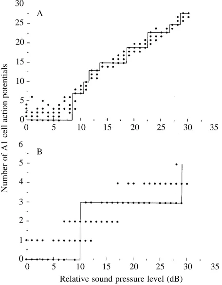
function drawn through the number of action potentials with increasing stimulus level, two examples of which are illustrated in Fig. 3A,B. The line is drawn such that all points along the horizontal step can be distinguished from those on the next step (Zimmerman, 1978). The maximum dynamic range and the real dynamic range with stimulus duration are given in Table 1. The real resolvable dynamic range is about half that calculated from the number of action potentials alone. For 3.1 ms and 1.5 ms stimuli, the real dynamic range is only 1 bit, indicating that for stimuli shorter than 3.1 ms the A1 cell can only encode, with 100 % accuracy, the presence or absence of a stimulus over a 30 dB range.
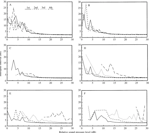
At low stimulus levels, the interspike interval between stimulus-locked A1 cell action potentials is highly variable, only becoming regular at more than 10 dB over threshold (Fig. 4A–F). Stimulus-locked action potentials could be identified with large interspike intervals for long-duration stimuli, but not for short-duration stimuli. The stimulus level required to produce regular A1 cell spike trains was greater with short-duration stimuli than with long-duration ones.
Repetition rate information encoding
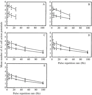
[image:4.609.55.283.400.692.2]The effect of increasing the stimulus repetition rate on the mean number of action potentials per stimulus is shown in Fig. 5A–E. Data are the mean number of action potentials per stimulus over a 1 s stimulus period and are the mean value from four individuals. At stimulus presentation rates above 100 Hz, no spike train could be identified as being stimulus-locked, an apparently continuous discharge being produced. Both the low-intensity stimulus set (2–3 action potentials threshold criterion) and the high-intensity set (+10 dB over low set) show similar results. In both cases, the mean number of action potentials per stimulus decreases as the pulse repetition rate is increased above 10 Hz. This effect appears to be independent of stimulus duration. Since the values presented are mean values over a number of pulses, it can be argued that the reduction in number of action potentials with higher repetition rates is due to higher rates of fatigue or to adaptation
Fig. 3. Minimum number of 100 % distinguishable energy levels for two representative durations (N=3 individuals, N=5 stimulus presentations per individual per stimulus level). (A) 50 ms stimulus; (B) 1.5 ms stimulus.
A 30 25 20 15 10 5 0
Number of A1 cell action potentials
0 5 10 15 20 25 30 35
Relative sound pressure level (dB) B
6 5 4 3 2 1 0
0 5 10 15 20 25 30 35
Table 1. Maximum and real information transfer by the A1 cell for a given stimulus duration over a 30 dB stimulus
range
Stimulus Maximum Real
duration dynamic range dynamic range
(ms) (bits) (bits)
50 4.7±0.13 2.9±0.11
25 3.9±0.23 2.4±0.34
12.5 3.3±0.17 1.7±0.24
6.25 2.7±0.13 1.7±0.24
3.125 2.5±0.15 1±0.00
1.5 2.2±0.19 1±0.00
Data are from three individuals with five stimulus presentations per stimulus level.
[image:4.609.312.560.589.697.2]at higher stimulus presentation rates. If this effect is significant, it should be apparent in a plot of the number of action potentials per stimulus with time. Such a plot is shown in Fig. 6, giving mean data from four individuals stimulated at 100 Hz with a 1.5 ms pulse (duty cycle 15 %) using the +10 dB stimulus set. These data represent the highest-intensity, highest-duty-cycle set for which data were available. As can be seen, the mean number of action potentials per stimulus does decrease rapidly within the first 100 ms of stimulation, but after this remains relatively stable with time. The data suggest that the reduction in mean number of action potentials with increasing repetition rate is a function of rapid adaptation or fatigue caused by the first few stimulus
presentations. As presentation rates increase, the ability of the A1 cell to differentiate two stimulus levels separated by 10 dB is reduced. For the 0.375 and 0.75 ms stimuli, the two stimulus levels are statistically differentiable at presentation rates between 3 Hz and 50 Hz, but not at 100 Hz. At all other presentation rate/stimulus duration combinations, the two stimulus levels can still be separated (Wilcoxon’s test P<0.05).
Adaptation rates
The initial A1 cell action potential trains at the start of the 2 s stimulus are shown in Fig. 7A–D. Mean data from three individuals of discharge rates for the A1 cell with increasing stimulus intensity are shown in Fig. 8. For stimulation with
A 30 25 20 15 10 5 0
0 5 10 15 20 25 30
1st 2nd 3rd 4th
C 30 25 20 15 10 5 0
0 5 10 15 20 25 30
E 30 25 20 15 10 5 0
0 5 10 15 20 25 30
Interspike interval (ms)
B 30 25 20 15 10 5 0
0 5 10 15 20 25 30
D 30 25 20 15 10 5 0
0 5 10 15 20 25 30
F 30 25 20 15 10 5 0
[image:5.609.56.566.73.527.2]0 5 10 15 20 25 30
Fig. 4. Mean interspike interval between the first four stimulus-locked A1 action potentials with increasing stimulus intensity (N=3 individuals, N=5 presentations per individual). (A) 50 ms stimulus; (B) 25 ms stimulus; (C) 12.5 ms stimulus; (D) 6.25 ms stimulus; (E) 3.125 ms stimulus; (F) 1.5 ms stimulus.
a 2 s sine wave, the A1 cell shows a rapid adaptation or fatigue effect, following an approximate exponential decay. At the +20 dB (70 dB SPL) and +30 dB (80 dB SPL) stimulus levels, the A1 cell appears to have reached saturation as no increase in discharge rate could be produced. The discharge rate of the A1 cell is lower at +30 dB (80 dB SPL) than at +20 dB (70 dB SPL) (Wilcoxon’s test P<0.05). The difficulty in confusing A1 with A2 action potentials from extracellular recordings is apparent in Figs 1 and 7. While it is possible to
identify A2 spikes when they occur between consecutive A1 spikes, the number may be underestimated, at least when the discharge rate is high, and the data for the +30 dB (80 dB SPL) presentation are consequently less reliable than those at lower stimulus levels.
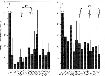
Spontaneous generation of A1 action potentials The rates of spontaneous A1 cell action potentials in each 5 s time window with time are shown in Fig. 9. There are no consistent effects with time, the measured discharge rate appearing to be random throughout the measuring period. A histogram showing all interspike intervals from all time periods in all individuals is shown in Fig. 10. The distribution of interspike intervals is log–normal, with a modal value of 9 ms when viewed with a bin width of 1 ms. The median discharge rate is 7.4 Hz (10 measurements in each of six individuals). The ratio of variance of interspike interval/mean interspike interval is 196.6, suggesting a clustered temporal distribution. By adopting a specific number of action potentials within a certain time period as a recognition criterion that a bat is present, it is possible to calculate the rate at which the moth would mistakenly identify a bat as being present when one is not solely on the basis of the occurrence of spontaneous A1 action potentials. The 50 s sampling window for each individual (1035 s spread throughout the 30 min experiment) was searched for two or three consecutive action potentials within a certain criterion time, incremented consecutively by 2 ms. The time to
A 6
5 4 3 2 1 0
Mean number of A1 cell action potentials
0 20 40 60 80 100
C 6
5 4 3 2 1 0
0 20 40 60 80 100
E 6
5 4 3 2 1 0
0 20 40 60 80 100
B 6
5 4 3 2 1 0
0 20 40 60 80 100
D 6
5 4 3 2 1 0
0 20 40 60 80 100
Pulse repetition rate (Hz)
[image:6.609.253.561.71.389.2]Pulse repetition rate (Hz)
Fig. 5. Mean ±S.D. number of A1 action potentials per
stimulus with increasing pulse repetition rate. Low-(lower line) and high- (+10 dB, upper line) intensity sets are shown (N=4 individuals). (A) 6.25 ms stimulus; (B) 3.125 ms stimulus; (C) 1.5 ms stimulus; (D) 0.75 ms stimulus; (E) 0.375 ms stimulus.
Fig. 6. Mean ±S.D. number of A1 action potentials with time during
stimulation with a 1.5 ms stimulus at 100 Hz repetition rate using the high-intensity (+10 dB) presentation set in Fig. 5 (N=4 individuals).
4
3
2
1
0
Number of action potentials per stimulus
0 200 400 600 800 1000
[image:6.609.45.291.543.701.2]mistakenly identify a bat based on this criterion was then calculated using the rate of occurrence of the criterion action potentials/interspike intervals combination. Data for a two-spike and a three-two-spike criterion are presented in Fig. 11A,B. For interspike intervals of less than 3 ms, the moth would not
make a mistaken identification within the 50 s sampled period for both two- and three-spike criteria. For interspike intervals greater than 3 ms, the moth would make a mistaken identification every few seconds. The time to make a mistake is greater using a three-spike criterion. The significance of this observation is discussed below.
B
C
D A
1 mV
[image:7.609.351.536.72.346.2]2.5 ms
Fig. 7. Initial A1 cell responses to a 2 s 50 kHz stimulus at increasing stimulus levels. (A) 50 dB SPL; (B) 60 dB SPL; (C) 70 dB SPL; (D) 80 dB SPL. In D, the apparently chaotic trace is produced by coincident A1 and A2 cell spikes.
600
500
400
300
200
100
0
A1 cell discharge rates (Hz)
0 500 1000 1500 2000
50 dB SPL 60 dB SPL 70 dB SPL 80 dB SPL
[image:7.609.49.295.73.412.2]Time (ms)
Fig. 8. Adaptation of the A1 cell to a continuous 2 s 50 kHz sine wave at four sound pressure levels. Data are mean ±S.D., N=3 individual
moths.
20
15
10
5
0
Spontaneous A1 cell discharge rate (Hz)
0 200 400 600 800
[image:7.609.330.558.402.604.2]Time (s)
Fig. 9. Spontaneous discharge rates of the A1 cell in six individuals with time. Each value is the mean rate in a sample window of 5 s at each time period.
0.05
0.04
0.03
0.02
0.01
Proportion
0 40 80 120 160 200
Interspike interval (ms)
[image:7.609.49.300.485.629.2]Discussion
For a noctuid moth flying under the risk of predation by bats, the moth must interpret information from its auditory system to provide a reliable indicator of the presence or absence of bats in its vicinity. If a bat is detected, a decision must be reached as to the best timing and vector of an escape response (Altes and Anderson, 1980). The moth must therefore be able to distinguish the echolocation calls of a foraging bat from acoustic noise within the environment, acoustic noise generated by its own movements (Waters and Jones, 1994) and informational noise produced by the spontaneous generation of action potentials within its own auditory system (Fullard, 1987). From the data presented here, it is clear that between threshold and +10 dB stimuli, the response of the A1 cell is erratic. For long-duration stimuli, such as 50 ms, the number of action potentials per stimulus increases rapidly, even though the variable interspike intervals do not produce a regular spike train. For short-duration stimuli, not only are fewer action potentials produced, but the interspike intervals are also more variable over a greater range of stimulus intensities. The inaccurate coding of the same stimulus level on consecutive presentations of the stimulus degrades the moth’s ability to distinguish discrete stimulus levels. For a moth attempting to judge the range of an approaching bat, the lowered ability to judge stimulus level will lead to a reduced ability to determine range and, hence, the most appropriate escape strategy. For 3.125 and 1.5 ms stimuli, the ability to determine stimulus level by the A1 cell is reduced to 1 bit, equivalent to the moth distinguishing either the presence or absence of a bat over a 30 dB stimulus range. If we accept the paradigm of the two-stage escape behaviour, then this is the only requirement of the sensory system. The A1 cell would simply detect a foraging
bat and trigger a negative phonotactic escape behaviour. However, given that inaccurate coding and spontaneous activity mean that more A1 action potentials would be required reliably to establish that a bat was present, the bat could approach the moth considerably more closely before eliciting enough A1 action potentials for the moth to be 100 % certain of its presence. For bats using low-frequency, high-intensity calls, the distances at which they are initially detected can be large, in some cases up to 40 m (Roeder, 1966). However, for bats using short-duration, low-intensity calls, these distances can be as little as 0.2 m (Fenton and Fullard, 1979). Clearly, the loss of information due to filtering of the non-bat information and inaccurate coding would be small if the moth detected low-frequency, high-intensity bats, but could be very costly if it were trying to detect low-intensity bats.
The main problem in distinguishing a bat from a non-bat signal at low intensity levels is the spontaneous generation of A1 action potentials, which occur at a median rate of 7.4 Hz. In principle, coincident A1 action potentials from both ears may serve as a reliable indicator of a real source, but this would only be possible if the moth were head-on or tail-on to the source. Away from these angles, the body of the moth diffracts ultrasound, resulting in a sound shadow on the contralateral side of between 20 and 30 dB of attenuation (Payne et al. 1966; Madsen and Miller, 1987). There is little published information on the spontaneous generation of A1 action potentials. Adams (1971) reports a quiescent level of 20–40 A1 action potentials per second, while Faure et al. (1990) report ‘>1 per second’. Coro et al. (1994) have found that spontaneous activity levels are positively correlated with temperature, rising from a rate of 15 Hz at 18 °C to 80 Hz at 34 °C. While the ambient temperature will almost certainly have a bearing on the results
B 60
50
40
30
20
10
0
6–1010–1414–1818–2222–2626–3030–3434–3838–4242–4646–5050–5454–5858–62
ISI 50
A 60
50
40
30
20
10
0
1–33–55–77–99–1111–1313–1515–1717–1919–2121–2323–2525–2727–2929–31
Type 1 error mistake time (s)
[image:8.609.193.562.71.340.2]ISI 50
Fig. 11. Mean time for an individual of Agrotis segetum to mistakenly identify that a bat is present owing to the spontaneous discharge of its A1 cell (mean + S.D., N=6). (A) Criterion
reported here, the internal temperature of the tympanic organ of free-flying moths is unknown. In winter-flying moths, the heat from the thoracic muscles during flight is not conducted or convected into the posterior part of the thorax because of the heat exchangers and insulation between the abdomen and thorax (Heinrich, 1987). Additionally, the tympanic organ sits within a number of vented air-sacs (Treat, 1959), which are likely to maintain the tympanum temperature close to that of the atmosphere. Another problem is whether the spontaneous A1 cell activity levels represent a ‘real’ phenomenon, or whether activity levels were affected by the dissection and recording techniques. Roeder and Treat (1957) and Treat and Roeder (1959) noted that stretching the tympanic nerve causes an increase in discharge rate of the non-auditory B cell, which is probably a development of the chordotonal system to monitor the flexion and position of the thoracic segments, and may encode stress within the tympanal frame (Yack and Fullard, 1993). No significant audible rise in the discharge rate of the B cell was noted during the present experiments, which suggests that the stress relationships within the tympanic region were unaffected by the recording procedure.
Spontaneous A1 cell action potentials are obviously a problem for the moth. An interneurone ‘noise filter’ has been proposed by Boyan and Fullard (1988), who found that the first-order interneurone IN 501 shows a 1:1 spiking relationship with the A1 afferent only at A1 discharge rates above 258–276 Hz, corresponding to an interspike interval of 3.6–3.9 ms. This corresponds to the lowest value found in the time-interval histogram of spontaneously generated A1 action potentials (Fig. 10). If the moth accepted this interspike interval criterion alone to trigger an escape behaviour, an enormous loss of sensitivity would occur since single action potentials elicited by a bat and pairs of action potentials with interspike intervals longer than 3.6 ms would be filtered out. This would mean a consequent reduction in maximum detection range, particularly in response to short-duration calls. The only possible mechanism by which sufficient information could be obtained by the moth to detect bats at large distances would be to look for regular temporal patterns in trains of single A1 cell action potentials. So far, no interneurones which follow the A1 spiking patterns at low discharge rates have been identified.
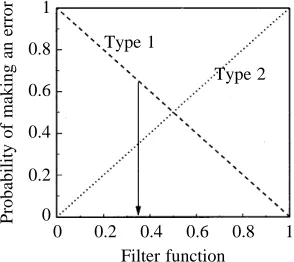
If the moth accepted all A1 cell information as valid, then, owing to spontaneous A1 activity, it would make a mistaken identification of a bat every few hundred milliseconds. A mistakenly activated escape behaviour would be physiologically expensive for the moth and might take it away from feeding sites or make it lose the pheromone trail of a mate. If the criterion is tightened to two or three action potentials within a certain time frame, the risk of mistakenly identifying a bat is reduced, but the chances of not reacting to a bat when one is present are increased. This can be expressed by analogy to statistics, defining a Type 1 error as the moth mistakenly deciding that a bat is present, and a Type 2 error as the moth mistakenly deciding a bat is not present. The amount of A1 cell information which is discarded can be defined by an
arbitrary filter function. This concept is illustrated in Fig. 12: as the filter function is tightened, more information is discarded and the probability of making a Type 2 error increases. The value of the filter function should be an evolutionary optimum given the relative costs of both error types. It has been shown that the selective advantage of reacting over non-reacting moths in encounters with bats is 44 % per encounter (Roeder and Treat, 1962), so clearly the costs of making a Type 2 error are high. The costs of making a Type 1 error are probably lower when considering the negative phonotactic behaviour, since the moth may simply change direction, but may be much higher with the expression of an erroneous complex behaviour. Dives and uncontrolled loops may mean that the moth falls to the ground where there are predators, onto the surface of a pond, or into vegetation where it may encounter the webs of spiders. Initiation of complex escape behaviour by a less-sensitive A2 cell may reduce the Type 1 error rate of the complex behaviour through lower spontaneous activity and a lowered likelihood of stimulation by environmental noise. A two-cell detection system, offset in sensitivity by 20 dB, may thus not only increase the useful dynamic range of the system, particularly for short-duration stimuli, but also minimise the overall Type 1 error rate.
[image:9.609.368.515.526.657.2]From the experiments to investigate adaptation rate, it was found that A1 cell discharge rate apparently decreases at 30 dB above threshold. This effect has been also observed by Coro and Pérez (1984), who propose a mechanism of lateral inhibition of the A1 cell over the A2 cell. Since this effect was not observed in the incremental stimulus increase experiments reported here, the mechanism or function of this phenomenon is unclear. The difficulty in distinguishing A1 and A2 cell spikes at high discharge rates may mean that the A1 cell spike rate is underestimated, and the A2 cell spike rate correspondingly overestimated, which could account for the results presented here. It is possible that the reduction in A1 cell spike rate is due to the tympanic membrane vibrating in a
Fig. 12. The two error types associated with selecting an escape strategy as a function of the information filtering at the interneurone level. A Type 1 error represents the moth mistakenly deciding that a bat is present. A Type 2 error represents the moth mistakenly deciding that a bat is not present. The filter function is arbitrary, a higher value representing more information being discarded by the central nervous system.
1 0.8 0.6 0.4 0.2 0
Probability of making an error
0 0.2 0.4 0.6 0.8 1 Type 1
Type 2
higher mode, where the attachment of the sensillum may correspond to a node and thus experience a reduced vibrational amplitude. At 30 dB above A1 threshold, the response of the A1 cell has probably served its purpose in alerting the moth to the long-distance presence of a bat. For short-duration stimuli, the extra energy required to produce another A1 cell action potential becomes large and so is of limited use in determining a decrease in range of the bat from the moth. At these stimulus levels, the A2 cell begins to fire and could provide a more reliable measure of bat proximity than the A1 cell. Notodontid moths possess only one A cell (Surlykke, 1984), and this does raise the question as to the functional significance of the A2 cell in noctuids. In addition, it is known that call intensity is reduced as the bat homes in on the target through echo-levelling (Kobler et al. 1985) or during the approach phase and terminal buzz (Schnitzler et al. 1988; Kalko and Schnitzler, 1989). For some species of bats which use high-frequency or low-intensity calls, this may mean that the incident sound level at the moth is never sufficient to excite the A2 cell.
A. segetum is able accurately to encode repetition rates of stimuli up to 100 Hz; above that, a continuous A1 cell discharge is produced. High repetition rates produce a reduction in the ability to distinguish different stimulus levels and hence a reduced ability to determine the range of the bat. A reduced ability to determine the distance to the bat may be counteracted by cues from the bat which are range-dependent. As echolocating bats approach targets, the call repetition rate increases during the approach phase, increasing further during the terminal buzz (Griffin et al. 1960). Search-phase echolocation repetition rates appear to be dependent on the bat’s wingbeat frequency, which is in turn dependent on body size (Jones, 1993). Search-phase calls are typically between 5 and 15 Hz (Jones, 1994). During the approach phase, repetition rates rise to between 30 and 70 Hz, corresponding to distances of 1.1–1.6 m from the target in Pipistrellus kuhli and 0.73–1.28 m in Myotis daubentoni (Schnitzler et al. 1988; Kalko and Schnitzler, 1989). During the terminal buzz, rates rise further to a maximum of 170–284 Hz at 0.3–0.7 m from the target (Jones and Rayner, 1988; Schnitzler et al. 1988; Kalko and Schnitzler, 1989). It has been suggested that tympanate moths may use the increased rate of echolocation as a cue for defensive behaviour (Roeder, 1964), as has been demonstrated for one phase of the escape behaviour of lacewings (Miller and Olesen, 1979). The arctiid moth Cycnia tenera is behaviourally maximally responsive to stimuli delivered at 30–50 Hz (Fullard, 1984), although the stimulus levels used were sufficient to trigger both the A1 and A2 cells. Roeder (1966) proposed a scheme of ‘repeater’ and ‘pulse coder’ interneurones to code repetition rate information, but recent studies have revealed the scheme to be more complex (Boyan and Fullard, 1986). Paul (1974) described ‘stable followers’, capable of following repetition rates greater than 10 Hz, and ‘labile followers’ which encoded stimuli at repetition rates lower than 3 Hz. The latter were proposed to encode non-regular stimuli, such as ambient noise in the environment, so that it did not initiate a behavioural response.
Specific interneurones have been identified which appear to be able to differentiate search-, approach- and terminal-phase information from the A1 cell discharge rate (Boyan and Miller, 1991). It has been argued that stimulation of the A2 cell at high stimulus levels is necessary to elicit the complex escape behaviour, but this does not explain why the ability to encode even the terminal phase is exhibited by interneurones making synaptic connections with the A1 cell. As discussed earlier, bats reduce emitted call intensity when closing on a target, and may alter the emitted SPL so that the intensity incident upon the target is constant (Hartley et al. 1989). The actual SPL received by the moth may therefore fall below that necessary to stimulate the A2 cell, resulting in the repetition rate cue encoded by the A1 cell being a viable trigger for the complex escape behaviour.
While an increase in pulse repetition rate is a valid measure of the distance of a bat from the moth, it is arguable that this gives the moth enough time to escape. Flight speeds in bats vary considerably depending on wing loading and aspect ratio (Norberg and Rayner, 1987), but a general range of values is 2–8 m s21. If the approach phase began a few metres from the
target, this would only give the moth a few hundred milliseconds to initiate an escape response and to move far enough away from the bat’s trajectory to evade capture. Altes and Anderson (1980) calculated that the moth’s best strategy is a dive normal to the bat’s velocity vector, initiated at 30 cm from the bat. Behavioural latencies of the escape behaviours are not known precisely. Under laboratory conditions, Roeder (1967b) measured a behavioural latency of the negative phonotactic behaviour of approximately 50 ms. The reaction time of the more complex behaviour appears to be longer, with reported minimum values between 80 ms (Treat, 1955) and 200 ms (Roeder, 1962). For these latency periods, the moths would only have time to initiate escape behaviours using the start of the approach phase as a cue. Although the range-determining properties of the A1 cell decrease with increasing repetition rate, the repetition rate itself, accurately encoded, is a measure of the range of the bat. The detection of the initiation of the approach phase would allow the moth enough time to initiate an escape behaviour and to move a distance away from the bat’s velocity vector. The detection of the initiation of the terminal buzz would, however, be too late to allow the moth to escape.
modulations in the echo (Schnitzler and Flieger, 1983). The combination of greater apparency of these calls from their increased energy content and greater signal-to-noise ratio in the peripheral auditory system of moths would result in an increased apparency. The selection imposed on the bats by this may have pushed these calls into higher (allotonic) frequencies, where noctuid moths are less sensitive (Fenton and Fullard, 1979), allowing these bats successfully to prey upon tympanate moths (Jones, 1992). Conversely, bats using very short duration calls may not encode enough information in the A1 cell to boost the signal-to-noise ratio above unity, making discrimination of the calls impossible, even though the calls are above physiological threshold levels. Such a response has been demonstrated to the short-duration calls of Myotis septentrionalis during gleaning attacks (Faure et al. 1993), where echolocation calls seldom produced more than one action potential from the A1 cell. Gleaning bats may also switch off echolocation prior to prey capture and localise the prey by passive acoustic cues (Anderson and Racey, 1991).
If the detection of bats is so reliant on the detection of a regular repetition rate, it raises the intriguing possibility that bats may avoid early detection by moths using an irregular call emission rate or by not producing feeding buzzes. Most bats emit one call per wingbeat, but certain groups, particularly gleaning bats, can emit clusters of calls (Jones, 1993) or fail to produce a feeding buzz prior to aerial prey capture (Anderson and Racey, 1991). Gleaning bats also tend to forage on noctuid moths. The short-duration calls emitted by these species, coupled with an irregular presentation rate or lack of feeding buzz, may render them inconspicuous to the central nervous system of the moth, even though the information is detected at the peripheral level.
D.A.W was supported by a Science and Engineering Research Council studentship. Moths were kindly supplied by DowElanco Europe Ltd. Comments from two anonymous referees and Professor Malcolm Burrows considerably improved this manuscript.
References
ADAMS, W, (1971). Intensity characteristics of the noctuid acoustic
receptor. J. gen. Physiol. 58, 562–579.
AGEE, H. R. (1967). Response of the acoustic sense cell of the bollworm and tobacco budworm to ultrasound. J. Econ. Ent. 60, 366–369.
ALTES, R. A. ANDANDERSON, G. M. (1980). Binaural estimation of
cross-range velocity and optimum escape manoeuvres by moths. In Animal Sonar Systems (ed. R. G. Busnel and J. F. Fish), pp. 851–852. New York: Plenum.
ANDERSON, M. E. AND RACEY, P. A. (1991). Feeding behaviour of captive brown long eared bats Plecotus auritus. Anim. Behav. 42, 489–493.
BOYAN, G. S. ANDFULLARD, J. H. (1986). Interneurones responding to sound in the tobacco budworm moth Heliothis virescens (Noctuidae): morphological and physiological characteristics. J. comp. Physiol. A 158, 391–404.
BOYAN, G. S. ANDFULLARD, J. H. (1988). Information processing at a central synapse suggests a noise filter in the auditory pathway of the noctuid moth. J. comp. Physiol. A 164, 251–258.
BOYAN, G. S. AND MILLER, L. A. (1991). Parallel processing of
afferent input by identified interneurones in the auditory pathway of the noctuid moth Noctua pronuba (L.). J. comp. Physiol. A 168, 727–738.
CORO, F. AND PÉREZ, M. (1984). Intensity coding by auditory receptors in Empyreume pugione (Lepidoptera, Ctenuchidae). J. comp. Physiol. 154, 287–295.
CORO, F., PÉREZ, M. ANDMACHADO, A. (1994). Effects of temperature
on a moth auditory receptor. J. comp. Physiol. A 174, 517–525. DOUGLASS, J. K., WILKENS, L., PANTAZELOU, E. ANDMOSS, F. (1993).
Noise enhancement of information transfer in crayfish mechanoreceptors by stochastic resonance. Nature 365, 337–340. FAURE, P. A., FULLARD, J. H. ANDBARCLAY, R. M. R. (1990). The
response of tympanate moths to the echolocation calls of a substrate gleaning bat, Myotis evotis. J. comp. Physiol. A 166, 843–849. FAURE, P. A., FULLARD, J. H. AND DAWSON, J. W. (1993). The
gleaning attacks of the northern long-eared bat, Myotis septentrionalis, are relatively inaudible to moths. J. exp. Biol. 178, 173–189.
FENTON, M. B. ANDFULLARD, J. H. (1979). The influence of moth hearing on bat echolocation strategies. J. comp. Physiol. 132, 77–86.
FIELDEN, A. (1960). Transmission through the last abdominal ganglion of the dragonfly nymph Anax imperator. J. exp. Biol. 37, 832–844.
FULLARD, J. H. (1984). Listening for bats: pulse repetition rate as a
cue for defensive behaviour in Cycnia tenera (Lepidoptera: Arctiidae). J. comp. Physiol. A 154, 249–252.
FULLARD, J. H. (1987). Sensory ecology and neuroethology of moths and bats: interactions in a global perspective. In Recent Advances in the Study of Bats (ed. M. B. Fenton, P. A Racey and J. M. V. Rayner), pp. 244–272. Cambridge: Cambridge University Press. GRIFFIN, D. R., WEBSTER, F. A. AND MICHAEL, C. R. (1960). The
echolocation of flying insects by bats. Anim. Behav. 8, 3–4. HARTLEY, D. J., CAMPBELL, K. A. ANDSUTHERS, R. A. (1989). The
acoustic behaviour of the fish-catching bat, Noctilio leporinus, during prey capture. J. acoust. Soc. Am. 86, 8–27.
HEINRICH, B. (1987). Thermoregulation by winter-flying endothermic moths. J. exp. Biol. 127, 313–332.
JONES, G. (1992). Bats vs moths: studies on the diets of rhinolophid and hipposiderid bats support the allotonic frequency hypothesis. In Prague Studies in Mammalogy (ed. I. Horácek and V. Vohralik), pp. 87–92. Praha: Charles University Press.
JONES, G. (1993). Flight and echolocation in bats: coupling and constraints on optimal design. Trends comp. Biochem. Physiol. 1, 595–606.
JONES, G. (1994). Scaling of wingbeat and echolocation pulse emission rates in bats: why are aerial insectivorous bats so small? Funct. Ecol. 8, 450–457.
JONES, G. ANDRAYNER, J. M. V. (1988). Flight performance, foraging
tactics and echolocation in free-living Daubenton’s bats Myotis daubentoni (Chiroptera: Vespertillionidae). J. Zool., Lond. 215, 113–132.
JONES, G. AND RAYNER, J. M. V. (1989). Foraging behaviour and echolocation of wild horseshoe bats Rhinolophus ferrumequinum and R. hipposideros (Chiroptera, Rhinolophidae). Behav. Ecol. Sociobiol. 25, 183–191.
and hunting behaviour of Daubenton’s bat Myotis daubentoni. Behav. Ecol. Sociobiol. 24, 225–238.
KOBLER, J. B., WILSON, B. S., HENSON, O. W. AND BISHOP, A. L. (1985). Echo intensity compensation by the echolocating bat Pteronotus parnelli. Hearing Res. 20, 99–108.
MADSEN, B. M. ANDMILLER, L. A. (1987). Auditory input to motor
neurones of the dorsal longitudinal flight muscles of the noctuid moth (Barathra brassicae L.). J. comp. Physiol. A 160, 23–31. MILLER, L. A. ANDOLESEN, J. (1979). Avoidance behaviour in green
lacewings. I. Behaviour of free flying green lacewings to hunting bats and ultrasound. J. comp. Physiol. 131, 113–120.
NORBERG, U. M. AND RAYNER, J. M. V. (1987). Ecological
morphology and flight in bats (Mammalia: Chiroptera): wing adaptations, flight performance, foraging strategy and echolocation. Phil. Trans. R. Soc. Lond. B 316, 335–427.
PAUL, D. H. (1974). Responses to acoustic stimulation of thoracic
interneurones in noctuid moths. J. Insect Physiol. 20, 2205–2218. PAYNE, R. S., ROEDER, K. D. ANDWALLMAN, J. (1966). Directional
sensitivity of the ears of noctuid moths. J. exp. Biol. 44, 17–31. PÉREZ, M. ANDCORO, F. (1985). Physiological characteristics of the
tympanic organ of noctuoid moths. II. Responses to 45 ms and 5 s acoustic stimuli. J. comp. Physiol. A 156, 689–696.
ROEDER, K. D. (1962). The behaviour of free flying moths in the presence of artificial ultrasonic pulses. Anim. Behav. 10, 300–304.
ROEDER, K. D. (1964). Aspects of the noctuid tympanic nerve
response having significance in the avoidance of bats. J. Insect Physiol. 10, 529–546.
ROEDER, K. D. (1966). Acoustic sensitivity of the noctuid acoustic organ and its range for the cries of bats. J. Insect Physiol. 12, 843–859.
ROEDER, K. D. (1967a). Nerve Cells and Insect Behaviour.
Cambridge, MA: Harvard University Press.
ROEDER, K. D. (1967b). Turning tendency of moths exposed to
ultrasound while in stationary flight. J. Insect Physiol. 13, 873–888. ROEDER, K. D. (1975). Neural factors and evitability in insect
behaviours. J. exp. Zool. 194, 75–88.
ROEDER, K. D. ANDTREAT, A. E. (1957). Ultrasonic reception by the
tympanic organ of noctuid moths. J. exp. Zool. 134, 127–157.
ROEDER, K. D. ANDTREAT, A. E. (1962). The acoustic detection of bats by moths. In Proceedings of the 11th International Entomological Conference, Wien, vol. 3, pp. 7–11.
SCHNITZLER, H.-U. ANDFLIEGER, E. (1983). Detection of oscillating
target movements by echolocation in the greater horseshoe bat. J. comp. Physiol. A 135, 385–392.
SCHNITZLER, H.-U., KALKO, E. K. V., MILLER, L. ANDSURLYKKE, A. (1988). How the bat, Pipistrellus kuhli, hunts for insects. In Animal Sonar, Processes and Performance (ed. P. E. Nachtigall and P. W. B. Moore), pp. 619–624. NATO ASI Series A, Life Sciences, vol. 156. New York: Plenum.
SURLYKKE, A. (1984). Hearing in notodontid moths: a tympanic organ
with a single auditory neurone. J. exp. Biol. 113, 323–335. SURLYKKE, A. (1988). Interaction between echolocating bats and their
prey. In Animal Sonar: Processes and Performance (ed. P. E. Nachtigall and P. W. B. Moore), pp. 551–566. NATO ASI Series A, Life Sciences, vol. 156. New York: Plenum.
SURLYKKE, A., LARSEN, O. N. ANDMICHELSEN, A. (1988). Temporal
coding in the auditory receptor of the moth ear. J. comp. Physiol. A 162, 367–374.
TREAT, A. E. (1955). The response to sound in certain Lepidoptera. Ann. ent. Soc. Am. 48, 272–284.
TREAT, A. E. (1959). The metathoracic musculature of Crymodes devastator (Brace) (Noctuidae) with special reference to the tympanic organ. In Studies in Insect Morphology. Smithson. misc. Collns 137, 365–377.
TREAT, A. E. AND ROEDER, K. D. (1959). A nervous element of unknown function in the tympanic organs of moths. J. Insect Physiol. 3, 262–270.
WATERS, D. A. ANDJONES, G. (1994). Wingbeat-generated ultrasound
in noctuid moths increases the discharge rate of the bat-detecting A1 cell. Proc. R. Soc. Lond. B 258, 41–46.
YACK, D. D. ANDFULLARD, J. H. (1993). Proprioceptive activity of the wing-hinge receptor in Manduca sexta and other atympanate moths: a study of the noctuoid moth ear B cell homologue. J. comp. Physiol. A 173, 301–307.