Our current understanding of the chemoreceptive control of ventilation in larval Rana catesbeiana is derived indirectly from studies on their intermediate and long-term ventilatory responses to changes of aquatic PO∑(for reviews, see Burggren and Just, 1992; Burggren and Infantino, 1994). In early developmental stages, bullfrog larvae are strictly water breathers, respiring with gills and skin. Their branchial performance and response to hypoxia are comparable to those of adult teleost fishes in that gill ventilation frequency and branchial water flow increase to maintain oxygen uptake when bullfrog larvae are exposed to aquatic hypoxia (Burggren and West, 1982; West and Burggren, 1982; Burggren and Doyle, 1986). In later developmental stages, when the respiration of larvae becomes bimodal, the patterns of respiratory regulation become more complex. Aquatic hypoxia stimulates lung ventilation but becomes progressively less effective at stimulating gill ventilation (Burggren and Doyle, 1986; Infantino, 1992). Hyperoxia decreases gill ventilation in these larvae. In contrast, changes in environmental CO2levels have little effect on either gill or lung ventilation in stage III–X larvae (Infantino, 1990; Walker et al. 1990), indicating that aquatic PO∑ is the major factor in the respiratory control of larval Rana catesbeiana, particularly in early stages.
It has been widely assumed that O2-sensitive chemoreceptors are involved in reflexively adjusting both gill and lung ventilation in response to changes in aquatic PO∑and that these adjustments vary with developmental stage. Boutilier (1990) indicates that externally located chemoreceptors in aquatic animals would provide a means for effectively matching the mode of gas exchange to the PO∑of the respective medium, since PO∑ in the aquatic environment is subject to considerable fluctuation. The rapid branchial responses of fish to sudden changes in environmental PO∑have been interpreted as evidence that there are ‘water-facing’ chemoreceptors monitoring the animal’s external environment (for references, see Smatresk, 1988; Burleson and Smatresk, 1990; Burleson and Milsom, 1993) which complement the more centrally located O2-sensitive receptors common to virtually all vertebrates. There are, however, no reports on the location and in vivo characteristics of O2-sensitive chemoreceptors in larval amphibians, although physiological evidence suggests their presence. Peripheral receptors could be associated with the gills, in which case they could be externally oriented to monitor the PO∑of water flowing over the gills or internally oriented to monitor blood PO∑within the gills, or the receptors could occur in the efferent branchial arteries immediately downstream from JEB0854
The time course of reflex changes in gill ventilation frequency, buccal pressure, branchial stroke volume and minute ventilation caused by a step-wise change in PO2 of
inspired water and by the introduction of NaCN into the inspired water was measured in unanesthetized larval bullfrogs (Rana catesbeiana) at developmental stages TK V–VII, IX–XI and XVII–XIX. The inspiration of hypoxic or hyperoxic water caused significant increases in gill ventilation within 7 s, while hyperoxic water had the opposite effect. Introduction of NaCN into the inspired water stream caused nearly instantaneous respiratory responses (<3 s). Early and middle stage larvae showed a significantly faster
and more pronounced reflex response than the older group in all experiments. The rapid changes in gill ventilation indicate the presence of a population of branchial receptors monitoring inspired water O2 levels or possibly blood O2
levels in the efferent branchial circulation. The progressive decrease in both the amplitude and rapidity of onset of the responses with development are consistent with the progressive degeneration of the gills and a reduction in the importance of branchial gas exchange in late larval stages.
Key words: chemoreceptors, gill ventilation, hypoxia, bullfrog, larva,
Rana catesbeiana. Summary
Introduction
DEVELOPMENTAL CHANGES IN CHEMORECEPTIVE CONTROL OF GILL
VENTILATION IN LARVAL BULLFROGS (RANA CATESBEIANA)
I. REFLEX VENTILATORY RESPONSES TO AMBIENT HYPEROXIA, HYPOXIA AND NaCN
XI XI JIA1 ANDWARREN W. BURGGREN2,*
1Department of Biology, Emory University, 1510 Clifton Road, Atlanta, GA 30345, USA and 2Department of
Biological Sciences, University of Nevada, Las Vegas, 4505 Maryland Parkway, Las Vegas, NV 89154-4004, USA
Accepted 9 June 1997
the gills. More centrally located receptors could be either in the central circulation (e.g. major central arteries or veins) or in the central nervous system.
In this study, we assess whether peripheral O2-sensitive chemoreceptors exist in larval bullfrogs by (1) analyzing the time course of reflex changes in gill ventilation stimulated by a sudden step-wise change in inspiration of hypoxic or hyperoxic water, and (2) using sodium cyanide (NaCN) introduced into inspired water flowing over the gills as a chemical probe for O2-sensitive elements influencing gill ventilation. Both of these techniques assume that very rapid changes (within a few seconds) indicate peripheral receptors, while slower responses indicate more centrally located receptors. We have also analyzed how the ventilatory reflexes evoked by a step-wise alteration in PO∑and by NaCN exposure change during the course of larval development in the bullfrog.
Materials and methods
Animals
Larval Rana catesbeiana were collected from ponds in western Massachusetts and maintained in holding tanks at 20–25 °C on a 14 h:10 h light:dark photoperiod. The animals were fed with boiled spinach ad libitum. Immediately before each experiment, animals were weighed and then staged according to Taylor and Kollros (1946). Three developmental groups were examined: stage V–VII, stage IX–XI and stage XVII–XIX. Stage V–VII (‘early’) larvae ventilate internal gills, with lungs present but rarely ventilated. In stage IX–XI (‘middle’) larvae, lung ventilation is common, but little O2is obtained through this route, and the gills and skin still serve as the major site of gas exchange. Stage XVII–XIX (‘late’) larvae are obligate bimodal breathers with lung ventilation being important for O2 uptake during aquatic hypoxia. The mean body mass (± 1 S.E.M.) of the three developmental groups for the hypoxia experiments was: stage V–VII, 5.92±0.23 g (N=8);
stage IX–XI, 7.38±0.33 g (N=9); and stage XVII–XIX, 9.33±0.49 g (N=8). The mean body mass ± 1 S.E.M. of the three developmental groups for the NaCN experiments was: stage V–VII, 4.50±0.28 g (N=28); stage IX–XI, 10.39±0.93 g (N=26); and stage XVII–XIX, 13.18±0.96 g (N=24). The relationship beween body mass and developmental stage in larval Rana catesbeiana depends on a variety of factors (e.g. season, population differences) (Burggren and Just, 1992) and consequently, for example, stage XV larvae may weigh more or less than stage X larvae. However, the morphologically based developmental stages are rigorous indicators of physiological maturity, which is largely unaffected by body mass variation within populations.
Gill ventilatory responses to ambient oxygen Apparatus
The apparatus used in these experiments is shown in Fig. 1. The experimental chamber was filled with flowing normoxic water (PO∑=20 kPa, 22–24 °C) via a water inflow port at the rear of the chamber. Water exited the chamber from a water outflow port at the front of the chamber. This produced a forward direction of water flow, so that inflowing water from the reservoirs did not diffuse backward towards any region of the body surface. A second, much smaller, water inflow port was located on the bottom of the chamber towards its front. The larva was placed in the chamber underneath a wire screen, which prevented it from roaming within the chamber. When confined by the screen, the mouth of the larva was located immediately above the smaller ventral water inflow port, with a gap of only 1–2 mm between the port opening and the larva’s lips. Three-way taps connected the ventral inflow port to three reservoirs containing hypoxic, hyperoxic or normoxic water. Water in the three reservoirs was bubbled continuously with one of the following gas mixtures: 7 % O2/93 % N2(hypoxia), 80 % O2/20 % N2 (hyperoxia) or air (normoxia). Manual switching of the three-way taps allowed the PO∑of inflowing
Pressure transducer
Restraining screen
Normoxic water in
Normoxic water Hypoxic water
Control water (with food color) Hyperoxic water
Chamber water out
Drop transducer Biotachometers
Chart recorder (PB, fG, VS, V·W)
water at the larva’s mouth to be changed nearly instantaneously between normoxic, hyperoxic and hypoxic water. A distinctive food coloring in each reservoir allowed (1) visual confirmation that all water inspired by the larva entered the chamber through the ventral inflow port, and (2) precise measurement of the time at which water from any given reservoir entered the mouth. Preliminary experiments with normoxic water and normoxic water containing food coloring showed that the addition of coloring to inspired water had no discernible affect on gill ventilation frequency, buccal pressure or stroke volume.
Larvae were anesthetized for opercular cannulation using MS-222 (1:5000 parts of water, buffered to pH 7.3). The opercular spout was cannulated after the method of Burggren and West (1982) for measurement of total branchial water flow (V˙W). A 15 cm water-filled PE-50 cannula was placed into the buccal cavity through one narial opening for measurement of gill ventilation frequency (fG) and buccal pressure (PB). This cannula was attached to a Narco pressure transducer, writing out on a Narco DMP-4B physiograph. The stroke volume of the branchial chamber (VS, in ml cycle−1) was calculated from measurements of fGand V˙Wusing the formula VS=V˙W/fG.
After cannulation, the larva was transferred to the experimental chamber filled with normoxic water at PO∑=20 kPa and 23±1 °C. The larva was confined in the chamber by a screen so that its mouth was immediately above the water inflow port, and it was unable to breathe air. Intervals between voluntary air breaths in unrestrained older larvae may be 30–60 min or longer (for references, see Burggren and Just, 1992). Each experimental measurement was completed within 30 min, and it was therefore assumed that larvae were not being denied access to air unreasonably during the course of these acute experiments. Moreover, there were no obvious attempts to struggle or swim to the surface for breathing during the very brief exposure to hypoxia or NaCN (see below), and hyperoxic exposure would actually decrease the normal tendency for air breathing (West and Burggren, 1982).
Experimental protocol
After allowing complete recovery from anesthesia in the chamber, each larva inspired normoxic water (without food color) for 20–30 min. The chamber inflow tap situated beneath the mouth of the larva was then switched to colored control (normoxic) water for 20 s to determine any effect of tap switching (almost always negligible), followed by a return to uncolored normoxic water. The taps were then switched so that the larva inspired hyperoxic water (PO∑>80 kPa). The time courses of the responses of fG, PB, VS and V˙W to hyperoxia were recorded for 40–50 s. Preliminary experiments showed very rapid changes in gill ventilation, usually developing within 5–15 s. Animals had not reached steady state during this recording period, but the initial response, which was the point of these experiments, was recorded within the first 20 s of exposure. Consequently, it was this initial 20 s period that was analyzed statistically. After 40–50 s of hyperoxic exposure, the inspiratory water was changed back to normoxia for 10 min, thus completing a measurement cycle.
After three sequential measurement cycles with hyperoxic water, the larva was allowed to recover for 20 min. The response times of fG, PBand V˙Wto hypoxia (PO∑=6.8 kPa) were then recorded, following the same procedure outlined above for hyperoxia.
Ventilatory responses to NaCN
Cyanide (NaCN) is a potent stimulant of O2-sensitive chemoreceptors. The effect of cyanide on cellular metabolism is to block the transfer of electrons from cytochrome a3 to oxygen. Cytochrome a3 is thought to be involved in O2 -chemoreception in some mammalian cells (Coburn, 1989). Thus, the use of NaCN functionally mimics a lack of oxygen at the intracellular level.
After allowing complete recovery following opercular cannulation, each larva was transferred to the experimental chamber filled with normoxic water and allowed to recover from anesthesia. The larva was confined in the chamber so that its mouth was immediately above the ventral water inflow port that, as in previous experiments, varied the PO∑ of inspired water. Each animal inspired normoxic water for 20–30 min. Colored control water (50µl) was injected into the water leading to the ventral inflow port as a control, to eliminate the possibility that either the food color or the injection could change respiratory parameters. Then, 50µl of NaCN solution (0.5 % w/v solution) with food color was injected into the inspired water. The presence of the food color allowed precise measurement of the time at which the injected solutions entered the mouth. The time interval (in seconds) from the onset of NaCN injection to the first noticeable response of the measured variables (fG and PB) was considered to be the response time and was determined directly from the recordings. The response times of fG and PBto both control (normoxic water) and NaCN were recorded.
Data analysis
Control values of fG, PBand V˙Wwere determined by taking the mean value during the 10 s period immediately before giving the larva a step-wise change in PO∑of inspired water. Values of fG, PBand V˙Wwere determined at each second for 20 s from the beginning of a step-wise exposure to a different PO∑ of inspired water. Each larva was subjected to three exposures at each PO∑level, as described above. The values at each second of the three successive exposures for each larva were then averaged to give a single value for that time for each variable.
one-way ANOVA (non-repeated measures) to test whether there were significant differences among the three developmental groups in normoxic water.
The response times of gill ventilation to NaCN exposure at different developmental stage were analysed by two-way ANOVA. This analysis determined whether the response times of gill ventilation to NaCN were different among the three development groups. Significant differences revealed by ANOVA were evaluated using the SNK multiple-range test to compare group means (Damon and Harvey, 1987). The minimum level of significance for all analyses was P<0.05.
Results
Gill ventilation in normoxia
Values for fG, PB, VSand V˙Win normoxic conditions at three different developmental stages are presented in Table 1. These values are comparable to those previously measured for larval R. catesbeiana (Burggren and West, 1982). There were no significant effects (P>0.1) of developmental stage on any of the four measured variables, with the exception of VSat stage V–VII, which was significantly lower (P<0.05) than at the two older stages.
Gill ventilatory responses to hyperoxia
Fig. 2 shows a pair of representative recordings during a brief, step-wise increase or decrease in inspired PO∑in a stage VI larva. A statistical compilation of the ventilatory responses of all three larval stages to hyperoxic water is shown in Fig. 3. Inspiring hyperoxic water caused a significant decrease of fG in all three larval stages. Early and middle stage larvae showed a faster and more pronounced reflex response in gill ventilation frequency than that of older larvae. The mean response times (times to the first significant response) were 7 s in larvae of early (V–VII) and middle (IX–XI) developmental stages, and 11 s in larvae of the oldest (XVII–XIX) stage. Twenty seconds after the introduction of hyperoxic water, fGhad decreased to 85 % of control values in both early and middle stage larvae, and to 89 % of the control value in late stage larvae (Fig. 3).
Inspiring hyperoxic water caused a significant decrease in buccal pressure (PB) in all three larval groups (Fig. 3). The mean response times were 6 s in early larvae and 7 s in middle stage larvae, but 20 s in the oldest larvae. PBdecreased to 72 % of control values in larvae of stage V–VII and to 74 % of control values in stage IX–XI, but only to 86 % of the control values in older larvae. Thus, PB decreased faster and to a greater extent in response to hyperoxia in early and middle stage larvae than in older larvae (Fig. 3). In stage XVII–XIX larvae, the response time to a change in buccal pressure following inspiration of hyperoxic water was slower than that of gill ventilation frequency.
Water flow over the gills was decreased by the inspiration of hyperoxic water (Fig. 3). Among the four measured variables, V˙W decreased the most, decreasing by 20 s after hyperoxic exposure to 63 % of control value in early larvae, to 70 % in middle stage larvae and to 73 % in later larvae. The mean response times were 8 s for early stage, 9 s for middle stage and 12 s for late larval groups.
[image:4.609.315.561.385.676.2]The reflex decrease in branchial stroke volume in response to a stepwise increase in inspired PO∑ was stronger in early larvae than in the middle and late stages. The response time to a change in VSduring hyperoxic exposure was 11 s in larvae of early developmental stage and 15 s in larvae of the older developmental stage. In middle stage (IX–XI) larvae, however,
Table 1. Gill ventilation variables in normoxic larval Rana catesbeiana
Stage V–VII Stage IX–XI Stage XVII–XIX
Variable (N=8) (N=9) (N=8)
fG(beats min−1) 88.8±3.0 84.4±3.7 88.9±5.9 PB(kPa) 0.13±0.01 0.15±0.01 0.17±0.01 VS(ml cycle−1) 0.029±0.002* 0.034±0.002 0.039±0.003 V˙W(ml min−1) 2.6±0.3 2.9±0.2 3.4±0.3
fG, gill ventilation frequency; PB, buccal pressure; VS, branchial stroke volume; V˙W, total branchial water flow.
*There were no significant effects (P>0.1) of developmental stage on any of the four measured variables, with the exception of VSat stage V–VII, which was significantly lower (P<0.05) than at either older stage.
B A 100
70
0 0.1
1 2 3
100
70
2 3 4
f
G
(beats min
−
1)
P
B
(kPa)
V
· W
(ml
min
−
1)
f
G
(beats min
−
1)
P
B
(kPa)
V
· W
(ml
min
−
1)
Control water
Control water
Hyperoxic water
Hypoxic water 5 s
5 s 0.1
[image:4.609.38.292.584.662.2]0
VS did not decrease as much as in younger larvae, and there was no significant change until at the end of 20 s exposure to hyperoxic water. By t=20 s, VS had decreased to 74 % of control values in stage V–VII larvae, to 82 % of control values in stage IX–XI larvae and to 81 % of control values in stage XVII–XIX larvae.
Among the four variables measured, fGdecreased the least following hyperoxic exposure and V˙Wdecreased the most in all three developmental groups. By the late larval stages (XVII–XIX), gill ventilation still responded to hyperoxia, but the magnitude of the response was smaller and the response was delayed.
Gill ventilatory responses to hypoxia
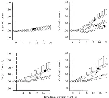
Ventilatory responses of larval bullfrogs to hypoxia are shown in Fig. 4. Inspiring hypoxic water caused a significant increase in fG in both early and middle stage larvae (Fig. 4), with response times of 10 s for the younger larvae and 11 s for the middle stage larvae. No significant change in fGoccurred within 20 s in stage XVII–XIX larvae. Twenty seconds after inspiring hypoxic water, fGwas significantly elevated to 106 % of the control value in both early and middle stage larvae.
Inspiring hypoxic water significantly increased PB in all stages of larval bullfrogs examined (Fig. 4). The significant change in PBoccurred at 14 s for early stage larvae and at 13 s for middle stage larvae. In the oldest larvae, however, a significant change in PBdid not occur until 19 s of exposure to
hypoxia. PBshowed a more pronounced response to hypoxia in stage IX–XI larvae than in younger larvae. By 20 s after inspiring hypoxic water, PB had increased by 122 % of the control value in stage V–VII larvae and by only 106 % of the control value in older larvae.
Hypoxia caused a significant increase of V˙W in all three developmental groups, the response times being 14 s for younger larvae, 15 s for the middle stage larvae and 18 s for late stage larvae. By the twentieth second of hypoxic stimulation, V˙Whad increased to 125 % of control values in larvae of both early and middle stages, and to 110 % of control values in late larvae (Fig. 4).
At 20 s of exposure to hypoxia, VS increased to 117 % of control values in early stages and 118 % in middle stages (Fig. 4). The significant change occurred at 17 s in larvae of both early and middle developmental stages. However, there was no significant change in VSof older larvae within 20 s. As in the case of exposure to hyperoxia, in all three developmental groups, V˙Wincreased the most and fG increased the least in response to hypoxic water.
Comparing the three developmental stages, the degree of reflex response of gill ventilation to hypoxia in older larvae (XVII–XIX) was reduced and the response to hypoxic stimulation was delayed.
Gill ventilatory responses to NaCN
There was no gill ventilation response to the introduction of 90
100
60 70 80
P
B
(% of control)
0 4 8 12 16 20
0 4 8 12 16 20
100
60 70 80 90
f
G
(% of control)
100
60 70 80 90
V
S
(% of control)
100
60 70 80 90
V
· W
(% of control)
0 4 8 12 16 20
0 4 8 12 16 20
[image:5.609.212.554.76.380.2]Time from stimulus onset (s) Fig. 3. Comparison of the time course of
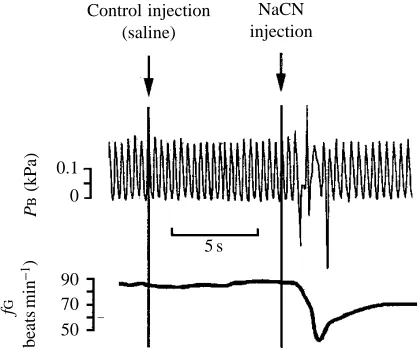
normoxic colored water as a control in any individuals from any of the developmental groups. However, a step-wise change from normoxic inspired water to water containing NaCN almost instantly caused a marked response in PB, best typified as rhythmic buccal pumping being replaced by several erratic deep gasps (Fig. 5) and, within a second or two, fG was normally seen to decrease in all three developmental groups. The initial response time of PBwas 0.8±0.3 s (mean ±S.E.M., N=28) in stage V–VII larvae, which was not significantly different from the response time of 0.6±0.2 s (N=26) in stage IX–XI larvae. The response time of 2.7±0.4 s (N=24) in stage XVII–XIX larvae, however, was significantly (P<0.05) longer than in both other stages.
Discussion
Gill ventilation responses to changes in inspired water PO∑ Our findings of an overall reduction in gill ventilation by larval bullfrogs inspiring hyperoxic water and an increase while inspiring hypoxic water in the present study indicates that O2is involved in the regulation of gill ventilation in both aquatic and bimodally breathing bullfrog larvae and confirm the findings of other authors (Burggren and West, 1982; West and Burggren, 1982; Burggren and Doyle, 1986; Infantino, 1992). The ventilatory changes in response to step-wise changes in PO∑ in the present study, which is the first to examine the time course of these events, were extremely rapid in all three groups. The ventilatory changes were also complex
and reflected clear developmental differences between the three larval groups.
In response to changes of inspired water PO∑, the reflex changes in fGand PBwere faster than the changes in V˙Wand VSin early and middle stage larvae. This response was different in older larvae, where the reflex response of PBwas slower than that of V˙W. In other words, the changes in V˙Winitially resulted from the change in fGand PBbut not from the change in VSin early and middle stage larvae, since the response time of VSwas slower than that of the other three measured variables. However, the change in V˙Wwas not directly associated with the change in PB for stage XVII–XIX larvae. The relationships among branchial pressure, buccal pumping frequency and total branchial water flow in anuran larvae are complex (Gradwell and Pasztor, 1968; Burggren and West, 1982). The pattern of change in the short-term hypoxic and hyperoxic responses of gill ventilation variables in the present study suggests that there are as yet unknown ontogenetic differences in the the reflex control of gill ventilation variables and the fluid dynamics of branchial pumping.
The reflex response to changes in PO∑of inspired water is more complex in older stage (XVII–XIX) larvae. As reported by other authors for older stage larvae, aquatic hyperoxia causes a decrease in fG, while aquatic hypoxia has no effect on fG (West and Burggren, 1982; Burggren and Doyle, 1986; Infantino, 1992). In the present study, no reflex response of fG to aquatic hypoxia in stage XVII–XIX larvae occurred within 20 s, even though PBhad changed. However, in response to
130 140
100
90 110 120
P
B
(% of control)
0 4 8 12 16 20
0 4 8 12 16 20
140
100
90 110 120 130
f
G
(% of control)
140
100
90 110 120 130
V
S
(% of control)
140
100
90 110 120 130
V
· W
(% of control)
0 4 8 12 16 20
0 4 8 12 16 20
[image:6.609.208.558.73.389.2]Time from stimulus onset (s) Fig. 4. Comparison of the time course of
reflex responses of gill ventilation to a step-wise shift from normoxia to hypoxia (at time 0, vertical broken line) in three stages of larval bullfrogs. Mean values ±
hyperoxia, fG decreased more rapidly than PB (11 s versus 20 s). These data suggest that a separate control of the frequency and pressure of buccal pumping exists in older larvae, although the mechanism is currently unknown.
When PB, V˙Wand VSresponses of the three groups at 20 s after inspiration of hyperoxic or hypoxic water are compared, the magnitude of the changes in gill ventilation as well as the time course differed between stages. In all three of these measured variables, the responses in stage XVII–XIX larvae were as great as in stage V–VII larvae (Figs 3, 4). Burggren and Doyle (1986) and Infantino (1992) found that progressive larval development is accompanied by a decline in reflex regulation of fG, since hypoxia increased fGin younger larvae up to developmental stage XIV but had no effect in older larvae. The present study shows that not only did the reflex response of fGto hyperoxia decline with development, but so too did the PB, V˙Wand VSresponses (Fig. 3).
The effects of cyanide on gill ventilation responses Cyanide exposure mimics a lack of oxygen and has been widely used in a variety of vertebrates for detecting and localizing O2-sensitive chemoreceptors (Benchetrit et al. 1977; Biscoe and Duchen, 1990; Bouverot and Leitner, 1972; Burleson and Smatresk, 1990; Denjean et al. 1991; Ishii et al. 1985; Lillo, 1980; Mulligan et al. 1981; Smatresk, 1986; Smatresk et al. 1986; Van Vliet and West, 1986; Burleson and Milsom, 1993). The extremely fast responses to externally applied NaCN suggest that the rapidly responding O2-sensitive chemoreceptors are probably monitoring PO∑ in branchial water in these larvae. The O2-sensitive chemoreceptors could be located on the gills, in the branchial chamber or in the efferent branchial vasculature.
Functional interpretation of rapid ventilatory responses The presence of peripheral O2-sensitive chemoreceptors has
been implied in fish because of their rapid responses to changes in aquatic PO∑. Irrigating the gills of rainbow trout (Oncorhynchus mykiss) with hypoxic water caused bradycardia within 30 s, and the O2-sensitive chemoreceptors mediating this response were, therefore, considered to be located on or in the gills (Smith and Jones, 1978; Daxboeck and Holeton, 1978). Compelling evidence for peripherally located chemoreceptors in gar (Lepisosteus osseus) comes from experiments in which increases in the PO∑of water flowing over the gills of artificially ventilated animals led to an immediate increase in opercular pressure (Smatresk et al. 1986).
To our knowledge, there have been no measurements of the response time of gill ventilation to step-wise changes in aquatic PO∑ or to NaCN exposure in larval amphibians. Significant changes in gill ventilation were slower in response to hypoxia than to hyperoxia in each developmental group. This may result in part from a large variation in reflexive response between animals. It may also result in part from a mix of inspired water with water already resident in the buccal, pharyngeal and branchial cavities. This mixture may elevate the PO∑ of water over the gills and could make the applied hypoxic stimulus quite small compared with the elevation in PO∑ following the inspiration of hyperoxic water. Therefore, inspiration of hyperoxic water would present a much larger signal to O2-sensitive receptors than would inspiration of hypoxic water.
Regardless of why hyperoxia more rapidly modified gill ventilation, the ventilatory response to a change of aquatic PO∑ and to NaCN was faster in early and middle stage larvae than in late stage larvae. The rapid gill ventilation responses (8 s) to changes in inspired water PO∑in early and middle larval stages strongly suggest that there is an O2-sensitive receptor, probably located on the gills or in the efferent branchial arteries immediately downstream from the gills.
In late larval stages, the ventilatory response time to both hyperoxia and hypoxia became distinctly slower, and the overall response at 20 s decreased. Similarly, the response to NaCN was muted in older larvae. This could result from a progressive decrease in the sensitivity of the the O2receptors responsible for reflexively modulating gill ventilation as larval development progresses. Alternatively, the function of peripherally located branchial receptors might be replaced by centrally located receptors that monitor blood O2 levels. Progressive degeneration of the branchial receptors would be a natural result of the progressive degeneration of the gills themselves. Internal receptors that are more remote from the gills (and therefore that would respond more slowly to changes in inspired water PO∑) could either first come ‘on line’ or could merely have their activity become relatively more prominent with the decrease in activity from peripheral receptors, either event potentially accounting for our observations. On the basis of manipulation of lung volumes and gas composition, West and Burggren (1982) proposed a population of receptors, possibly in the lungs or in a blood pathway associated with the lungs, that induce an inhibition of gill ventilation when blood PO∑is elevated by air breathing or hyperoxic water.
0.1
0
50 70 90
P
B
(kPa)
f
G
(beats
min
−
1)
5 s Control injection
(saline)
[image:7.609.68.278.74.248.2]NaCN injection
The present study has described and characterized extremely rapid changes in gill ventilation in the larva of Rana catesbeiana following a step-wise change in environmental PO∑ or exposure to NaCN and shown how these responses change during development. These data indicate a population of branchial receptors monitoring inspired water oxygen levels or possibly blood oxygen levels in the branchial circulation. In our companion study (Jia and Burggren, 1997), we use surgical ablation of gill arches and NaCN injection in the circulation to identify specific sites for O2receptors.
This study was supported by an NSF operating grant.
References
BENCHETRIT, G., ARMAND, J. AND DEJOURS, P. (1977). Ventilatory chemoreflex drive in the tortoise. Respir. Physiol. 31, 183–191. BISCOE, T. J. ANDDUCHEN, M. R. (1990). Monitoring PO∑ by the
carotid chemoreceptor. News physiol. Sci. 5, 229–233.
BOUTILIER, R. G. (1990). Control and co-ordination of gas exchange in bimodal breathers. In Advances in Comparative and Environmental Physiology, vol. 6 (ed. R. G. Boutilier), pp. 279–345. Berlin: Springer-Verlag.
BOUVEROT, P. ANDLEITNER, L. (1972). Arterial chemoreceptors in domestic fowl. Respir. Physiol. 15, 310–320.
BURGGREN, W. W. ANDDOYLE, M. (1986). Ontogeny of regulation of gill and lung ventilation in the bullfrog, Rana catesbeiana. Respir. Physiol. 66, 279–291.
BURGGREN, W. W. ANDINFANTINO, R. L., JR(1994). The respiratory transition from water to air breathing during amphibian metamorphosis. Am. Zool. 34, 238–246.
BURGGREN, W. W. ANDJUST, J. J. (1992). Developmental changes in amphibian physiological systems. In Environmental Physiology of the Amphibia (ed. M. E. Feder and W. W. Burggren), pp. 467–530. Chicago: University of Chicago Press.
BURGGREN, W. W. ANDWEST, N. H. (1982). Changing respiratory importance of gills, lungs and skin during metamorphosis in the bullfrog Rana catesbeiana. Respir. Physiol. 47, 151–164. BURLESON, M. L. ANDMILSOM, W. K. (1993). Sensory receptors in
the first gill arch of rainbow trout. Respir. Physiol. 93, 97–110. BURLESON, M. L. AND SMATRESK, N. J. (1990). Evidence for two
oxygen-sensitive chemoreceptor loci in channel catfish, Ictalurus punctatus. Physiol. Zool. 63, 208–221.
COBURN, R. F. (1989). ATP-sensing reactions and oxygen chemoreception. In Chemoreceptors and Chemoreflexes in breathing – Cellular and Molecular Aspects (ed. S. Ahiri, R. E. Forster II, R. O. Davies and A. I. Pack), pp. 184–196. London: Oxford University Press.
DAMON, R. A. AND HARVEY, W. R. (1987). Experimental Design, ANOVA and Regression. New York: Harper & Row.
DAXBOECK, C. ANDHOLETON, G. F. (1978). Oxygen receptors in the rainbow trout, Salmo gairdneri. Can. J. Zool. 56, 1254–1259. DENJEAN, A., CANET, E., PRAND, J. P., GAULTIER, C. ANDBUREAU, M.
(1991). Hypoxia-induced bronchial responsiveness in awake sheep: role of carotid chemoreceptors. Respir. Physiol. 83, 201–210. GRADWELL, N. ANDPASZTOR, V. M. (1968). Hydrostatic pressures
during normal ventilation in the bullfrog tadpole. Can. J. Zool. 46, 1169–1174.
INFANTINO, R. L. (1990). Ventilatory responses to inspired gas variation in larval bullfrogs (Abstract). Physiologist 33, A35. INFANTINO, R. L. (1992). Ontogeny of ventilatory regulation in the
bullfrog Rana catesbeiana. PhD dissertation, University of Massachusetts, Amherst, MA, USA.
ISHII, K., ISHII, K. AND KUSAKABE, T. (1985). Chemo- and baroreceptor innervation of the aortic trunk of the toad Bufo vulgaris. Respir. Physiol. 60, 365–375.
JIA, X. X. ANDBURGGREN, W. W. (1997). Developmental changes in chemoreceptive control of gill ventilation in larval bullfrogs (Rana catesbeiana). II. Sites of O2-sensitive chemoreceptors. J. exp. Biol.
200, 2237–2248.
LILLO, R. S. (1980). Localization of chemoreceptors which may cause diving bradycardia in bullfrogs. Can. J. Zool. 52, 931–936. MULLIGAN, E., LAHIRI, S. ANDSTOREY, B. T. (1981). Carotid body O2
chemoreception and mitochondrial oxidative phosphorylation. J. appl. Physiol. 51, 438–446.
SMATRESK, N. J. (1986). Ventilation and cardiac reflex responses to hypoxia and NaCN in Lepisosteus osseus, an air-breathing fish. Physiol. Zool. 59, 385–397.
SMATRESK, N. J. (1988). Control of the respiratory mode in air-breathing fishes. Can. J. Zool. 66, 144–151.
SMATRESK, N. J., BURLESON, M. L. AND AZIZI, S. Q. (1986). Chemoreflexive responses to hypoxia and NaCN in longnose gar: evidence for two chemoreceptor loci. Am. J. Physiol. 251, R116–R125.
SMITH, F. M. AND JONES, D. R. (1978). Localization of receptors causing hypoxic bradycardia in trout (Salmo gairdneri). Can. J. Zool. 66, 144–151.
TAYLOR, A. C. AND KOLLROS, J. J. (1946). Stages in the normal development of Rana pipiens larvae. Anat. Rec. 94, 7–24. VAN VLIET, B. N. AND WEST, N. H. (1986). Cardiovascular
responses to electrical stimulation of the recurrent laryngeal nerve in conscious toad (Bufo marinus). J. comp. Physiol. 156B, 363–375.
WALKER, R., GALANTE, R. J., FISHMAN, A. P. AND PACK, A. I. (1990). Effect of GABA on gill and lung ventilation in an in vitro isolated brainstem preparation in the tadpole. Physiologist 33, A35.