Angiographic Comparison
Robert P. Ochi, Pedro T. Vieco, and Cordell E. Gross
Summary: Two patients with subarachnoid hemorrhage under-went CT angiography and conventional angiography at presen-tation. In each patient, both studies were repeated after the onset of intracranial vasospasm. In both cases, CT angiograms were able to demonstrate convincingly the conventional angio-graphic findings. CT angiography may prove useful in the evalu-ation of vasospasm in patients with subarachnoid hemorrhage.
Index terms: Computed tomography, three-dimensional; Vaso-spasm
An estimated 28 000 ruptured intracranial aneurysms occur each year in North America; approximately 8500 of these patients survive to receive treatment (1). Among patients with in-tracranial aneurysms, roughly 2% per year will experience subarachnoid hemorrhage resulting from aneurysmal rupture (2). The most debili-tating complication of aneurysmal subarach-noid hemorrhage is cerebral vasospasm, ac-counting for 14% of deaths or severe disability in those treated (3).
Conventional angiography is one method of diagnosing this complication. However, the risk of performing this procedure in the critically ill patient can limit its application. Transcranial Doppler sonography is noninvasive and rapidly performed, but does not provide anatomic in-formation and is limited to a small acoustic window (4).
Computed tomographic (CT) angiography has recently been used to evaluate the vessels in the region of the circle of Willis (5–7). This technique offers the potential for a rapid, mini-mally invasive means of accurately diagnosing and monitoring this complication. We report our findings in two patients with clinical evidence of cerebral vasospasm shown by both CT angiog-raphy and conventional angiogangiog-raphy.
Case Reports Case 1
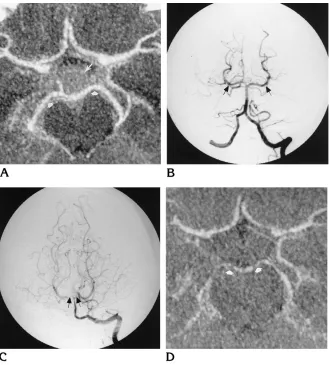
A 50-year-old man with a history of hypertension pre-sented with acute onset of a severe frontal headache. CT scans showed subarachnoid hemorrhage involving the su-prasellar and ambient cisterns, Hunt-Hess class I. A CT angiogram and a conventional angiogram (Fig 1A and B) did not reveal the source of hemorrhage. Eleven days after admission, clinical signs and symptoms of cerebral vaso-spasm were noted. Findings at transcranial Doppler sonography, performed just prior to angiography, were interpreted as normal. However, vasospasm involving the basilar artery, the posterior cerebral arteries bilaterally, and the left M1 segment of the middle cerebral artery, was identified at catheter angiography. The P1 and P2 seg-ments of the posterior cerebral arteries (Fig 1C) were most severely affected. Findings at CT angiography performed 2 days later (Fig 1D) were concordant with the above findings. Vasospasm was successfully treated with volume expansion. The patient was ultimately discharged neuro-logically intact; the source of his subarachnoid hemor-rhage was never identified.
Case 2
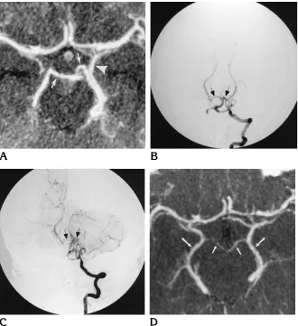
A 40-year-old woman presented with sudden onset of a posterior occipital headache. CT examination and lumbar puncture were confirmatory for subarachnoid hemor-rhage, Hunt-Hess class I. CT angiography and conven-tional angiography (Fig 2A and B) were performed. A left posteroinferior cerebellar artery aneurysm was identified at conventional angiography; surgical repair was per-formed without incident. On the 10th postoperative day, severe vasospasm of the basilar artery, P1 segments of the posterior cerebral arteries bilaterally, right M1 segment of the middle cerebral artery, and supraclinoid portion of the left internal carotid artery was shown by conventional an-giography (Fig 2C). The remainder of the vessels studied were unremarkable. CT angiography repeated 2 days later showed similar findings (Fig 2D). Of interest, findings at serial transcranial Doppler sonography performed
concor-Received December 2, 1995; accepted after revision March 22, 1996.
From the Departments of Radiology (R.P.O., P.T.V.) and Surgery (C.E.G.), University of Vermont College of Medicine, Burlington.
Address reprint requests to Pedro T. Vieco, Department of Diagnostic Imaging, Evergreen Hospital Medical Center, 12040 NE 128th St, Kirkland, WA 98034.
AJNR 18:265–269, Feb 1997 0195-6108/97/1802–0265©American Society of Neuroradiology
dantly during this period were interpreted as normal. The patient’s vasospasm was treated effectively with volume expansion, hemodilution, and inotropic support.
Both patients were studied with CT angiography and digital subtraction angiography. Each was evaluated at presentation and later at the clinical onset of vasospasm. CT angiography was performed using a Hi-speed helical scanner (General Electric Medical Systems, Milwaukee, Wis). Imaging parameters included 120 kV, 280 mA, 1 second, 1-mm collimation, 1:1 pitch, with a 12.8-cm field of view. Sixty axial source images were obtained from the foramen magnum through the circle of Willis after intrave-nous administration of contrast material (Omnipaque 300, Sanofi-Winthrop, Barcelona, Puerto Rico), delivered via an antecubital vein at 2 mL/s for a total of 100 mL. Acquisi-tion of source images was started 30 seconds after the start of contrast injection. Images were transferred to a GE Advantage Windows Workstation (General Electric Medi-cal Systems). Each data set was then reconstructed using a maximum-intensity-projection (MIP) algorithm. Image reconstruction and interpretation were performed by a neuroradiologist. Conventional digital subtraction angiog-raphy was performed via femoral catheterization using a 0.3-mm focal spot and a 1024 3 1024 matrix (Model
DFP-60A, Toshiba Corp., Kawasaki, Japan). Selective bi-lateral carotid and vertebral injections were performed in the anteroposterior and lateral projections. Magnetic res-onance (MR) angiography was not performed in either patient.
Discussion
Cerebral vasospasm is the leading cause of morbidity and mortality in patients with aneu-rysmal subarachnoid hemorrhage (1, 3). Time of onset ranges from 4 to 9 days after hemor-rhage, with a peak at the seventh day (3). From 40% to 70% of affected patients have some re-duction in caliber of one or more of the arteries of the circle of Willis (8). The diagnosis is often based on clinical findings and the exclusion of other complications such as recurrent hemor-rhage, hematoma, hydrocephalus, or metabolic derangement. Correct diagnosis of vasospasm is useful before embarking on complicated, po-tentially hazardous therapy (3). At our
institu-Fig 1. Case 1: 50-year-old man with subarachnoid hemorrhage followed by vasospasm.
A, CT angiogram (MIP display) ob-tained at presentation shows normal ves-sels of the circle of Willis. Note the nor-mal caliber of the posterior cerebral arteries bilaterally (short arrows) and the presence of recent subarachnoid hemor-rhage within the suprasellar cistern (long arrow).
B, Conventional digital subtraction angiogram, left vertebral injection, pos-teroanterior view. The basilar artery and posterior cerebral arteries (arrows) are normal.
C, Conventional digital subtraction angiogram, left vertebral injection, pos-teroanterior view, obtained after onset of vasospasm, shows the spastic appear-ance of the basilar artery and P1 seg-ments bilaterally (arrows).
[image:2.587.218.548.82.447.2]tion, treatment of these patients generally depends on the severity of vasospasm encoun-tered. Severely affected patients are treated with hemodilution, induced hypertension, and hypervolemia. Less ill patients are treated by intravascular volume-expanding agents. Diag-nosis is made clinically and confirmed predom-inantly by transcranial Doppler sonography. Catheter angiography is reserved for patients in whom the clinical and sonographic findings are discordant.
The radiologic diagnosis of this phenomenon is typically made by conventional angiography, a procedure that is not without risks. Central nervous system complications from this proce-dure are reported to be as high as 0.9% in healthy persons (9). Additionally, less serious but more common complications of angiogra-phy such as nausea, vomiting, puncture site hematoma, hypotension, arrhythmia, and heart failure may have more clinical significance in the critically ill patient.
CT angiography has recently been applied in the evaluation of the circle of Willis (5–7, 10, 11), mostly for the detection of berry aneu-rysms. CT angiography offers the ability to show three-dimensional detail of the intracra-nial vasculature in a rapid, easily reproducible fashion, and has proved useful in screening for intracranial aneurysms (5, 10), evaluating acute subarachnoid hemorrhage (6), planning surgical therapy, and assessing aneurysmal clip placement (7).
[image:3.587.50.380.83.444.2]Disadvantages of CT angiography include ra-diation exposure and contrast administration. Moreover, the images are of poorer resolution than those obtained by conventional angiogra-phy. Vessel anatomy adjacent to bone or cal-cium density, such as at the skull base, may be difficult to evaluate (6). Venous opacification may also impair evaluation of arterial anatomy; the cavernous portion of the internal carotid artery being one such example (6). Beam-hard-ening artifacts caused by dental amalgam or
Fig 2. Case 2: 40-year-old woman with subarachnoid hemorrhage followed by vasospasm.
A, CT angiogram (MIP display) ob-tained at presentation shows normal ves-sels of the circle of Willis. Note the nor-mal caliber of the P1 segments of the posterior cerebral arteries bilaterally ( ar-rows) and the basal vein of Rosenthal (arrowhead).
B, Conventional digital subtraction angiogram, left vertebral injection, pos-teroanterior view. The basilar artery and P1 segments of the posterior cerebral ar-teries (arrows) are normal.
C, Conventional digital subtraction angiogram, left vertebral injection, pos-teroanterior view, obtained after onset of vasospasm, shows the spastic appear-ance of the basilar artery and P1 seg-ments bilaterally (arrows).
other metallic objects can also degrade image quality. Postprocessing requires a finite amount of physician or technologist time to provide the most useful images, and, as with any technique, there will be interobserver variability in both image preparation and interpretation. The inter-preter must be familiar with the variant arterial and venous anatomy of the region of the circle of Willis to avoid misdiagnosis. For example, the inexperienced observer could mistake the basal veins of Rosenthal (Fig 2C) for the poste-rior cerebral arteries. In our two patients, MIP reconstructions were used instead of shaded surface displays. While there are advantages to each of these techniques (12), we chose to use the MIP images because of the diminished cal-iber of the vasospastic vessels. Shaded surface displays may inadvertently exclude small ves-sels of the circle of Willis (12).
MR angiography has been used in the diag-nosis of intracranial aneurysms (10, 13–16), and it shares the advantage of multiplanar post-processing capabilities of CT angiography. Re-ported sensitivities in diagnosing intracranial aneurysms range from 67% to 95%, with a spa-tial resolution comparable to that of CT angiog-raphy (13–15). Other advantages include lack of exposure to ionizing radiation and no require-ment for intravenous contrast administration. However, for obtunded, often mechanically ventilated, critically ill patients, MR imaging may not be ideal owing to relatively prolonged acquisition times and limited patient access. Electric and metallic devices, such as aneurys-mal clips, also preclude it routine use in the postoperative patient (17). To our knowledge, MR angiography has not been shown to be use-ful in the examination of patients with vaso-spasm after subarachnoid hemorrhage.
Transcranial Doppler sonography is often used to monitor patients during the posthemor-rhagic period (18). Advantages of this tech-nique include its rapidity and noninvasiveness, allowing frequent monitoring of the intracranial circulation. This technique, however, is limited by the relatively small area of intracranial vas-culature that is sonographically accessible (4). Additionally, it requires training and experience to perform and is inherently operator-depen-dent. Moreover, the velocities reported must be considered in clinical context. Relying purely on the assumption that lower velocities reflect wider vessel caliber ignores the possibility that elevated intracranial pressure limits the cerebral
perfusion pressure, and hence the measured velocity (19). In these patients, the possibility exists for falsely excluding vasospasm in a mor-ibund patient with elevated intracranial pres-sure. Transcranial Doppler sonography may be useful for frequent monitoring, but it does not offer the anatomic detail of CT angiography.
CT angiography provides several advantages over MR angiography for evaluating vaso-spasm. The acquisition of data (60 seconds) is much faster than with MR angiography at present (6). Motion artifacts are also reduced, and the critically ill patient is not left relatively isolated from support staff for as long a time. The vascular anatomy shown corresponds to opacification by intravenous contrast material, and is independent of slow or complex flow. The flow-dependent imaging characteristics inher-ent to MR angiography may theoretically limit the evaluation of vasospasm.
In summary, CT angiography shows promise as a rapid, minimally invasive method of eval-uating vasospasm of the intracranial circula-tion. While the use of CT angiography did not alter the management of the patients reported here, this technique may prove to be a suitable alternative to catheter angiography for the diag-nosis and treatment of patients with intracranial vasospasm.
References
1. Drake CG. Management of cerebral aneurysm.Stroke1981;12: 273–283
2. Heiserman JE, Bird CR. Cerebral aneurysms.Neuroimaging Clin N Am1994;4:799 – 822
3. Kassell NF, Sasaki T, Colohan ART, Nazar G. Cerebral vasospasm following aneurysmal subarachnoid hemorrhage.Stroke1985;16: 562–581
4. Fontaine S, Lafortune M, Lebrune LH, Couillard P. Le Doppler transcranien.Can Assoc Radiol J1991;42:389 –396
5. Alberico RA, Patel M, Casey S, Jacobs B, Maguire W, Decker R. Evaluation of the circle of Willis with three-dimensional CT an-giography in patients with suspected intracranial aneurysms. AJNR Am J Neuroradiol1995;16:1571–1578
6. Vieco PT, Shuman WP, Alsofrom GF, Gross CE. Detection of circle of Willis aneurysms in patients with acute subarachnoid hemor-rhage: a comparison of CT angiography and digital subtraction angiography.AJR Am J Roentgenol1995;165:425– 430 7. Zeman RK, Silverman PM, Vieco PT, Costello P. CT angiography.
AJR Am J Roentgenol1995;165:1079 –1088
8. Saito I, Sano K. Vasospasm after rupture: incidence, onset, and course. In: Wilkins RH, ed.Cerebral Arterial Spasm. Baltimore, Md: Williams & Wilkins; 1980:294 –301
9. Waugh JR, Sacharias N. Arteriographic complications in the DSA era.Radiology1992;182:243–246
cerebral aneurysms with helical CT: correlation with conventional angiography and MR angiography.Radiology1994;192:717–722 11. Vieco PT, Morin EE III, Gross CE. CT angiography in the exami-nation of patients with aneurysm clips.AJNR Am J Neuroradiol 1996;17:455– 457
12. Vieco PT, Zeman RK, Silverman PM, Costello P. Reply.AJR Am J Roentgenol1996;166:1228 –1229
13. Ross JS, Masaryk TJ, Modic MT, Ruggieri PM, Haacke EM, Sel-man WR. Intracranial aneurysms: evaluation by MR angiography. AJNR Am J Neuroradiol1990;11:449 – 455
14. Gouliamos A, Gotsis E, Vlahos L, et al. Magnetic resonance an-giography compared to intra-arterial digital subtraction angiogra-phy in patients with subarachnoid haemorrhage.Neuroradiology 1992;35:46 – 49
15. Ronkainen A, Puranen MI, Hernesniemi JA, et al. Intracranial
aneurysms: MR angiographic screening in 400 asymptomatic in-dividuals with increased familial risk.Radiology1995;195:35– 40 16. Schuierer G, Huk WJ, Laub G. Magnetic resonance angiography of intracranial aneurysms: comparison with intra-arterial digital subtraction angiography.Neuroradiology1992;35:50 –54 17. Klucznik RP, Carrier DA, Pyka R, Haid RW. Placement of a
ferro-magnetic intracerebral aneurysm clip in a ferro-magnetic field with a fatal outcome.Radiology1993;187:855– 856
18. Aaslid R, Huber P, Nornes H. Evaluation of cerebrovascular spasm with transcranial Doppler ultrasound.J Neurosurg1984; 60:37– 41