With 9 text-figures Printed in Great Britain
AN ELECTROPHYSIOLOGICAL STUDY OF THE
TENTACLE-REGULATING POTENTIALS IN NOCTILUCA
BY TAKAO SIBAOKA* AND ROGER ECKERT
Department of Zoology, Syracuse University, Syracuse, New York, and Marine Biological Laboratory, Woods Hole, Massachusetts
(Received 26 June 1967)
INTRODUCTION
In the preceding paper the movements of the food-gathering tentacle of the marine dinoflagellate Noctiluca mtUaris were shown to be temporally related to spontaneous potential patterns in a manner which indicates that the potential changes are involved in the control of tentacle movements (Eckert & Sibaoka, 1967). The spontaneous potentials were therefore termed tentacle-regulating potentials (TRPs) to distinguish them from the action potential which triggers light emission from organelles in the perivacuolar cytoplasm (Eckert, 1965 a, b; Eckert & Reynolds, 1967).
The basic TRP, recorded from the vacuole, consists of a positive-going pre-spike wave which reaches a peak of about —10 mV before undergoing a slow negative shift which terminates in the negative-going TR spike. A post-spike d.c. level is maintained for variable durations at — 45 to — 60 mV. The vacuolar potential alter-nates cyclically and spontaneously between the pre- and post-spike levels.
In this paper the nature and origin of the TRP are explored by examining the influence of passive and active potential changes on the potential wave pattern, by measurements of local impedance changes and of recordings of electrical potential from the surface, the vacuole and the cytoplasm. The evidence indicates that the TRP complex consists of two basic components: a pacemaker potential and a pro-longed action potential. Both originate in or near the base of the tentacle. Unlike the flash-triggering action potential, TRP activity does not propagate over the peri-vacuolar cytoplasm.
MATERIALS AND METHODS
Experiments were performed on the luminescent form of N. miliaris largely under the conditions and with the methods described in the preceding paper. Specimens in filtered natural sea water were held to the end of a suction pipette which was used also as an external electrode for impedance measurements and for potential recordings at the cell surface. Experiments were performed at ambient temperatures of 20-250 C. The TRPs were routinely recorded from the vacuole with conventional 3 M-KC1-fllled glass capillary microelectrodes and a unity-gain electrometer with anti-capaci-tance feedback.
For measurement of regional cellular impedance changes a relatively large
water-filled suction pipette (internal diameter of tip, about 100 /i) was used to hold the specimen and served as one of a pair of electrodes in the unknown arm of an a.c. bridge, the other being a calomel electrode in the sea-water bath (Fig. 5). The total impedance to 2 kHz. current in the unknown arm was determined primarily by that portion of cell surface covered with the pipette, since the ratio of its area to that of the whole cell was approximately 1 to 100. Due, perhaps, to the interposed pellicle, the leakage resistance of the pipette was low (< 1-2 x io8 £2), and so the sensitivity of the method was limited. Only recordings made with leakage resistances of 100 kfi or above were used for comparisons of regional impedance changes. The calibrated sensitivity of the a.c. bridge was such that an increase or decrease of 0-5 % in measured impedance resulted in a 1 cm. increase in width on each side of the a.c. envelope of the impedance trace.
Surface recordings from the pellicle were made with a small suction pipette (in-ternal diameter of tip, about 40 fi) which also served to hold the specimen. The pipette was filled with sea water and was connected to the amplifier through an Ag-AgCl wire. The range of frequency response (— 3 db. frequency limits) of the amplifier was from o-1-60 cyc./sec. The time delay introduced by filtering was barely detectable for the frequency components in question at the sweep rates employed. Potentials recorded externally were IR drops produced along a leakage resistance (< io5 D.) between the
pipette and the cell surface by a current which flowed across the part of the cell surface covered with the pipette. An outward current across that part therefore caused the electrode to become positive. Changes in surface-recorded potential associated with active (primarily resistive) current through the membrane under the surface electrode should show a polarity opposite to that of the intracellular recording. On the other hand, capacitative currents across the local membrane were expected to give external recordings of the same sign (Freygang, 1958; Werman, 1963).
RESULTS
Modifications of TRP duration and repetition rate
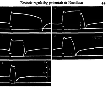
As reported previously (Eckert & Sibaoka, 1967, fig. 6D), TR spikes were elicited directly during passage of an inward current pulse provided the cell was in the pre-spike state. To examine the influence of the negative-going flash-triggering potential on the TRP the former was elicited by a 0-5 msec, inward current during different portions of the pre-spike wave of the evoked TRP (Fig. 1). Although subthreshold stimuli had no effect on the TRP, the pre-spike wave was abruptly terminated by the flash-triggering response, which, in turn, was followed by the TR spike. (Fig. iB-E). When the flash-triggering potential was evoked during the post-spike state the com-plete TRP complex (pre-spike wave followed by spike and post-spike level) followed the flash-triggering action potential just as it follows the termination of a passive, negative-going displacement evoked by an inward current pulse (Eckert & Sibaoka, 1967, fig. 6H). The influence of the flash-triggering potential wave on the TRP therefore closely resembles the effect of passive vacuolar potential deflexions due to applied current pulse. In both cases the d.c. level of the vacuole switches from the existing quasi-stable potential level to the other.
449
Fig. i. Interaction of flash-triggering action potential with the basic TRP. Upper trace: vacuolar potential recordings. Lower trace: polarizing current monitor and — 8omV. level of vacuolar potential. Outward current pulse, ioo msec duration, 5-5 x io~f A. in each case.
A, Control recording, an outward pulse from an intravacuolar polarizing electrode directly
evoked a positive deflection followed by a pre-spike wave, and TR spike. B-E, Flash-triggering action potential elicited by brief inward pulse (0-5 msec.) through the holding pipette during the pre-epike wave. Deviations in the vacuolar potential prior to the current pulse are due to spontaneous TR activity.
Fig. 2. Modification of rapid cyclic TRP pattern by applied current pulses. Upper trace: vacuolar potential recordings; calibration pulse at the beginning of each trace is iomV.
Middle trace: current trace positioned at the — 80 mV level. Lower trace: potentials recorded at
[image:3.451.44.380.421.588.2]450
during the post-spike state, spontaneous TRPs were inhibited when the flash-trigger-ing potential was elicited at 5 sec. to 20 sec. intervals.
The cyclic pattern of rapidly recurring TRP wave forms (without post-spike d.c. levels; see fig. 5 of previous paper) was modified in a graded manner by the applica-tion of outward or inward current pulses (Fig. 2). The small effect of the outward current pulse of A in driving the pre-spike wave more positively and delaying the subsequent spike was increased with stronger current in B. A similar response was obtained with the application of an inward pulse of sufficient strength to evoke a spike directly (Fig. zD). With weaker inward current the potential was displaced passively and the following spike was delayed for a period significantly longer than the duration of the current pulse (Fig. zC). In all cases a positive correlation is seen between the amplitude of the pre-spike wave in the positive direction and the peak negativity of the subsequent spike.
0
- 4 0
- 8 0 mV.
0
- 4 0
- 8 0 mV.
Fig. 3. Influence of polarizing current on spontaneous TR activity. Lower panel is a continua-tion of upper panel. Upper trace: level of current delivered with polarizing electrode in the vacuole. Lower trace: vacuolar potential recorded with second electrode. Upper panel begins 170 sec. after two electrodes were inserted into vacuole. The initial spike following recovery from electrode insertion is indicated by arrow, and occurred when the vacuolar d.c. level was — 28 mV. Toward the end of the upper panel an inward current of 2 x io~* A. was applied. At the middle of the lower panel the current was increased to 4 x io~* A. Time marks, 1 sec. apart.
level attained by the pre-spike wave. Since the ' resting' potential level is undefined in the rapidly recurring TRP wave forms, the vacuolar potential was measured as the most positive excursion of the pre-spike wave in these experiments. The positive correlation which normally exists between the maximum height of a pre-spike wave and the peak negativity of the subsequent spike is also seen in this figure.
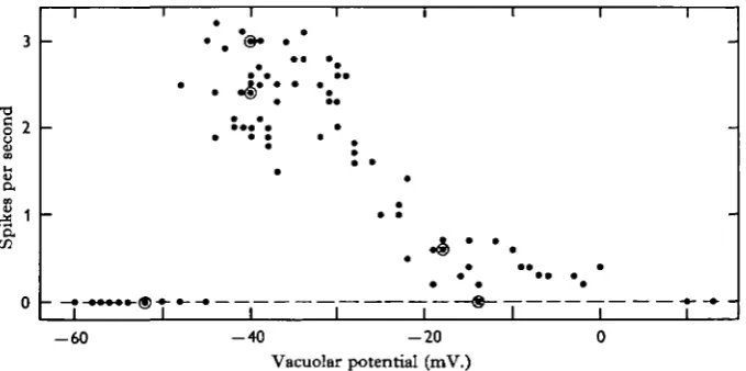
As shown in Fig. 4, there was a nearly linear relationship between spike frequency and general level of vacuolar potential between about o and — 50 mV. The spike frequency abruptly dropped to zero when the pre-spike wave became more negative than — 50 mV. This potential level corresponds to that of the post-spike negative level in the slowly recurring TRP (Eckert & Siboaka, 1967, fig. 4), and also to the potential level which is maintained nearly constant for a period of several seconds following increases in the strength of polarizing current (Fig. 3). The abolition of spontaneous TRPs by inward current was also noted by Watanabe and Hagiwara (Bullock & Horridge, 1965).
3 •o c 2 CO pe r Spike s 0 i i -• -• -• • 1 i • • • • i 1 I • 1 1 * * | I * • 1 I -1
- 6 0 - 4 0 - 2 0
[image:5.451.55.395.255.424.2]Vacuolar potential (mV.)
Fig. 4. Relationship of spike frequency to vacuolar potential. The abscissa indicates the most positive vacuolar potential attained during the TRP. The vacuolar potential was shifted by application of polarizing current through a microelectrode in the vacuole. Points plotted are from five specimens. Concentric circles indicate two coincident points.
TRPs in specimens •without tentacles
Specimens can be found in various stages of multiple fission leading to zoospore formation (Pratje, 1921). Those in the early stages of zoospore formation were nearly round and had no external tentacle structure,* whereas their cytoplasmic strands and food vacuoles remained normal in appearance. Although the tentacle was either absent or vestigial, spontaneous and evoked TRPs were readily observed, as well as normal vacuolar d.c. negativity and membrane resistance, Externally recorded poten-tial changes (see section on surface recording) could not be distinguished from those recorded from specimens having tentacles.
Similar results were obtained with ordinary specimens from which the tentacles had to a large extent been surgically removed. Both vacuolar negativity and membrane
452
resistance dropped gradually with time as zoospore formation progressed. This was associated first with a loss of spontaneity, and finally with a failure of response to current pulse stimulation.
Impedance changes during the TRP
Impedance measurements were made as indicated in Fig. 5 primarily with the intention of localizing electrically active regions. The imbalance of the bridge reached its maximum at the peak of the spike and was related in both shape and amplitude to
Fig. 5. Impedance changes associated with the spike during three TRP patterns in one specimen. Upper trace: vacuolar potential. Lower trace: bridge balance. Increments in the trace width are proportional to the recorded impedance change. Schematic of experimental set-up is shown in the bottom right: h, holding pipette having an internal tip diameter of about ico /*;
e, recording microelectrode; c, calomel electrode in sea-water bath; p, neutralized capacity
unity-gain electrometer; /, high-cut filter, a, due. amplifier used for potential recordings, b, a.c. differential amplifier used for recording bridge balance. A 2000 Hz. sine wave was applied to the bridge.
the spike. No impedance changes were detected during other portions of the TRP complex. The bridge imbalance observed was toward decreased impedance. A de-crease in impedance also occurs during the flash-triggering action potential (Chang,
i960).
453
External recordings of the TRP
Recordings from the surface were made together with those from the vacuole, as another method of relating the TRP recorded from the vacuole to activity of an excitable membrane. When the pipette electrode was located close to the base of the tentacle, an external negativity (increase in inward or decrease in outward current) was recorded during the positive deflexion of the pre-spike wave, which slowly decreased during the pre-spike wave (Fig. 7 A, B). Since the external negativity of the
Fig. 6. Impedance changes during the spike at different locations on the cell surface. Locations of holding pipette in each case, A, B, and C, are shown in diagram. A was obtained from the same specimen as Fig. 5. It was not feasible to record from more than one site on any one specimen; however, these samples are representative of numerous recordings. Note diat the leading edge of the spike in all three frames is of similar steepness, and compare with Fig. 5.
recording site was associated with an increase in vacuolar positivity, the recording site is inferred to have undergone an active conductance change. A brief decrease in inward current (or increase in outward current) was coincident with the peak of the spike, also indicative of activity at the recording site. This was followed by a marked inward current transient (Fig. jB). The similar polarities of the internal and external record-ings during the falling portion of the spike suggest active current flow in a region other than that directly under the recording electrode.
were recorded except for a small inward transient coincident with the peak of the spike. Attempts at direct recording of either internal or external potentials from the tentacle were unsuccessful.
TRPs recorded from the perinuclear cytoplasm
The internal recordings of the TRP discussed thus far were made from the vacuole, and were therefore extracytoplasmic. To avoid some of the problems of interpretation inherent in such recordings, it was desirable to record from the cytoplasm. Attempts
Fig. 7. Intracellular (upper trace) and surface (lower trace) recordings of TRPs. A sea-water-filled pipette (internal diameter of tip, about 40 ft) which held the specimen with mild suction also served as a surface recording electrode. A, B and C were obtained from one specimen; hold-ing pipette was located close to the base of the tentacle with the distal three-fourths of the tentacle in the pipette. D, a second specimen; E and F, a third specimen. The nearest inner edge of the pipette was located less than 30 degrees from the base of the tentacle in D, and 120 degrees in E and F. Calibration line in the lower right of A indicates 0-02 mV. for C, 0-05 mV. for all of the rest.
in the arm of a Wheatstone bridge as shown diagrammatically in Fig. 8. Both potential recordings were with reference to the grounded bath.
[image:9.451.60.394.190.377.2]As shown in Fig. 8, the TRPs recorded from the perinuclear cytoplasm were attenuated facsimiles of those recorded from the vacuole, and were of the same polarity. The d.c. potentials of both nuclear cytoplasm and vacuole with respect to the external medium were similar in the resting, pre-spike state (Fig. 8^4, C). In the post-spike state, however, the d.c. potential in the vacuole was markedly more negative than that in the cytoplasmic mass (Fig. 8B, prior to the stimulus). The potential deflexions
Fig. 8. Simultaneous recordings of potential changes from the vacuole (trace v) and from the perinuclear cytoplasm (trace c). Experimental arrangement shown in the bottom right. A microelectrode, e,, inserted into vacuole, was also used to apply the stimulating current pulse. Potential changes across the electrode resistance due to stimulating current flow were balanced by the bridge circuit. A simple recording electrode, ec, was inserted into the peri-nuclear cytoplasmic mass. Both microelectrodes were led into neutralized-capacity high-impedance d.c. amplifiers. er, Calomel electrode in the sea-water bath. Calibration at the beginning of each trace, 10 mV., and 20 msec. A, Passive potential changes in response to outward current passed from the vacuole through the cytoplasm to the external medium, and also the first spontaneous spike, which occurred 210 sec. after insertion of the electrodes. B, Evoked TRP in another specimen. C, Spontaneous spike in a third specimen. Ratio of spike amplitudes, v to c, were 2-6 in A, 4-1 in B, and 2-2 in C. Potential levels in millivolts negative are indicated at the beginning of each trace.
elicited by an outward current pulse passing from the vacuole through both membranes (vacuole-limiting and plasmalemma) and pellicle to the external medium showed a potential drop between the perivacuolar cytoplasm and the sea water of about one-half the magnitude of that between the vacuole and sea-water reference (Fig. 8 A). Un-certainties regarding the relative surface areas of membrane bounding the perinuclear cytoplasm, and regarding the cable properties of cytoplasmic strands which constitute much of the continuity between perinuclear and perivacuolar cytoplasms, hinder a comparison of the resting resistances of the plasmalemma and the membrane which limits the vacuole.
456
DISCUSSION
The TRP is a complex pattern generated by a cell of intricate morphology. An interpretation of the potentials recorded from the vacuole is further complicated by the extracytoplasmic location of the recording electrode. Nevertheless, the data provide a basis for some deductions and meaningful speculation regarding the nature of the TRP.
A prominent feature of potentials recorded from the vacuole is the negative-going spike. The unorthodox polarity of the spike could result from a conductance increase
A . >
E
8 0
-Vacuole recording
I
Neg.Cytoplasm (?)
Spike
Plateau
Repolarization
Pacemaker potential
Fig. 9. Proposed relationship between TRP recorded from vacuole and potential changes in the cytoplasm of the active region. A, Typical wave form of TRP recorded from vacuole with re-spect to sea-water bath. Potential levels are indicated on the ordinate. B, The same potential pattern inverted and with relative potential scale indicated. The uncertainty of the zero poten-tial level in B is indicated by the dotted lines.
in a region of the membrane which faces the vacuole. Active flow of positive current from the vacuole into the cytoplasm should result in an increased negativity of the vacuole with respect to the sea-water reference. In the same context, experimentally imposed inward current from the sea water into the vacuole is, in effect, an outward current with respect to cytoplasm for the membrane facing the vacuole. A current pulse in this direction should, according to orthodox electrogenic behaviour, be effective for stimulation (Eckert, 19656). This was seen to be the case (Eckert & Sibaoka, 1967, fig. 6D). It is therefore assumed in the present discussion that the inner membrane is active. According to this view an increase in negativity of the vacuole due to active current flow should be accompanied by a decrease in negativity of the cytoplasm of the active region.
cytoplasm of the active region are illustrated in Figure 9. The 'pre-spike wave' bears a strong similarity to certain pacemaker and pacemaker-like potentials (e.g. cardiac pacemaker cells (Hutter & Trautwein, 1955); crayfish stretch receptor (Eyzaguirre & Kuffler, 1956); cardiac ganglion cells (Bullock & Terzuolo, 1957)), while the spike and post-spike levels resemble prolonged action potentials found in certain normal (Draper & Weidmann, 1951) or pharmacologically treated (Tasaki & Hagiwara, 1957; Werman, McCann & Grundfest, 1961) cells. The TRP is proposed to consist of these two basic components, a pre-spike pacemaker potential and an action potential having a post-spike plateau of variable duration.
The analogy between the TRP of Noctiluca and repetitive activity in metazoan cells can be extended. When the potential of the vacuole grows more negative (pre-sumably, the cytoplasm of the active region more positive), either spontaneously or in response to applied current, the maximum depolarized level in the pre-spike wave is decreased, the interval between spikes becomes shorter, and the post-spike d.c. level (plateau) becomes shorter (Eckert & Sibaoka, 1967, fig. 3). A similar, but less pro-nounced shortening of the plateau occurs in ventricular action potentials when the interval between action potentials is reduced (Brady & Woodbury, i960).
The linear relationship between the spike frequency and the vacuolar potential (Fig. 4) is similar to that observed in certain metazoan neurons (e.g. eccentric cells in
Limulus, Fuortes, 1959) and NiteUa internodes (Kishimoto, 1966). This can be
ex-pressed as the increment of spikes per second for each unit of transmembrane poten-tial change or of applied current (Bullock & Horridge, 1965, p. 157). The value calculated from Fig. 4 is about 0-08 spikes per sec. per mV. hyperpolarization, or, with an effective resistance of 10 MQ (Eckert & Sibaoka, 1967), about o-8 spikes/sec./ nanoampere.
In contrast to the relationship which exists between a cardiac potential cycle and cardiac contraction, the prolonged action potential of the TRP (Fig. 9, bottom) is associated with relaxation of the tentacle rather than contraction. That is to say, during the potential plateau (' post-spike d.c. level') the tentacle assumes the extended position characteristic of low-calcium paralysis (Eckert & Sibaoka, 1967, Plate III). The data give no clue as to whether the pacemaker and action potential originate in the same area of membrane. The measurements of impedance change and the surface recordings both indicate, however, that activity is restricted in the soma to the vicinity of the base of the tentacle. Unlike the flash-triggering action potential, the TR spike does not propagate extensively through the perivacuolar cytoplasm. The external recordings of the spike, suggestive of an active-passive current sequence (Fig. 7) are most easily explained by the propagation of activity away from the record-ing site. While it would be reasonable to associate this with propagation into the tentacle, surface recordings from specimens having no external tentacle structure show the same pattern as recordings made from normal specimens.
The recordings made from the perinuclear cytoplasm are difficult to interpret. Much of the difficulty is presumably due to morphology. The perinuclear cytoplasm is connected to the perivacuolar complex by numerous fine (< 1 /i thick) strands of cytoplasm, which penetrate the floatation vacuole. Furthermore, it is unlikely that the perinuclear cytoplasm is in close electrotonic continuity with the active region, for the potentials recorded from it are of the same polarity as those recorded from the
vacuole, and of a smaller amplitude. The active region appears to be coupled directly to the vacuole, while coupled only capacitatively to the perinuclear cytoplasm via the vacuole. Consistent with this interpretation is the effectiveness of a.c. coupling between vacuole and perinuclear cytoplasm in contrast to poor d.c. coupling (Fig. 8). The relatively low resting potential recorded from the perinuclear cytoplasm need not, according to this view, be representative of the cytoplasmic resting potential in the region of activity. Cytoplasmic potential differentials and concomitant steady current flow present no conceptual difficulties since they may, in fact, be involved in pace-maker activity.
The observations presented above plus the presence of TRPs in specimens without discernible tentacles suggest that the TRPs originate in a limited portion of membrane facing the vacuole in the soma near the base of the tentacle. The relatively high resis-tance existing between vacuole and sea water limits the load imposed on the active area of membrane, 1 or 2 nanoamperes being sufficient to cause a vacuolar potential displacement equivalent to the difference between pre- and post-spike levels (Eckert & Sibaoka, 1967, fig. 6E, G).
SUMMARY
1. The duration and frequency of tentacle-regulating potentials (TRPs) recorded from the vacuole of Noctiluca miUaris were modified by both steady and abrupt changes in the vacuolar potential. The spike frequency increased linearly with an increase in the vacuolar negativity over the range of o to — 50 mV.
2. A transient decrease of impedance across the soma occurs coincidentally with the negative-going TR spike recorded from the vacuole. Local measurements of impedance changes as well as local surface potential recordings indicate that the TR spike does not propagate over the remainder of the cell from its region of origin near the base of the tentacle. Specimens without visible tentacles exhibited TRPs of normal shape.
3. Concurrent potential recordings from the vacuole and the perinuclear cytoplasm indicate that the vacuolar membrane bordering that portion of the cell is inactive. Except for a relatively more pronounced post-spike hyperpolarization the TRP shape and polarity in the perinuclear cytoplasm, referred to the sea-water bath, are similar to those recorded from the vacuole, but are of smaller amplitude.
4. The TRP appears to consist of a pre-spike pacemaker potential and an action potential having a prolonged post-spike plateau, the pattern recorded from the vacuole being the approximate inverse of that which would be recorded if a microelectrode could be introduced into the cytoplasm adjacent to the electrically active portion of membrane. Several of the findings indicate that the active membrane faces the vacuole and is electrotonically isolated from the perinuclear cytoplasm except through the vacuole.
We are grateful to the staff of the Biologische Anstalt, Helgoland, Germany, for providing the initial specimens of our Noctiluca culture, and to Dr Luigi Provosoli of Haskins Laboratories, New York City, for providing a culture of the food organism,
Dttnaliella. The excellent condition of our cultures, and hence the success of these
Woods Hole, for maintaining a subculture of Noctiluca. Finally, we are indebted to Dr Martin Mendelson for a critical reading of the manuscript. Support was provided by the National Science Foundation and the National Institutes of Health, U.S.D.H.E.W., and in part by a grant to the Marine Biological Laboratory by the Office of Naval Research.
REFERENCES
BRADY, A. J. & WOODBURY, J. W. (i960). The sodium-potassium hypothesis as the basis of electrical activity in frog ventricle. J. Physiol. 154, 385-407.
BULLOCK, T. H. & HORRIDGE, G. A. (1965). Structure and Function in the Nervous Systems of
Inverte-brates, Vol. 1. San Francisco and London: Freeman.
BULLOCK, T. H. & TERZUOLO, C. A. (1957). Diverse forms of activity in the somata of spontaneous and integrating ganglion cells. J. Physiol. 138, 341-64.
CHANG, J. J. (i960). Electrophysiological studies of a non-luminescent form of the dinoflagellate
Nocti-luca miliaris. J. Cell Comp. Physiol. 56, 33-42.
DRAPER, M. H. & WEIDMANN, S. (1951). Cardiac resting and action potentials recorded with an intra-cellular electrode. J. Physiol. 115, 74-94.
ECKKRT, R. (1965 a). Bioelectric control of bioluminescence in the dinoflagellate Noctiluca. I. Specific nature of triggering events. Science 147, 1140-2.
ECKERT, R. (1965ft). Bioelectric control of bioluminescence in the dinoflagellate Noctiluca. II. Asyn-chronous flash initiation by a propagated triggering potential. Science 147, 1143-5.
ECKERT, R. & REYNOLDS, G. T. (1967). The subcellular origin of luminescence in Noctiluca miliaris.
J. gen. Pkysiol. 50, 1429-58.
ECKERT, R. & SIBAOKA, T. (1967). Bioelectric regulation of tentacle movement in a dinoflagellate. J. exp.
Biol. 47, 433-46.
EYZAGUTRRE, C. & KUFFLER, S. W. (1956). Processes of excitation in the dendrites and in the soma of single isolated sensory nerve cells of the lobster and crayfish. J. gen. Pkysiol. 39, 87-119.
FREYGANG, W. H. Jr. (1958). An analysis of extracellular potentials from single neurons in the lateral geniculate nucleus of the cat. J. gen. Physiol. 41, 543-64.
FUORTES, M, G. F. (1959). Initiation of impulses in visual cells of Limulus. J. Physiol. 148, 14-28. HISADA, M. (1957). Membrane resting and action potentials from a protozoan, Noctiluca scintillans. J.
cell. comp. Physiol. 50, 57-71.
HuTTER, O. F. & TRAUTWEIN, W. (1955). Vagal and sympathetic effects on the pacemaker fibers in the sinus venosus of the heart. J. gen. Physiol. 39, 715—33.
KISHIMOTO, U. (1966). Repetitive action potentials in Nitella internodes. Plant Cell Physiol. 7, 547-58. PRATJE, A. (1921). Noctiluca miliaris Suriray. Beitrflge zur Morphologie, Physiologie und Cytologie. I.
Morphologie und Physiologie. Arch. Protistenk. 4a, 1-95.
TASAKI, I. & HAGIWARA, S, (1957). Demonstration of two stable states in the squid giant axon under tetraethylammonium chloride. J. gen. Physiol. 40, 859—85.
WERMAN, R. (1963). Electrical inexcitability of the frog neuromuscular syBtem. J. gen. Physiol. 46, 517-31.
WERMAN, R., MCCANN, F. V. & GRUNDFEST, H. (1961). Graded and all-or-none electrogenesis in
arthropod muscle. I. The effects of alkali-earth cations on the neuromuscular system of Romalea