Printed in Great Britain © The Company of Biologists Limited 1984
EARLY VISUAL PROCESSING IN INSECTS
BY S. R. SHAW
Psychology Department, Life Sciences Centre, Dalhousie University, Halifax, Nova Scotia, Canada B3H 4JJ
INTRODUCTION
The present account reviews some of the progress made recently towards under-standing better the processing of visual information in the peripheral visual system of insects. To limit the scope, it concentrates mainly upon the group that has been the subject of the most intensive recent work, and that is better understood from most aspects: the Diptera, or true flies. The region discussed is the retina proper and first synaptic neuropile, the lamina, and thus encompasses visual processing up to the level of third-order visual neurones. A number of reviews give either wider or fuller accounts of the same area: those of Laughlin (1980) and Jarvilehto (1984) are particularly wide-ranging, Shaw (1981) discusses the neural connections in detail, whilst Meinertzhagen & Frohlich (1983) and Meinertzhagen (1984) provide introductions to aspects of neural development. A recent NATO conference volume features this area (Ali, 1984).
Much of the recent advance in knowledge has come from detailed application of electron-microscopic (EM) methods to the anatomy of the neuropile. To illuminate the function of the microanatomical circuits, the optics and general layout of the eye are first reviewed briefly; several recent accounts of this heavily researched area may be consulted for more detail, for instance Kirschfeld (1976), Stavenga (1979), Land
(1980) and Ali (1984).
OPTICS OF THE VISUAL SYSTEM
The compound eyes of insects are unusual amongst the eyes of advanced animals, in having developed as multilens devices. In the higher Diptera, most of which are diurnal animals, the number of lenslets or facets runs from as few as 1—3 in nycteribiids, ectoparasites of bats, up to tens of thousands in larger flies. This type of multifacetted eye loses the singular advantage of the camera eye of vertebrates, cephalopods and spiders, by having a much lower visual resolution, caused by the severe effect of optical diffraction at the small lens aperture. Facet size commonly varies over the eye, but even in the specialized frontal 'foveal' zone of a large male calliphorid fly, does not exceed about 70 /im. The small lens aperture also reduces the light gathering power for radiation from a point source such as a star, but, as Kirschfeld (1976) points out, does not affect the detectability of an extended object. In some flies and other arthropods, most of the optical power of the lens system is concentrated at the front surface of each facet, due to the extreme curvature of the cornea there, which has a refractive index of 1-4—1*5. This can produce a high
magnification lens with a focal length of only 50jum (Mclntyre & Kirschfeld, 1982Ji In turn, this can provide some compensatory advantage for this kind of compound eye, which can thus form a good image even at very short object distances, of 1 mm or less (Pick, 1977), usefully allowing a small insect, for instance, to see and perhaps identify a conspecific at very short range. Another advantage is that the transparency of the cornea and the short path length allow greater penetration of short wavelength ultraviolet light, which some insects use for instance for orientation and identifying flowers.
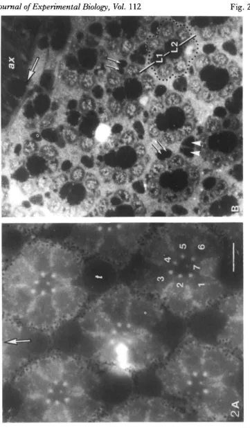
The facetted substructure of the eye leads to one of the system's main attractions for experimentalists from several disciplines. Not only do the optics repeat across the eye's surface, but the light they supply falls upon a sensory receiving structure under-neath that repeats in a like fashion (Fig. 2A). This is a cluster of photoreceptors (usually eight), glial and optical support cells called collectively the ommatidium (little eye). With certain important reservations, the ommatidium is known to repeat itself with anatomical precision across the eye. The attractive possibility arises that despite the thousands of cells even in the peripheral part of the visual system, there may be only a limited range of cell types, so that an overall understanding can be reached of what these are doing, through repeated sampling of identifiable cells. Of course other systems such as the vertebrate retina contain regularly repeating arrays of neurones, but the modular columns of cells are not so clearly distinct from one another.
The light from the lens passes through an optically transparent zone, to focus on the distal tip of the ommatidium directly beneath, on the end of a thin light conducting and absorbing rod, the rhabdom. In most insect groups, the rhabdom is made from microvilli put out by each of the photoreceptor cells, pressed together as a single fused structure running down the centre of each ommatidium. The microvilli may be thought of as a specialization to increase the surface area of the cell on which to spread a high concentration of visual pigment, at the same time increasing the pigment's exposure to the incoming light. This optical effect occurs because the high concentra-tion of membrane in a small area 1-2 pun across increases the collective refractive index of the rhabdom over the surrounding watery eye medium. Light focused on the end of such a structure conducts down inside it, trapped by its waveguide property. For the appropriate wavelengths, light is gradually and almost completely absorbed by the visual pigment that resides there. In the higher Diptera, the optics are slightly different, in that the rhabdomeres in one ommatidium are separate from one another. Since their tips lie near the back focal plane of the lens, each has a slightly different
Journal of Experimental Biology, Vol. 112
Fig. 1
Journal of Experimental Biology, Vol. 112
Fig. 2
[image:4.451.49.411.48.667.2]line of sight out through the lens from that of its neighbours, by about 1—2 degrees. The curvature of the eye serves to diverge the axes of neighbouring ommatidia through a similar amount, with the consequence that certain receptors in neighbour-ing ommatidia share a common visual axis. These cells in fact form a precise pattern, and as Kirschfeld (1967) discovered, a major function of the neural wiring is to recombine these concordant cells in the subsequent synaptic zone.
This columnar structure in the compound eye repeats throughout the deeper visual neuropiles, but the individual columns are nowhere clearer than in the first neuropile of flies, the lamina ganglionaris, or lamina. The columns or cartridges there are readily resolvable because they are surrounded and separated by a certain type of glial cell, that retains its texturally distinct appearance with the histological methods com-monly used (Figs 1, 2B).
The photoreceptor system in flies is reviewed briefly next, as a necessary prelude to describing the neuronal connections in the cartridge, that form the basis for discuss-ing what neuronal processdiscuss-ing might or might not occur at the level of the lamina. It appears that glial cells also have a prominent part to play in this.
THE RETINA AND RETINA-LAMINA CONNECTIONS
At the gross anatomical level, two types of photoreceptor can be recognized in the higher flies (the Brachycera, distinguished from the older group with a different structure of the retina, the Nematocera). Six larger, peripherally located photorecep-tors, Rl—R6, run the length of each ommatidium, become axons at the basement membrane (a basal lamina) under the eye, and make synaptic terminations a short distance below this in the cartridges of the lamina (Figs 1,2). The photoreceptors in an ommatidium can be named uniquely because their rhabdomeres are arranged in an asymmetrical fashion in all the higher flies, with that of one of the six (R3) displaced out of the basic hexagonal plan (Fig. 2). The vacant location in the hexagon is filled by the soma of the second subtype of photoreceptor, either R7 or R8. In the flies examined carefully (only the two neighbouring families, muscids and calliphorids), R7 is the longer of the two and projects its rhabdomere into the centre of the hexagon on a thin cytoplasmic stalk, to occupy the central optical axis of the ommatidium (Fig. 2). R8 is a shorter photoreceptor that lies proximal to R7 at the base of the ommatidium. Its rhabdomere lies in line behind that of R7, and is thus screened optically by absorption by R7. In other families of Brachycera, generally the more phylogenetically primitive, the contributions of R7 and R8 to the central compound rhabdomere alternate along its length (Wada, 1975). In all the Brachycera so far
examined, there is extreme conservatism of the peculiar asymmetrical arrangemer« of rhabdomeres: the same pattern illustrated in Fig. 2 for Lucilia is found throughouf the suborder (Wada, 1974, 1975). The pattern can be detected externally in the corneal pseudopupil in living flies (review: Stavenga, 1979); the pseudopupil in a variety of families confirms the invariance of the pattern (S. R. Shaw, in preparation). The eye is not completely invariant, however: the soma of R8 can occupy one of two positions (Wada, 1975).
The axons from one ommatidium grow as a group during adult development, down to a point above the presumptive lamina. At a particular time, the subgroup Rl-6 begins to diverge so that each axon grows into a different but nearby cartridge, in a highly stereotyped pattern in which there are very few errors made (Meinertzhagen, 1972). The corollary is that each cartridge receives the synaptic terminals from six photoreceptors, one each from R1-R6 from a different ommatidium. The elegant result of the particular projection pattern is that the outputs of only those photorecep-tors which have identical optical axes, are collected from six neighbouring ommatidia into a single cartridge. The cartridge is therefore the site for pooling optical input from one point in external space, through six different lenslets: the light gathering power is increased, but the visual field of the cartridge remains narrow, helping maintain the eye's relatively high acuity (Kirschfeld, 1967). A benefit to the experimenter is that the inputs to one cartridge run from different lenslets, so that the visual stimulation of individual photoreceptor axons of one cartridge is possible.
Optically, the combined rhabdomere of R7 plus R8 in a central ommatidium of the cluster also points along the same visual axis, so that in principle, a seventh facet could be added advantageously to the scheme above. In most of the eye, however, the axons of R7 and R8 run together alongside but outside the cartridge in question (Figs 1,2), to synapse in the next visual neuropile beyond the lamina, the medulla. An exception to this rule occurs in the medio-dorsal part of the eyes of male calliphorids and muscids, a region used for detection of flying females. There, R7 takes on the form and spectral sensitivity of Rl-6, and synapses as a short fibre in the appropriate cartridge (Hardie, Franceschini, Ribi & Kirschfeld, 1981; Hardie, 1983). We now also have reason to doubt the absolute separation of the two subgroups Rl-6 and R7,8, in the rest of the retina, as discussed below.
THE CONNECTIONS IN THE CARTRIDGE
The photoreceptors from the ommatidia and the neurones of a cartridge comprise a total of fifteen identified neuronal types, most of which recur in each structural subunit (Fig. 3). All of the connections between all of these neurones are believed to be known, from an anatomical standpoint. This comprises a primary resource un-matched in most other neural systems, even in invertebrates. The reader wishing to delve deeper in this area is advised initially to avoid the primary anatomical literature, which can be confusing and inconsistent. Laughlin (1980) gives a balanced account, and an earlier review of mine gives a detailed summary and Table of the connections (Shaw, 1981). The latter was written when I had little first-hand knowledge of the ultrastructure in Diptera. The present account is tempered by recent experience examining the connections in the lamina in two very similar calliphorid flies, Lucilia cuprina andL. sericata. Despite the seemingly complete description at both light- and electronmicroscope level, new connections and even a new cell type have been found, and some of the supposedly established connections now appear to be wrong. The evidence for some types of connection is stronger than for others, so that a degree of scepticism is warranted in some cases. Fig. 4 summarizes my current view of the status of the known connections.
Neuronal types in the lamina
A cartridge is made up of five monopolar cell types, L1-L5, all of which send an axon on to the next neuropile, the medulla. Three of these, LI, L2 and L3, are directly postsynaptic to photoreceptors Rl—6. So is an amacrine cell, that sends one or two of its sinuous alpha processes along the cartridge, between pairs of receptor terminals. Since there are six amacrine processes per cartridge, these are thought to come from several different amacrine neurones. The amacrine is the only neurone in the lamina that does not have an axon. One of its several branches associates closely with one of the dendritic processes of the basket fibre T l , that runs to the medulla. Monopolars LI and L2 also associate closely at the centre of the cartridge (Fig. 1), and so do L3 and L4, and R7 and R8. Perhaps these paired associations have some developmental significance, from the time that the cartridge was forming. Most of the synapses form on spines extended from the dendrites, so the close associations do not appear to have any functional significance. Monopolars L4 and L5, and basket cell T l are all third-order fibres, with no direct synaptic input from the photoreceptors. The complement of neurones is completed by no less than five efferents. From their dendritic arborizations in both the lamina and medulla (Fig. 3), efferents C2 and C3 would appear to be narrow-field units confined to one cartridge. Conversely, efferents TAN 1 and TAN 2 appear to be wide-field elements running to many cartridges (Strausfeld & Campos-Ortega, 1977). Recently, a fifth efferent TAN 3 has been described (Nassel, Hagberg & Seyan, 1983). This is a serotonergic cell that runs above the lamina and may release its transmitter non-synaptically, at varicosities.
Photoreceptor synapses
TAN 3
Lamina
TAN1
Medulla
Fig. 3. The fifteen types of neurone in the lamina of a calliphorid or muscid fly, with their termina-tions in the medulla. Based mainly on Golgi preparatermina-tions, after Fig. 38 of Strausfeld & NSssel (1980), but rearranged to match a similar composite prepared for Drosophila (K. F. Fischbach, in prepara-tion), kindly loaned by Dr Fischbach. Despite the phylogenetic distance, comparison reveals a close resemblance in most of the neurones between families, but with a few differences (K. F. Fischbach, personal communication).
• o m monopolar cell LI, and its matching partner from L2. The two remaining end positions are more varied, and are usually occupied by extensions from an alpha process of an amacrine cell. Occasionally, one end position is occupied by monopolar cell L3, or a glial process. Since the epithelial glial cells (EGC) of the lamina seem to insinuate into vacant spaces in the cartridge, their presence at this postsynaptic location does not necessarily mean that they are active participants at this synapse, though this is not ruled out.
In freeze-fracture replicas, the P-face of the terminals carries numerous characteris-tic bow-shaped arrays of parcharacteris-ticles, that were initially incorrectly equated with the 'close appositions' where receptor axons touch each other (Chi & Carlson, 1980a). A. Frohlich & I. A. Meinertzhagen (personal communication) had concluded from their own replicas from Musca that these bows were instead the presynaptic sites of the tetrads, and we were able to confirm this also for Lucilia (Shaw & Stowe, 1982a). Particularly in illuminated preparations, small depressions occur around the bow arrays, presumably either vesicle release or uptake sites, similar to those from the vertebrate neuromuscular junction; this reinforces the identification of the bow arrays as synaptic sites (Saint Marie & Carlson, 1982). So far, the bow is the only known freeze-fracture correlate of a synapse, from amongst the many types of synapse known from sections of the lamina.
All the recent estimates of the number of tetrads are uniformly high, indicating about 200 per Rl-6 terminal, or 1200 per cartridge. The number appears to be regulated developmentally (Nicol & Meinertzhagen, I982a,b; Meinertzhagen & Frohlich, 1983). It is not clear what is achieved by having multi-element synapses of this type, as opposed to simple monads, except that with certain types of rules of connection, automatic balancing of the synaptic drive on to different elements should be achieved (Meinertzhagen & Frohlich, 1983).
No clear example of a synapse from photoreceptors directly on to any other neurone has been demonstrated within the lamina. This includes the basket cell T l and its beta processes (e.g. Campos-Ortega & Strausfeld, 1973). In particular, no synapses in-volving receptors and T l have turned up in extensive serial sectioning studies from Musca cited above, or from Lucilia. The suggestion in the important review of Strausfeld & Nassel (1980), that this connection to T l is an extremely common one (up to 150 sites per cartridge), appears to be an error. There is no convincing evidence that T l is a second-order neurone.
Other synapses and synapses involving glia
Table 1. Categories and relative numbers of synapses from the lateral eye region oy Lucilia, in serial sections from two cartridges sampled from the proximal and middle
lamina (S. R. Shaw, in preparation)
Synaptic contributors
Relative number of synapses Pre Post (Wxl)»
100
24-3
10-1 6-1
4-3J
u(C3?) — >L2/1 1-2
0-5
0-2 0-3
• Relative numbers (N) of each synaptic type, weighted by the mean length of synaptic contact (/), normalized to 100 for the first category, the main afferent tetrad. Each of the other numbers in the column is expressed relative to these 100 tetrads; it is not a percentage. The sample contained 337 tetrads.
•(•These two synapses are shown linked, since they frequently occur together.
I These synapses occur only at the bottom the cartridge, and are therefore overrepresented in this sample. Other synapses such as a—* L4 occur only at the top of the cartridge, and are therefore missing here.
u, unidentified process or axon.
(Table 1), and not foreshadowed by earlier analyses of collected photographs (Boschek, 1971).
I
.verage observed length of the synapse by its observed frequency. This results in aentative 'rating' for the synaptic drive on to the glial cell, of almost 25 % of that of the tetrad. There is no reason to suspect that these synapses on to glia are not functional, and thus there is expected to be a relatively strong, somewhat delayed effect of light on EGCs: this arises because both alpha and beta processes are driven directly or indirectly by light, from photoreceptors Rl-6.The EGC might therefore be suspected of being a closet neurone, recapitulating the history of the vertebrate retina's horizontal cell, but in fact the EGC possesses no output synapses. It does behave as a glial cell in exhibiting phagocytic activity (Stark & Carlson, 1982; S. R. Shaw, unpublished data). Evidence is presented below that the EGCs control and block the extracellular route between cartridges, and could form an intracellular route for trans-lamina current, perhaps taking on some of the functions of the horizontal cell. If this is so, some means to shunt the lateral flow of current is required to explain the physiological results of Dubs (1982), that indicate a change in effectiveness of the inhibition in the lamina with change in light intensity. The numerous synapses on to the EGC might be the way this is achieved, by changing the input conductance of the EGC membrane, during light activation.
The only two other fairly common synaptic types in my sample were both predominantly feedback synapses engaging photoreceptor terminals. The more com-mon (about 10 % of the 'rating' of the tetrads) was the reciprocal synapse from alpha elements back to Rl-6. Frequently this was a monadic connection, but often it also incorporated a small spine from the beta element of T l , making a dyad. Large numbers of synaptic vesicles, and prominent postsynaptic cisternae are associated with this connection, which can extend over considerable distances, making it the most noticeable synapse in the cartridge. I suggested earlier (Shaw, 1981) that the reported difference between muscids and calliphorids in possession of this synapse might be an oversight, but this suggestion was incorrect. In fact the synapse is com-mon in calliphorids, but there is no sign of it in Musca, confirming the opinion of Campos-Ortega & Strausfeld (1973).
The other common feedback connection runs from L2 back to a receptor terminal, but incorporates 1—3 spines at its edge, usually forming a triad synapse. Its rating is about 6 % of that of the tetrad. The usually paired lateral processes are the thinnest of all those for which tracings have been attempted, and are often lost. All cases traced led to the beta process of T l . This feedback synapse is confined to the lower half of the cartridge. This confirms the account above, that T l is a third order neurone, whose major input in Lucilia comes from two second-order fibres, amacrines and L2. The synapse is also present inMusca (see Fig. IB of Nicol & Meinertzhagen, 1982a; in one of their unpublished pictures, a beta process can be seen to reach almost to the postsynaptic site before running out of the plane of section).
numerical superiority would be expected to allow them to dominate. This becomes important below, in discussing possible sources of lateral inhibition at cartridge
leveft-The evidence for neunmal connections between cartridges
Neuronal connections between cartridges have been stressed as the source of lateral inhibitory effects, for instance in the review by Strausfeld & Campos-Ortega (1977). There are only two potential neuronal routes. The first involves the proximal connections between the intercartridge extensions of third-order monopolar L4 (Figs 3, 4). The synaptic sites have been examined in serial section by Braitenberg & Debbage (1974), and reveal reciprocal synapses between processes from different L4s, along with feedback to LI and L2. The two large monopolars LI and L2 are the cells from which the best evidence has emerged for lateral inhibition (Zettler & Jarvilehto, 1972; Zettler & Autrum, 1975; Zettler & Weiler, 1976; Shaw, 1981; Dubs, 1982), and the branched L4 network is the only intercartridge pathway that is presynaptic to them. As discussed earlier (Shaw, 1981), the problem is that only a small number of L4—>Ll/2 synapses are found in each cartridge (Braitenberg & Debbage, 1974), such that this circuit is expected to be out-gunned by about 100:1, by the 1200 or so receptor tetrads that feed both LI and L2 directly. This would not matter if the L4—» L l / 2 synapses were part of a powerful high gain network, but the circumstantial evidence indicates the opposite: L4 is probably the spiking fibre recorded extracellularly by Arnett (1972), and shows a response-intensity relation of a low gain system - it is the tetrad synapse that possesses high gain characteristics (see Shaw, 1981). Given the imbalance of synaptic drive, it is difficult to imagine what the function of the input back to L l / 2 might be. Perhaps this is a sort of relict neuronal fauna, to anticipate an evolutionary argument developed at the end of this chapter. The second lateral connection is that created by the amacrine cell (Fig. 3), which extends processes amongst several cartridges, but is not presynaptic to L l / 2 directly. Any lateral inhibitory output of this network would therefore have to be expressed again via L4, driven from the proximal contacts at which alphas synapse with L4. The argument developed above against involvement of L4 therefore applies in this case also. Thus it is unlikely that either of the two lateral neural connections can explain lateral inhibition involving L l / 2 .
orominent feedback notch that would fit with the anatomy discussed above of a prominent reciprocal synapse, R ^ alpha. Because of the convergence of six photoreceptors into each cartridge, no fibre in the lamina ought to show such a limited optical projection, except perhaps a 'disconnected' alpha process, fed by synaptic contact with just a single receptor terminal. The presence of electrical coupling between R l - 6 terminals (see below) may thwart this explanation; alternatively, the slow time constant of this coupling might fail to reveal a more extended projection, if the R—> a synapse has a transient response. Another potential objection is that the amacrines are believed to synapse with one another distal to the cartridge (shown queried, in Fig. 4). This would seem to be of no use, if no significant signal were being carried by the connecting fibres. However, there is no means to distinguish an amacrine process from the others at this level, and the synapses reported there (Campos-Ortega & Strausfeld, 1973) might belong to different neurones.
The detail above can be summarized by saying that there are strong arguments against the existence of powerful lateral inhibitory pathways in the lamina, that act via neural circuits. This points to non-neural pathways as the only plausible alternative source of lateral inhibition, as discussed later in this chapter.
Efferent pathways
There is no new information on the efferents from the medulla, mentioned earlier, except for the discovery of a new serotonergic cell TAN 3 by Nassel et al. (1983); before this neurone was properly characterized, it was believed to be a new type of amacrine by Strausfeld & Nassel (1980; and D. R. Nassel, personal communication). Apart from C2, the other.efferents synapse in the upper part of the cartridge, and neither they nor this region have been properly documented with EM methods. My own cursory surveys of single sections have failed to reveal any striking populations of synapses that are not found in the proximal cartridge, that might seriously challenge the emphases in Table 1, but such impressions are unreliable, and quantification of the connections in this part of the cartridge is badly needed. In particular, I have never observed examples of what could be efferent synapses ending on the photoreceptor terminals themselves, and have therefore queried such connections in Fig. 4. The responses and functions of the efferents are completely unknown.
ELECTRICAL COUPLING IN THE PERIPHERAL VISUAL SYSTEM
There is evidence, some anatomical, some physiological, to support the idea that several cell types in the retina and lamina may be electrically coupled. So far, most of the evidence relates to implied coupling between like types of cell.
electrical coupling. Extracellular responses are usually thought to be negligible, buj one of the several unusual features of the lamina zone, noticed by all authors, is tftB extremely large size of the extracellular positive potential evoked by light (up to about 40mV, e.g. Mote, 1970).
More direct evidence of coupling comes from fibre optic stimulation of single facets, whilst recording the intracellular response of single Rl—6 photoreceptors from the retina, using differential recording to eliminate electric field contamination (Shaw,
Am(a)-*
MSA/
Tl(/S)o o o o o
o o o o o o o o
. ^ Other
o o o o o
to medulla
TAN 2 C3
L4
C2
TAN 3
[image:14.451.40.409.168.552.2]•1981, 1982, and in preparation). When external stray light is eliminated, a facet map ?an be compiled that reveals miniature interactive responses, originating in the penetrated cell from stimulation of adjacent facets, in a particular pattern (Fig. 6). In most cases, this pattern is that expected from the known projection of Rl—6 into the cartridge, and the place of the penetrated cell within the pattern allows its identity to be discovered. The miniature responses are distinguished by having a maximum that becomes progressively phase-shifted as the fibre optic stimulates cells further removed from the penetrated receptor, in the ring of terminals in the cartridge.
5mV
[image:15.451.44.409.156.577.2]50 ms
238
R7/8
Rl
R2
5mV
20 mV
R5
60
R6 20
8
o
o
o
.o'
100 ms
0 1 2 3
No. of gap junctions away from R6 Fig. 6. (A) Tracing of signal averaged responses from the soma of a photoreceptor R6, identified from the position of its own facet in the map of electrically interacting responses plotted around it, using a roving fibre optic probe (Lucilia, lateral eye region). The responses in (A) are attenuated versions of those of the corresponding terminals around R6 in its cartridges, mapped in (B). Responses Rl—R5 illustrate the progressive reduction in the size of the response and the lengthening of time-to-peak, going away from R6 in both directions around the ring of terminals (shown graphically in C). The coupled responses all show a miniature depolarizing afterpotential. Terminals nearest the recorded cell exhibit a 'notch' early on their rising phases (arrowheads), not very prominent in this recording. The largest coupled response in this map came from the R7/R8 input to this cartridge. (D) The filtering effect in (A) and (C) can be reproduced if a real photoreceptor response (first trace) is injected into an electrical model of the terminals of one cartridge (see text). The three later traces come from the three progressively more distant 'somata' of the model, and have been normalized for comparison (from S. R. Shaw, in preparation).
[image:16.451.35.426.49.457.2]lafterpotential allows the degree of d.c. coupling to be estimated. On average, the ratio •of terminal coupling resistance to the input resistance of the axon cable comes out to be about 0-24, indicating very strong coupling at terminal level (S. R. Shaw, in preparation). The coupled responses can be reproduced to a first approximation by an electrical model of the terminals, provided that the membrane time-constant of an unstimulated photoreceptor is greater than about 50 ms. A very wide range of values can be recorded for the time constant, presumably reflecting degradation of the photoreceptor input resistance by a shunt leakage caused by the microelectrode, but some values higher than 50 ms have been obtained. These experiments also reveal non-linear interactions between terminals of the same cartridge, when two inputs to it are stimulated together, that are thought to result from terminal membrane rectifi-cation. This seems to run counter to an experiment by Scholes (1969), but re-examination of the test in question (his Fig. 15) shows that there also, non-linear response reductions of up to 35 % are occurring.
The coupling at cartridge level has an anatomical correlate in the occurrence of gap junctions between the terminals in one cartridge, in Lucilia. These were originally observed as 'close appositions' by Chi & Carlson (1976), who later argued that they did not resemble gap junctions (Chi & Carlson, 1980a). Ribi (1978) produced more persuasive evidence from sections, and Shaw & Stowe (1982a) were able to correlate these structures with freeze-fracture (FF) sites that are typical of arthropod gap junctions. There are some 28 junctions per pair of adjacent terminals on average, throughout the cartridge. They are distributed somewhat asymmetrically, but all adjacent terminals share junctions (Shaw, 1984). Whilst the FF appearance is un-mistakeable, the appearance of the junctions in sections is less striking. The only really definitive feature is the presence in the cytoplasm on either side of the narrowed extracellular cleft of a darkly staining fuzz, that appears to correspond to the zone of particles seen in FF.
Despite the estimated strong coupling and the presence of gap junctions, no detect-able dye coupling was ever observed between the terminals of one cartridge, using Lucifer Yellow (Shaw & Stowe, 1982a).
A major anomaly in the facet mapping (Fig. 6) was the presence of another apparent input to the projection from the facet containing the central pair of photoreceptors, R7 and R8, that run along outside the cartridge of interest. This input was found more often than not, was sometimes the strongest indirect input to the projection (Fig. 6), and was observed in the lateral region of the eye as well as in the 'male' area, where an input from the R7/8 facet is expected (Hardie, 1983). It may be relevant that we have recently discovered a small region in the cartridge where both R7 and R8 make a large area of contact with one of the shorter photoreceptors (usually R6). In some of these cases at least, gap junctions are made with this R6, judged by the criterion of dense subjunctional material described earlier. What is not yet clear is whether these gap junctions from R7/8 are present in every cartridge, and whether they are sufficiently extensive to be able to account for the strong inputs from R7/8 sometimes observed (S. R. Shaw, I. A. Meinertzhagen, A. Frohlich & G. Chernenko, in preparation).
Electrical coupling in the retina
Calliphora revealed a double projection, suggesting that two cells in an ommatidiurn* may be coupled on occasion. Likewise, a proportion of experiments in which dye is1 injected into one photoreceptor result in up to three cells stained, both in my hands (Fig. 7) and for others (R. C. Hardie, personal communication). Counts of the quantal absorptions by individual photoreceptors (Dubs, Laughlin & Srinivasan, 1981) have produced values that are too high for the catch expected through a single facet, suggesting some sort of pooling. Perhaps related, off-axis stimulation of a photoreceptor may produce a sub-population of 'slow' single photon responses, with the peaks phase-shifted like the coupled responses described above (Dubs, 1982). Finally, receptive field maps from the fly Sarcophaga show prominent star-like exten-sions, unlike those known for other species (Mimura, 1981). All these observations point to the possibility of electrical coupling within a single ommatidium in the fly. This would certainly degrade the response of the visual system to higher spatial frequencies, but the recent analysis of Srinivasan, Laughlin & Dubs (1982) indicates that such a degradation at an extreme distal level could provide an acceptable trade-off, for an increase in signal/noise discrimination at low light levels.
The alternative explanation is that this coupling is real in the experiments, but represents some relatively frequent artefact induced by the presence of the electrode. There is no direct information on this in the fly, but in other species, horseradish peroxidase (HRP) injected into one photoreceptor can cross into the next (in fish, Kaneko, Nishimura, Tauchi & Shimai, 1981; in the crustaceansDaphnia, Schehr & Macagno, 1983 and personal communication; and Squilla, H. Susuki, personal com-munication). HRP is much too large a molecule to cross gap junction channels, indicating that the coupling must be artefactual. Corroborating this interpretation, no gap junctions have turned up connecting the photoreceptor somata in flies, despite a number of TEM and FF surveys. As discussed below, there is no reliable evidence either for the presence of extracellular barriers in the ommatidial zone, that might couple cells.
Against this evidence suggestive of artefactual coupling stands the observation of Dubs (1982), that similar phenomena pointing to coupling may be observed also at the postsynaptic level. It is difficult to see how this could be caused by the microelectrode, which did not touch the photoreceptor zone in Dubs' preparation.
Thus no firm resolution of the question of intraommatidial coupling in the fly is possible at present. There is a strong suggestion that some coupling is artefactual, but there may in addition be a real, perhaps weaker effect, that could be rationalized as advantageous in dim light. The absence of obvious junctions that could mediate this type of coupling remains an obstacle to accepting this view.
Coupling between epithelial glial cells
Fig. 7. Fluorescence micrograph showing two photorcceptors, Rl and R2, stained equally with Lucifer Yellow, following intended intraccllular injection of a single cell. Such anomalous dye-coupling always involved cells in the ommatidium that directly abutted one another. R8 interpolates between Rl and R2 at the bottom of the ommatidium, but showed no staining. The axons of the cells stained here ran to different cartridges in the lamina, that were positioned as expected. Scale bar, 10 /an.
[image:19.451.42.414.52.454.2]Gap junctions between the somata of monopolar cells?
Recently, Carlson, Saint Marie & Chi (1983) have published evidence claiming that the clustered somata of the monopolar cell group L1-L4 have gap junctions interconnecting them. If this were so, the spatial frequency response of the main output from the lamina cartridge would be expected to be seriously degraded. The supposed gap junctional sites identified in their sections are not convincing, however, not being accompanied by the subjunctional dense material that identifies the gap junctions between Rl-6 terminals. Unfortunately, as we stressed earlier (Shaw & Stowe, 1982a), many places in the cartridge exhibit what we interpret to be collapsed extracellular spaces, that may have to do with the delayed penetration of the aldehyde fixatives across the blood-brain barrier around the optic lobe. The reason for believing this interpretation is that such 'tight' areas are common in TEM within the cartridge, for instance at extensive stretches of apposition between Rl-6 and LI or L2, where we can find neither gap or tight junctions in FF replicas. We therefore think that these are not junctional sites (Shaw & Stowe, 1982a).
Chi & Carlson (l980a,b) originally labelled some of these regions as tight junctions. This also appears to be incorrect, since true tight junctions are confined to certain classes of glial cell that surround the cartridges (see below). In general, it is unwise to rely solely upon the detection of a close approach of membranes to define any kind of junction in the lamina, if there are no other distinctive features.
The evidence for gap junctions in FF replicas from the region of monopolar somata is convincing (Carlson et al. 1983), but the identification of cell type is not. The junctions illustrated by Carlson et al. could equally well be connecting the somata or processes of the satellite glial cells that inhabit the same zone, and as stated already, coupling between glia would be no surprise.
Summarizing, there is no convincing evidence yet to suggest that monopolar cells are coupled by gap junctions, although this has not been excluded.
THE CARTRIDGE ENVIRONMENT: BLOOD-BRAIN BARRIER, RESISTANCE BARRIERS
The barrier around the optic lobe
barriers to diffusion in the retina, in addition to a distal barrier to lanthanum jus| below the basement membrane. My own observations are that neither direct in viva observation with fluorescent dyes, nor subsequent histology (LM and EM), reveal any sign of lateral barriers to diffusion in the retina, and confirm that tracers pass through the basement membrane but stop a short distance proximal to this (Shaw, 1983; and in preparation).
Thus there appears to be free diffusibility of the tracers used so far in the retina and in the haemocoel, but diffusion is blocked at the entrance to the optic lobe, both from the direction of the retina, and from the blood. This might suggest two discrete barriers, one at each point of tracer arrest, but the situation appears to be more complicated than this. When tracer (ionic lanthanum) is released between the two sites, 'behind' both supposed superficial barriers, it still fails to penetrate to any extent into the lamina (Shaw, 1983). The barrier, or some component of it, therefore appears to be an extensive structure, not a simple seal of tight junctions, as classically en-visaged (Lane, 198 la). These findings stand in contrast to the report of Chi & Carlson (1981), who imply that lanthanum can penetrate both through the overlying perineurium and into the lamina, to outline parts of the epithelial glial cells. This was not observed in my preparations except at the edge of the zone of damage; damage is apparent in the relevant illustration of Chi & Carlson (1981, their Fig. 9). Open access to lanthanum is also difficult to reconcile with an extensive network of occluding tight junctions which connects EGCs, as suggested by Saint Marie & Carlson (19836), who do not cite the earlier publication (see next section).
The nature of the barrier system around the optic lobe and eye is unclear, and somewhat controversial. Shaw (1978) failed to find any tight junctions, the usual explanation for the barrier in insects, at the points at which lanthanum was arrested on its approach towards the barrier. Clear evidence for tight junctions was presented by Chi & Carlson (19806) and Lane (19816) in FF replicas from fly optic lobe. Lane (19816) maintained that these junctions explain the presence of the retina's barrier to lanthanum, but did not identify the site of the junctions. Saint Marie & Carlson (19836) deny this claim, because they find only septate junctions at the point of tracer arrest, with tight junctions deeper along the receptor axons. I re-examined the zone of photoreceptor axons regarding the latter claim, and found that the particle arrays described as tight junctions by Saint Marie & Carlson are not junctional specializa-tions at all, but appear only to be arrays of intrinsic membrane particles (Shaw, 1983, 1984).
Summarizing, all parties seem to agree that a diffusion barrier lies just proximal to the basement membrane, but there is no evidence at all that this is made up of tight junctions. These are clearly present below this region, but at some distance removed from the zone of tracer arrest, in the lamina synaptic zone (see below). There is no clear evidence as to what causes the retinal barrier, but some variety of septate junction might be suspected.
To try to clarify the discrepancy between the lack of evidence for tight junctions Connected with the barrier in the eye, and the numerous proposals in the literature of their involvement in the barrier around the nerve cord (Lane, 1981a), we recently reverted to a study of the nerve cord of the cockroach. We discovered that it is surrounded by a monolayer of previously unrecognized thin flat cells (sheath cells), some six of which are sufficient to encircle the entire cord (Shaw & Henken, 1984; D. B. Henken & S. R. Shaw, in preparation). Overlying these are numerous small perineurial cells, but contrary to an extensive literature, these are everywhere circum-vented by tracer. Encirclement by the sheath cells leaves only about six points of extracellular entry at any one level of section, which are easy to recogni2e for analysis of the nature of the barrier. In a large sample using serial sections, the tracer always stopped within these clefts, but we only ever found septate junctions there, never tight junctions. In surveying the literature, we were also unable to find any convincing tight junctions from the blood-brain interface in the many published illustrations supposed to show them, from the nerve cord. We concluded that the barrier is not absolute, and may be explained largely by the very large dimensional change as the extracellular pathway runs through the septate junctions that cross-connect the sheath cells. The change in dimensions is sufficient to predict an extremely long diffusional half-time, of the order of several days (Shaw & Henken, 1984). There is no need to postulate true occluding junctions or other special effects.
A barrier system within the lamina
Cartridge-/
Fig. 8. Idealized scheme of the lamina, after Boschek (1971), illustrating how each cartridge is surrounded and isolated by three epithelial glial cells (EGCs, stippled). The only access routes from one cartridge to the next therefore run along the extracellular clefts leading to and from R7/8, at which site three different EGCs meet. Tight junctions (TJ) along these clefts therefore cut off extracellular access from one cartridge to the next. Photocurrents can be detected travelling laterally between cartridges, and must presumably pass transcellularly through the EGCs (arrows), using the gap junctions known to connect them. The geometry of cartridge packing suggests that currents should spread more readily in the horizontal than the vertical dimension, and this is observed. IMS, an extensive internal membrane system, invaginating from the EGC's surface membrane.
involved in the compartmentalization of the lamina, inside the outermost barrier zone. This is thought to form the substrate for a novel form of neural interaction (see below).
Extracellular resistance barriers
R
as that a zone of increased extracellular resistance extended from about the level of ie basement membrane, down at least to the border of the lamina; below this, interpretation became difficult because of the presence of many blood capillaries above the lamina, that shunt part of the measuring current around the optic lobe. The distribution of high resistance suggestively parallels the anatomical evidence for barriers and compartmentalization in the fly's lamina. Unfortunately, there is no detailed anatomy of a similar kind for the locust.There is indirect evidence for a zone of increased resistance between retina and lamina in flies, from the profile of the retinal standing potential, the origin of which is still unknown (Heisenberg, 1971; Zimmerman, 1978). Plots of extracellular resist-ance have been reported for the fly by Zimmerman (1978), using a single-electrode technique. There are several problems with the interpretation of this study, stemming partly from the inappropriate assumption that the impedance seen by the electrode is cumulative with distance from the reference electrode. The original data upon which analysis should be based show only a small, gradual increase in resistance not clearly related to the position of the lamina; this could perhaps reflect the method of taking measurements immediately whilst penetrating the eye, rather than after a delay whilst withdrawing, allowing for the healing of tissue damage. A powerful barrier emerges only when the data are weighted heavily by a non-linear correction procedure, to compensate for a supposed low resistance shunt along the electrode shank. The final differentiation of this corrected result to produce the axial impedance profile (Fig. 3 of Zimmerman, 1978) is inappropriate, and is responsible for the spurious appearance of a negative resistance region.
From the anatomy and the profile of the standing potential, it would be surprising if there were not a zone of high resistance in and around the fly lamina, but clearer primary impedance data are needed before this can be accepted.
LATERAL INHIBITION AND LIGHT ADAPTATION: RECENT RESULTS
;
[image:26.451.54.403.40.523.2]AMI
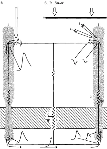
The mechanism proposed involves the passage of photocurrent from one receptor Sown its axon, into the extracellular space (ECS) near the cartridge, to influence the transmembrane potential and transmitter release of nearby, less active photo-receptors. The return circuit actually runs through the less active cell, up its axon into the soma, from which the photocurrent emerges (Shaw, 1975; Fig. 9). The appropriate transmembrane potential gradients can be detected at points along the way, except at the receptor terminals themselves. There, the extracellular position of a reference microelectrode tip is difficult to verify and the local field varies in a seemingly capricious and as yet undocumented manner.
A number of recent findings have reinforced this picture of electrical fields causing inhibition. MatiC (1983) has uncovered a similar but apparently stronger system of inhibitory interaction in the retina of a butterfly, and proposes a similar circuit to that of Fig. 9. The system in the butterfly appears to be concerned with interactions between photoreceptors having widely different spectral sensitivities (Horridge, Marcelja, Jahnke & MatiC, 1983; MatiC, 1983).
Dubs (1982) completed a wide range of tests on the lateral inhibitory system in the fly lamina, including one in which the presence of recurrent lateral inhibition was tested. The results were compatible with the field potential mechanism, but not with a neural process of recurrent inhibition, in line with the anatomical arguments presen-ted earlier here. Dubs' experiments with gratings demonstrate an asymmetrical spread of the field potential in the lamina, facilitated in the horizontal plane. The inhibitory surround of the monopolar cells shows a similar dependency. This can be shown more directly using single facet stimulation, which demonstrates the lateral asymmetrical spread of photocurrent between cartridges (S. R. Shaw, unpublished data). Since it was argued above that the extracellular route between cartridges is sealed off, the route by which this current travels must be intracellular. The anatomy of the epithelial glial barrier around the cartridge (Fig. 8) requires that the current must penetrate the EGCs and cross out into other cartridges again, to complete its return circuit to the retina. The numerous synapses on to the EGCs, identified here earlier, are therefore strategically positioned: they could affect the degree of lateral pooling of the presumed inhibitory signal, perhaps shunting the spread of current that would otherwise pass through the gap junctions between EGCs.
receptive fields of the monopolar cells fluctuate as expected with changes in ambienj light flux. Their arguments extend easily to the temporal domain, and again, corres^ ponding changes in temporal organization have been observed.
INTRACARTRIDGE FEEDBACK
The two prominent intracartridge feedback loops alpha—> R l - 6 and L2—• R l - 6 are now thought to explain a fast feedback effect that can be detected acting at the presynaptic terminals, using single facet stimulation (Shaw, 1982). The effect con-sists of a hyperpolarizing 'notch' early on the rising phase of the depolarizing response to light, that is not normally seen in response to direct illumination with an electrode in the cell soma (Fig. 6). This phenomenon was originally interpreted as another extracellular electrical interaction (Shaw, 1981), but the current knowledge of the positions of the barriers around the cartridges makes this interpretation improbable. The latency around the loop (1'5—2 ms) is compatible with a disynaptic latency, as required (e.g. R—* a—* R). The function of the synaptic feedback is not known from any other evidence, but is likely to be that of providing rapid negative feedback to the R l - 6 terminals, to produce a transient adapting output. The onset of the notch in Rl—6 corresponds in time with the down-swing of the monopolar neurone's initial transient, and may therefore be the origin of the extremely phasic response of the lamina monopolar neurones. In this interpretation, an inherently slow process of electrical presynaptic inhibition is preceded by a faster, transient phase of chemical synaptic inhibition, allowing the action to commence earlier. This provides an interesting reversal of the inhibitory sequence in the Mauthner cell, where electrical inhibition anticipates the slower chemically mediated effects.
AN EVOLUTIONARY PERSPECTIVE
Hut a few cases have been found where differing connections are made by the same •cell in different species. The most obvious is known already, the alpha—* Rl-6 synapse that is common in calliphorids but missing in muscids, two closely related families of flies.
A final speculative possibility along these lines is even more intriguing. It would seem to be a profligate strategy to develop a particular neural connection on a time scale of millions of years, only to have to abandon it completely if environmental conditions change. A way to escape this dilemma is suggested by experiments with the Falk & Fatt (1972) model of synaptic transmission, which confirm the obvious, that the relative effectiveness of a given connection can be reduced drastically by reducing the size of the synapse. Since the synapses in the lamina all appear to be modular structures of fixed area, a decrease in size could be realized merely by reducing the relative frequency of occurrence of the particular class of synapse. Thus in principle, a synaptic connection can be effectively 'turned off in this type of system, simply by controlling and reducing the number of modules, but without also having to sacrifice the basic genetic information.
Could this be the explanation of the small numbers of seemingly useless synapses mentioned earlier — relict populations saved in the genome, for a possible future awakening? Perhaps at some future date we might hope to find evidence for a dual grouping of the genetic controls for synapse formation, one set specifying the type of connection itself, and the other set that could vary independently, controlling the frequency of occurrence or size of each synaptic type. Studies of eye mutants might throw some light on these possibilities.
I am indebted to Ami Frohlich, Debbie Henken, Matti Jarvilehto and Ian Meinertz-hagen for many useful discussions on structure and function, to Dr K. Fischbach for information about Drosophila, to Garry Chernenko for expert assistance, and to NSERC (Canada) and NIH (U.S.A.) for financial support.
REFERENCES
ALI, M. (1984). Pkotoreception and Vision in Invertebrates: NATO-ASl (ed. M. AH). New York: Plenum Press. AKNETT, D. W. (1972). Spatial and temporal integration properties of units in the first optic ganglion of
dipterans. J. Neumphysiol. 35, 429-444.
BOSCHEK, C. B. (1971). On the fine structure of the peripheral retina and lamina ganglionaris of the fly, Musca
domestica. Z. Zellforsch. mikrotk. Anat. 118, 369-409.
BRAITENBESG, V. & DEBBAGE, P. (1974). A regular net of reciprocal synapses in the visual system of the fly
Musca domestica. J. comp. Physiol. 90, 25—31.
BURKHARDT, W. & BRAITENBERG, V. (1976). Some peculiar synaptic complexes in the first visual ganglion of the fly, Musca domestica. Cell Tissue Res. 173, 287-308.
CAMPOS-ORTEGA, J. A. & STRAUSFELD, N. J. (1973). Synaptic connections of intrinsic cells and basket arborizations in the external plexiform layer of the fly's eye. Brain Res. 59, 119-136.
CAJILSON, S. D., SAINT MARIE, R. L. & C H I , C. (1983). Interpretation of freeze-fracture replicas of insect nervous tissue. In Functional Neuroanatomy, (ed. N. J. Strausfeld), pp. 339—375. Berlin: Springer-Verlag.
CHI, C. & CARLSON, S. D. (1976). Close apposition of photoreceptor cell axons in the housefly. J. Insect Physiol. 22, 1153-1157.
CHI, C. & CARLSON, S. D. (1980a). Membrane specializations in the first neuropil of the housefly, Musca
domestica L. I. Junctions between neurones. J. Neurocytol. 9, 429—449.
CHI, C. & CARLSON, S. D. (19806). Membrane specializations in the first optic neuropil of the housefly, Musca
CHI, C. & CARLSON, S. D. (1981). Lanthanum and freeze fracture studies on the retinular cell junction in thjj compound eye of the housefly. Cell Tissue Res. 214, 541-552.
DUBS, A. (1982). The spatial integration of signals in the retina and lamina of the fly compound eye under different conditions of luminance. J. comp. Physiol. 146, 321-343.
DUBS, A., LAUGHLIN, S. B. & SRINIVASAN, M. V. (1981). Single photon signals in fly photoreceptors and first order interneurons at behavioural threshold. J. Physiol., Land. 317, 317-334.
FALK, G. & FATT, P. (1972). Physical changes induced by light in the rod outer segment of vertebrates. In
Handbook of Sensory Physiology, V I I / 1 , (ed. H. J. A. Dartnall), pp. 200-244. Berlin: Springer-Verlag.
FROHUCH, A. & MEINERTZHAGEN, I. A. (1982). Synaptogenesis in the first optic neuropile of the fly's visual system. J. Neurocytol. 11, 159-180.
HARDIE, R. C. (1979). Electrophysiological analysis of fly retina. I. Comparative properties of Rl—6 and R7 and 8.7. Comp. Physiol. 129, 19-23.
HARDIE, R. C. (1983). Projection and connectivity of gex-specific photoreceptors in the compound eye of the male housefly (Musca domestica). Cell Tissue Res. 233, 1—21.
HARDIE, R. C , FRANCESCHINI, N., RIBI, W. A. &KIRSCHFELD, K. (1981). Distribution and properties of
sex-specific photoreceptors in the fry Musca domestica. J. comp. Physiol. 145, 139-152.
HEISENBERG, M. (1971). Separation of receptor and lamina potentials in the electroretinogram of normal and mutant Drosophila. J. exp. Biol. 55, 85-100.
HEISENBERG, M. &BUCHNER, E. (1977). Role of retinula cell types in visual behaviour of Drosophila
melanogas-ter.J. comp. Physiol. 117, 127-163.
HORRIDGE, G. A., MARCELJA, L., JAHNKE, R. & MATIC, T . (1983). Single electrode studies on the retina of the butterfly Papilio. J. comp. Physiol. 150, 271-294.
JARVILEHTO, M. (1984). The eye: vision and perception. I n Comprehensive Insect Physiology, Biochemistry and
Pharmacology, Vol. 6, Chapter 7, (eds G. A. Kerkut & L. I. Gilbert). London: Pergamon Press (in press).
KANEKO, A., NISHIMURA, Y., TAUCHI, M. & SHIMAI, K. (1981). Morphological observation of retinal cells presumably made syncitial by an electrode penetration. J. Neurosci. Methods 4, 299-303.
KIRSCHFELD, K. (1967). Die Projection der optischen Umwelt auf das Raster der Rhabdomere im Komplexauge von Musca. Expl Brain Res. 3, 248-270.
KIRSCHFELD, K. (1976). The resolution of lens and compound eyes. In Neural Principles in Vision, (eds F. Zettler & R. Weiler), pp. 354-370. Berlin: Springer-Verlag.
KIRSCHFELD, K., FRANCESCHINI, N. SCMINKE, B. (1977). Evidence for a sensitising pigment in fly photorecep-tors. Nature, Land. 269, 386-390.
LAND, M. F. (1980). Optics and vision in invertebrates. In Handbook of Sensory Physiology, VII/6B, (ed. H. Autrum), pp. 471-592. Berlin: Springer-Verlag.
LANE, N. J. (1981a). Tight junctions in arthropods. Int. Rev. Cytol. 73, 243-318.
LANE, N. J. (19816). Vertebrate-like tight junctions in the insect eye. Expl Cell Res. 132, 482-488. LAUGHLIN, S. B. (1974). Neural integration in the first optic neuropile of dragonflies. I I . Receptor signal
interactions in the lamina. J. comp. Physiol. 92, 357—375.
LAUGHLIN, S. B. (1980). Neural principles in the visual system. lnHandbook of Sensory Physiology, VII/6B, (ed. H. Autrum), pp. 133-280. Berlin: Springer-Verlag.
LAUGHLIN, S. B. tc HARDIE, R. C. (1978). Common strategies for light adaptation in the peripheral visual systems of fly and dragonfly. J. comp. Physiol. 128, 319-340.
MCINTYRE, P. & KIRSCHFELD, K. (1982). Chromatic aberration of a dipteran corneal lens. J. comp. Physiol. 146, 493-500.
MATIC, T . (1983). Electrical inhibition in the retina of the butterfly Papilio. J. comp. Physiol. 152, 169-182. MEINERTZHAGEN, I. A. (1972). Erroneous projection of retinula axons beneath a dislocation in the retinal
equator of Calliphora. Brain Res. 41, 39-49.
MEINERTZHAGEN, I. A. (1984). The rules of synaptic assembly in the developing insect lamina. InNATO-ASI:
Pkotoreception and Vision in Invertebrates, (cd. M. Ali). New York: Plenum Press (in press).
MEINERTZHAGEN, I. A. & FROHUCH, A. (1983). The regulation of synapse formation in the fly's visual system.
Trends Neurosci. 6, 223-228.
MIMURA, K. (1981). Receptive field patterns in photoreceptors of the fly. J. comp. Physiol. 141, 349-362. MOTE, M. (1970). Focal recording of responses evoked by light in the lamina ganglionaris of the fly Sarcophaga
bullata.J. exp. Zool. 175, 149-158.
NASSEL, D. R., HAGBERG, M. & SEVAN, H. S. (1983). A new, possibly serotonergic neuron in the lamina of the blowfly optic lobe: an immunocytochemical and Golgi-EM study. Brain Res. 280, 361-367.
NICOL, D. 8c MEINERTZHAGEN, I. A. (1982a). An analysis of the number and composition of the synaptic populations formed by photoreceptors of the fly. J. comp. Neural. 207, 29—44.
NICOL, D. & MEINERTZHAGEN, I. A. (19826). Regulation of the fly photoreceptor synapses: the effects of alterations in the number of pre-synaptic cells. J . comp. Neural. 207, 45-60.
PICK, B. (1977). Specific misalignments of rhabdomere visual axes in the neural superposition eye of dipteran flies. Biol. Cybernetics TA, 215-224.
RIBI, W. A. (1978). Gap junctions coupling photoreceptor axons in the first optic ganglion of the fly. Cell Tissue
JNT MARIE, R. L. & CARLSON, S. D. (1982). Synaptic vesicle activity in stimulated and unstimulated photoreceptor axons in the housefly. A freeze-fracture study. J. Neurocytol. 11, 747—761.
SAINT MABJB, R. L. & CARLSON, S. D. (1983a). The fine structure of neuroglia in the lamina ganglionaris of the housefly, Musca domestica L.J. Neurocytol. 12, 213-241.
SAINT MARIE, R. L. & CARLSON, S. D. (19836). Glial membrane specializations and the compartmentalization of the lamina ganglionaris of the housefly compound eye. .7- Neurocytol. 12, 243-275.
SCHEHR, R. & MACAGNO, E. (1983). Possible tnchromacy in a crustacean visual system. Soc. Neurosci. Abstr. 9,325.
SCHOLES, J. H. (1969). The electrical responses of the retinal receptors and the lamina in the visual system of the fly Musca. Kybemetik 6, 149-162.
SHAW, S. R. (1972). Decremental conduction of the visual signal in barnacle lateral eye. J . Physiol., Land. 220, 145-175.
SHAW, S. R. (1975). Retinal resistance barriers and electrical lateral inhibition. Nature, Land. 255, 480-483. SHAW, S. R. (1977). Restricted diffusion and extracellular space in the insect retina. J. comp. Physiol. 113,
257-282.
SHAW, S. R. (1978). The extracellular space and blood-eye barrier in an insect retina: An ultrastructural study. Cell Tissue Res. 188,35-61.
SHAW, S. R. (1979). Signal transmission by graded slow potentials in the arthropod peripheral visual system. In The Neurosciences: Fourth Study Program, (eds F. O. Schmitt & F. G. Worden), pp. 275-295. Cambridge, Massachusetts: M I T Press.
SHAW, S. R. (1981). Anatomy and physiology of identified non-spiking cells in the photoreceptor-lamina complex of the compound eye of insects, especially Diptera. In Neurones without Impulses, (eds A. Roberts & B. M. H. Bush), pp. 61-116. Cambridge: Cambridge University Press.
SHAW, S. R. (1982). Synaptic gain control in insect photoreceptors. Soc. Neurosci. Abstr. 8, 44.
SHAW, S. R. (1983). Is the blood-brain barrier of insects just a single seal of tight junctions, a» in vertebrates?
Soc. Neurosci. Abstr. 9, 885.
SHAW, S. R. (1984). Asymmetric distribution of gap junctions amongst identified photoreceptor axons of
Lucitia cuprina (Diptera). 7. Cell Set. 66, 65-80.
SHAW, S. R. & HENKEN, D. B. (1984). The formation of the insect blood-brain: evidence from the cockroach nerve cord against the tight junction hypothesis. In Insect Neurochemistry and Neurophysiology, (eds A. B. Borkovec & T . J. Kelly), pp. 471-473. New York: Plenum Press.
SHAW, S. R. & STOWE, S. (1982a). Freeze-fracture evidence for gap junctions connecting the axon terminals of dipteran photoreceptors. J. Cell Sci. 53, 115-141.
SHAW, S. R. & STOWE, S. (19826). Photoreception in Crustacea. In The Biology of Crustacea, Vol. 3, (eds H. L. Atwood & D. C. Sandeman), pp. 291-367. New York: Academic Press.
SRINIVASAN, M., LAUCHUN, S. B. & DUBS, A. (1982). Predictive coding: a fresh view of inhibition in the retina. Proc. R. Soc. B 216, 427-459.
STARK, W. S. & CARLSON, S. D. (1982). Ultrastructural pathology of the compound eye and optic neuropiles of the retinal degeneration mutant (w rdg KS222) Drosophila melanogaster. Cell Tissue Res. 22S, 11-22. STAVENCA, D. G. (1979). Pseudopupils of compound eyes. In Handbook of Sensory Physiology, VII/6A, (ed.
H. Autrum), pp. 225—313. Berlin: Springer-Verlag.
STRAUSFELD, H. J. & CAMPOS-ORTEGA, J. A. (1977). Vision in insects: pathways possibly underlying neural adaptation and lateral inhibition. Science, N.Y. 195, 894-897.
STRAUSFELD, N. J. & NASSEL, D. R. (1980). Neuroarchitecture of brain regions that subserve the compound eyes of Crustacea and Insects. In Handbook of Sensory Physiology, VII/6B, (ed. H. Autrum), pp. 1-134. Berlin: Springer-Verlag.
TRUJILLO-CEN6Z, O. (1969). Some aspects of the structural organization of the medulla in muscoid flies.
J. Ultrastruct. Res. 27, 533-553.
VOGT, K. (1983). Is the fly visual pigment a rhodopsin? Z. Naturf. 38c, 329-333.
WADA, S. (1974). Spezielle randzonale Ommatidien der Fliegen (Diptera: Brachycera): Architektur und Verteilung in den Komplexaugen. Z. Morph. Okol. Tiere 77, 87-125.
WADA, S. (1975). Morphological duality of the retinal pattern in flies. Experientia 31, 921-923.
ZETTLER, F. & AUTRUM, H. (1975). Chromatic properties of lateral inhibition in the eye of a fly. J . comp.
Physiol. 97, 181-188.
ZETTLER, F. & JARVILEHTO, M. (1972). Lateral inhibition in an insect eye. Z. vergl. Physiol. 76, 233-244. ZETTLEK, F. & JARVILEHTO, M. (1973). Active and passive axonal propagation of nonspike signals in the retina
of Calliphora. J. comp. Physiol. 85, 89—104.
ZETTLER, F. & WEILER, R. (1976). Neuronal processing in the first optic neuropile of the compound eye of the fly. In Neural Principles in Vision, (eds F. Zettler & R. Weiler), pp. 227-237. Berlin: Springer-Verlag. ZIMMERMAN, R. P. (1978). Field potential analysis and the physiology of second-order neurones in the visual