With 3 text-figures rrinted in Great Britain
PHYSIOLOGY OF INSECT ECDYSIS
III. RELATIONSHIP BETWEEN
THE HORMONAL CONTROL OF ECLOSION AND OF TANNING IN THE TOBACCO HORNWORM, MANDUCA SEXTA
JAMES W. TRUMAN
The Biological Laboratories, Harvard University, Cambridge, Mass., 02138 U.S.A.
(Received 7 December 1972)
Ecdysis of insects involves a series of behavioural and physiological processes which are concerned with the escape of the insect from the confining exuviae and the expansion and tanning of the new cuticle. Cottrell (1962 a, b) and Fraenkel & Hsiao (1962) first reported that a blood-borne factor triggered the tanning of the cuticle of the newly emerged fly. This hormone, which was later termed bursicon (Fraenkel & Hsiao, 1965), is released after the newly emerged fly has dug its way out of the soil. In other insects bursicon secretion is more closely associated with ecdysis and occurs slightly before or during the shedding of the old skin (Cottrell, 19626; Fraenkel & Hsiao, 1965; Mills, Mathur & Guerra, 1965).
Subsequent studies on the giant silkmoth indicated that a second hormone was associated with adult emergence (Truman & Riddiford, 1970; Truman, 1971a). The eclosion hormone is released at a characteristic time of day from the brain of the pharate moth and acts on the nervous system to release the behaviour which leads to the ecdysis of the adult (Truman, 1971a; Truman & Sokolove, 1972).
Preliminary experiments by Fraenkel & Hsiao (1965) indicated that bursicon was released during eclosion of Galleria melonella moths. Thus, it was of interest to determine whether or not bursicon was secreted during adult emergence of other lepidopterans and, if so, to investigate the relationship between bursicon and the eclosion hormone.
MATERIALS AND METHODS 1. Experimental animals
The tobacco hornworms, Manduca sexta, used in these experiments were derived from insects obtained from Dr R. A. Bell, U.S.D.A., Fargo, North Dakota. Larvae were reared on an artificial diet (Yamamoto (1969), as modified by Bell, unpublished) under a 17L: 7D photoperiod regimen at 25 °C. Shortly after pupation the hornworms were transferred to a i2L:i2D regimen (photophase from 10.00 to 22.00 E.S.T.) under which they completed the remainder of their development.
822 JAMES W. TRUMAN
2. Preparation of homogenates
Parts of the nervous system were dissected from Manduca and homogenized in 20 or 40 {A of Ringer (Ephrussi & Beadle, 1936). If not assayed immediately, the tissue was rinsed in Ringer and then frozen. A few crystals of phenylthiourea were added to prevent blackening (Williams, 1959).
Blood samples were obtained by cutting off the abdomen and firmly squeezing the blood from both the thorax and the abdomen into a small dish which contained crystals of phenylthiourea. Haemolymph which was contaminated with gut contents was discarded. Samples were frozen until assayed.
3. The biological assays
Pharate Manduca in the last day of adult development were used for the bursicon assay. At this stage hornworms had characteristically digested most of the overlying pupal cuticle. Consequently, the abdomens of these animals were soft and flexible to the touch. This condition is in marked contrast to the hard, rigid covering which is present through most of adult development.
Homogenates which were to be tested for bursicon activity were injected into the thoracic tergum of the test hornworms. Routinely, assays were performed between 10.00 and 14.00 (7-11 h before the normal time of emergence which was approxi-mately 20.30 to 23.30 under this photoperiod regimen (Truman, 1971 b)). The assays were then scored 3-4 h after injection. At this time the forewings of the pharate test animals were severed at their base, coated with hot wax on the inside surface and placed in 95 % ethanol. After about 0-5 h the alcohol had hardened the wing. Removal of the wax then effectively removed the scales from the underside, thereby exposing the wing veins.
Positive responses ranged from a slight yellowing of the proximal ends of the sub-costal and radial veins to a dark yellow-brown colouring of all veins as well as part of the membranous area of the wing. Although the extent of tanning appeared to be related to dosage, the effectiveness of a homogenate was simply scored as positive or negative. The colour of the assay wings was stable for several weeks in 95 % ethanol; but after prolonged storage the wings showed a gradual nonspecific yellowing. Occasionally, a brownish-black coloration of the veins was seen in the assay wing. This appearance seemed analogous to the nonspecific injury-tanning described in Calliphora (Cottrell, 1962 a, b) and was scored as a negative assay except when a yellow colour was also present.
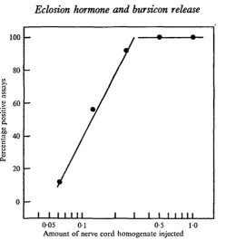
The bursicon assay was calibrated using abdominal nerve cords (including the peri-visceral organs) from pharate Manduca. Each nerve cord was homogenized and then serially diluted to yield doses which ranged down to one-sixteenth of the hormonal material contained in a pharate nerve cord. The various doses were then assayed to produce the dose-response curve shown in Fig. 1. The Manduca assay gave some positive responses at the lowest dose level tested. Doses containing only one-eighth of the material in a nerve cord yielded approximately 50 % positive scores.
Eclosion hormone assays employed pharate adults of the oak silkmoth, Antheraea pernyi. Details of the assay and of the scoring system are published elsewhere (Truman,
1973)-Eclosion hormone and bursicon release 823
[image:3.451.97.347.47.313.2]005 01 0-5 10 Amount of nerve cord homogenate injected
Fig. 1. A dose-response curve for bursicon activity using the Manduca assay. The various concentrations of bursicon were obtained by dilution of homogenates prepared from the abdominal nervous system of pharate moths. Each point represents approximately ten assays.
Table 1. Distribution of bursicon and the eclosion hormone in the nervous
system of pharate tobacco hornworm moths
Bursicon assay Eclosion hormone assay
Source of homogenate
Brain plus CC-CA complex Thoracic ganglia
Abdominal nerve cord (in-cluding perivisceral organs) Naked abdominal nerve
cord (without perivisceral organs)
Perivisceral organs
No.
1 0
8
16 13
9
% positive responses
0 0 1 0 0
2 4
88
No.
1 0 — 1 0
—
—
% positive responses
1 0 0
— 0
—
—
RESULTS
1. Distribution of eclosion hormone and of bursicon in the hornworm
nervous system
Various parts of the nervous system were dissected from pharate Manduca moths, homogenized, and tested for bursicon or eclosion hormone activity. As seen in Table i, eclosion hormone could be recovered from homogenates of the brain plus corpora cardiaca-corpora allata (CC-CA) complex. Homogenates prepared from the abdominal nerve cord yielded no eclosion hormone activity.
824 JAMES W. TRUMAN
abdominal nervous system. The brain plus CC-CA complex as well as the thoraci| ganglia consistently gave negative assays (Table 1).
In orthopteroid insects the abdominal ganglia contain neurohaemal organs which function in neurohormone storage and release (Dupont-Raabe, 1966; Brady & Maddrell, 1967). Examination of the hornworm abdominal nerve cord revealed a series of structures which were apparently homologous to the perivisceral organs (PVOs) of the Orthoptera. Each arose from a small median nerve which led from the posterior end of each ganglion, extended caudally between the ventral connectives, and then turned dorsally and penetrated the thick translucent sheath which runs the dorsal length of the nerve cord. Upon reaching the haemocoel each nerve split into two flattened branches which extended dorsally towards the heart. These branches had a faint bluish cast which is commonly associated with neurosecretory structures (Wigglesworth, 1965).
As shown in Table 1, when homogenates were prepared from only the PVOs of pharate moths hormonal activity was obtained in 8 of 9 cases. The 'naked* nerve cord (lacking the PVOs and dorsal sheath) produced positive responses in only three of thirteen assays. The abdominal PVOs therefore appear to be the principal storage site of bursicon in the pharate moth.
2. Timing of hormone release
The eclosion hormone triggers the sequence of acts of behaviour which results in the emergence of the moth from the pupal exuviae. Accordingly, the eclosion hormone has been detected in the blood of pharate moths prior to the act of emergence (Truman, 1973). It was therefore of interest to determine the time of bursicon release relative to the moment of adult eclosion.
Tobacco hornworm moths were allowed to emerge in small paper bags. Within approximately 15 min of ecdysis the moths showed the first signs of wing inflation and by about 80 min the fully spread and tanned wings were rotated at their bases and brought to lie in a tent-like fashion over the back and sides of the abdomen. Bursicon secretion was determined by assaying the level of hormonal activity present in the abdominal nerve cord at various times during this sequence.
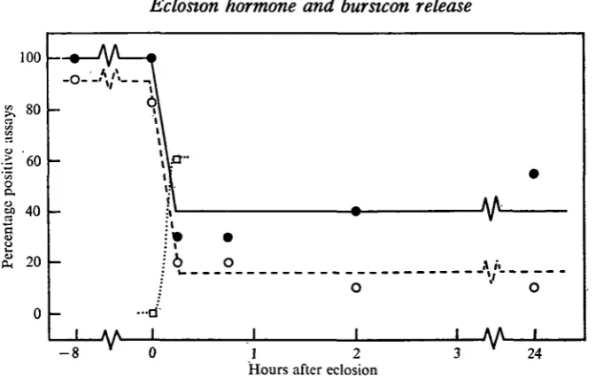
The adominal nerve cord (including the PVOs) was dissected from each moth, homogenized, and then diluted to yield doses containing one-half and one-quarter, respectively, of the material contained in the nerve cord. These homogenates were then tested in the Manduca assay. As seen in Fig. 2, at the moment of emergence the abdominal nerve cord contained an amount of bursicon which was equivalent to that seen in the pharate insect. By 15 min after eclosion, the amount of extractable hormone had dropped to a low level which represented an approximately 80% depletion of bursicon activity. The abdominal nerve cords from older moths showed a similar low level of hormone activity.
Eclosion hormone and bursicon release 825
[image:5.451.51.388.49.263.2]Hours after eclosion
Fig. 2. Changes in bursicon titre in Manduca moths during the period including adult eclosion. Newly emerged moths were immediately provided with a suitable wing-spreading site. Assays on the abdominal nervous system utilized 0-5 (•—•) or 0-25 (O — O) of the material contained in each nerve cord. Assays using the blood (D...D) were performed on 50 fil from each animal. Each point represents approximately ten assays.
of pharate PVOs had given 88% positive scores (Table 1). Thus, the abdominal PVOs also appear to be the main site of bursicon release. The depletion which was observed in the nerve cord probably represents transport of hormonal material from the ganglia into the peripheral neurohaemal organs.
The disappearance of bursicon from the abdominal nervous system coincided with the appearance of activity in the blood (Fig. 2). When blood was removed from moths at the moment of emergence and 50 fi\ were injected into each of ten pharate Manduca, no positive assays were obtained. By contrast, the same amount of blood taken from moths 15 min after eclosion gave positive responses in six of ten assays.
3. Peripheral influences upon bursicon release
(a) Effect of confining the newly emerged moth
In the blowfly, Calliphora, bursicon release and the onset of tanning can be delayed for considerable periods of time by forcing the newly emerged fly to dig through sand (Fraenkel, 1935; Cottrell, 1962a; Fraenkel & Hsiao, 1962). To determine whether similar treatment could also delay bursicon release in Manduca, newly emerged moths were transferred to shell vials. These confined animals performed intense digging movements and made no attempt to spread their wings. As seen in Fig. 3, abdominal nerve cords from moths which had been confined for up to 24 h still retained an amount of bursicon which was similar to that seen in the pharate animal. Thus, as in the blowfly, bursicon secretion can be delayed by depriving the moth of the proper environmental cues - i.e. a suitable site from which it can hang and spread its wings. When digging moths were transferred to paper bags after various durations of *«nfinement, bursicon release then occurred. Fig. 3 shows that moths which were
8z6
JAMES W. TRUMAN100
I 80
o
: ! 6 0
o
u> 40
&20
/•A S ' ~ \
A A
—1—V^-1 2 Hours after eclosion
24 25
Time of treatment*
— — -7h
-7h
- 8 h
' 1 "IH*I^ f\f t"*M"vi f\T7o 1 X 1I11C U l r C H l U V a l
of abdominal nerve cord*f
- 8 h
+ 2 h
-4h
+ I 2 h
-4h
TVT rnlar « f INUIIlDCr OI moths assayed 9 IO IO IO IO % positive A o-s of nerve cord IOO 4 0
1 0 0
3 0 4 0
scores 0-25 of nerve cord
1 0 0
1 0
1 0 0
1 0
1 0
Fig. 3. The effect of confinement in glass vials on bursicon secretion in Manduca. Circles represent tests on nerve cords from moths which had been confined in vials. Triangles indicate insects which had been released from the vials and placed in paper bags. Assay utilized 0-5 (closed symbols) or 0-25 (open symbols) of the material contained in each abdominal nervous system. The dotted line shows bursicon depletion which occurs in the nerve cord of an unrestrained moth (assayed at the 0-5 level). Each point represents approximately ten assays.
Table 2. Effect of various treatments on the secretion of bursicon
Treatment None
None
Pupal cuticle removed Pupal cuticle removed Injected with eclosion
hormone
• Time relative to the normal time of onset of adult eclosion (ca. 21.00). t Including the perivisceral organs.
transferred to bags after 1 h of' digging' released bursicon within 15 min after removal from the vials. After 24 h confinement in small vials the digging moths were generally in poor condition. Some had covered themselves with their meconium, while others had shredded their wings with their tibial spurs and had consequently lost a consider-able amount of blood. Of the ten moths tested, only three were consider-able to spread their wings fully after being placed in paper bags. Despite their poor overall condition, the moths showed substantial bursicon secretion soon after they were released from confinement.
(b) Effect of removal of the pupal cuticle from the pharate moth
Eclosion hormone and bursicon release 827 Manduca moths at 14.00 - approximately 7 h before the beginning of their eclosion gate. The peeled pharate moths were then placed in paper bags. Most of the moths suspended themselves from the sides of the bag but none showed any sign of wing spreading. Furthermore, when the amount of hormone in the nervous system at 3 h after peeling was determined (Table 2), the pharate amount of bursicon was found. Therefore, although these peeled moths had the stimuli which would normally provoke bursicon secretion, no release occurred.
Peeled moths do eventually release bursicon, but only after the arrival of their normal eclosion time. In the morning following their gate, all of 10 peeled moths showed fully spread wings. Moreover, their abdominal nervous system showed the low adult titre of bursicon (Table 2).
4. The effects of injection of eclosion hormone
The effect of eclosion-hormone injections on bursicon release was determined. Homogenates were prepared from the brain and corpora cardiaca of two pharate Manduca moths. This amount of material was then injected into the thoracic tergum of another pharate moth which was in its last day of adult development. Injections were performed between 12.00 and 13.00 (approximately 8 h before the onset of the Manduca eclosion gate).
Pharate Manduca proved to be more refractory to the eclosion hormone than are pharate Antheraea moths (Truman, 1973). Of 14 Manduca injected, only 10 emerged precociously. These moths typically underwent ecdysis within 4 h of injection, and were then immediately placed in paper bags. Each of the 10 was sacrificed 30 min after eclosion, the abdominal nervous system was removed, and the resulting homo-genate was tested for bursicon activity. As seen in Table 2, these moths showed a precocious release of bursicon. Thus, the action of the eclosion hormone in stimulating emergence is a prerequisite for the subsequent release of the tanning hormone.
DISCUSSION
In the fly Sarcophaga bullata, Fraenkel & Hsiao (1965) reported that bursicon activity was present in the thoracic ganglionic mass and, to a lesser extent, in the brain. In the ganglionic mass activity was confined to the portion which was formed from the fused abdominal ganglia. Manduca is similar in that bursicon is stored in the abdominal portion of the nervous system. But unlike the fly, the moth shows no demonstrable activity in the brain.
The principal release site for bursicon in insects also appears to be the abdominal ganglia. Mills et al. (1965) demonstrated that in the cockroach bursicon is secreted by the last abdominal ganglion. Similarly, in Manduca there is a clear loss of activity from the abdominal PVOs which is coincident with appearance of bursicon in the blood. But in Manduca I found no evidence for preferential release from the last ganglion. Although bursicon is presumably secreted from the fused ganglionic mass of the fly, Fraenkel & Hsiao (1965) were unable to demonstrate a depletion of activity from this structure during tanning. This failure was probably due to the storage of most of the hormone in peripheral neurosecretory terminals (such as described by Osborne, Finlayson & Rice, 1971) from whence it was released. These nerves would
828 JAMES W. TRUMAN
probably have been left in the carcass during a routine extirpation of the ganglionic mass.
The release of bursicon from the ganglionic mass of the fly is reportedly triggered by neural commands from the brain (Fraenkel & Hsiao, 1965). The neural aspects of the control of bursicon release in moths will be considered in a subsequent report (Truman & Endo, in preparation). Of interest to the present paper is the dependence of bursicon upon the eclosion hormone. In causing emergence the eclosion hormone triggers a sequence of behavioural acts which start with the pre-eclosion behaviour and which terminate with the spreading of the wings (Truman, 19710). Besides neural events which give rise to the overt acts of behaviour, the present study shows that a neurosecretory event - the release of bursicon - is also a part of this program. Thus a neurosecretory hormone from the brain acts directly on another portion of the nervous system (the chain of abdominal ganglia) to promote the release of a second neurosecretory hormone.
Under a 12I/12D regimen in conditions where suitable wing-spreading sites are readily available, bursicon secretion is confined to a portion of the day which encom-passes the lights-off signal (Truman, 19716). This apparent gating of bursicon release is not due to an independent timing of this secretory event but rather to the gated release of the eclosion hormone which, in turn, triggers bursicon secretion.
The data given above show that in the moth, as in the fly (Cottrell, 1962 a; Fraenkel & Hsiao, 1962), the secretion of bursicon can be delayed by forcing the newly emerged insect to dig. The neuroendocrine centres can apparently be inhibited by certain types of peripheral input. When this inhibition is finally removed, bursicon is swiftly released. Thus the secretion of bursicon in Manduca is controlled by an interaction between neural and neuroendocrine components. The eclosion hormone triggers a series of neural events, one of which is the secretion of bursicon. This neurohormone release can then be delayed by neural input associated with the lack of a proper wing-spreading site.
SUMMARY
1. In pharate Manduca sexta moths eclosion hormone activity was present in the brain and corpora cardiaca. Bursicon activity was confined to the abdominal nervous system, and was most concentrated in the abdominal perivisceral organs (PVOs).
2. When newly emerged moths were given access to suitable wing-spreading sites, bursicon activity was depleted from the PVOs and appeared in the blood within 15 min after eclosion. This hormone was responsible for the tanning and hardening of the wings.
3. Bursicon release could be delayed for at least 24 h by forcing the newly emerged moth to dig. Secretion then occurred swiftly upon giving the moth a suitable wing-spreading site.
4. The pupal cuticle was removed from pharate Manduca approximately 7 h before their normal eclosion gate, and the peeled moths were provided with a wing-spreading site. These moths did not then secrete bursicon until after their normal time of eclosion.
Eclosion hormone and bursicon release 829 6. It was concluded that bursicon release is regulated by both neural and hormonal factors. The eclosion hormone triggers a program of neural output which includes the secretion of bursicon. This release, however, can be delayed by neural input which is associated with the digging behaviour of the moth.
I thank Professor L. M. Riddiford for helpful comments during this investigation and for a critical reading of the manuscript. This work was supported by a grant from the Rockefeller Foundation and by the Harvard University Society of Fellows.
REFERENCES
BRADY, J. & MADDRELL, S. H. P. (1967). Neurohaemal organs in the median nervous system of insect.
Z. Zellforsch. 76, 389-404.
COTTRELL, C. B. (1962a). The imaginal ecdysis of blowflies. The control of cuticular hardening and darkening. J. exp. Biol. 39, 395-411.
COTTRELL, C. B. (19626). The imaginal ecdysis of blowflies. Detection of the blood-borne darkening factor and determination of some of its properties. J. exp. Biol. 39, 412—30.
DUPONT-RAABE, M. (1966). Etude des phenomenes de neurosecretion au niveau de la chaine nerveuse ventrale des phasmids. Bull. Soc. zool. FT. 90, 631-54.
EPHRUSSI, B. & BEADLE, G. W. (1936). A technique of transplantation for Drosophila. Am. Nat. 70,
218-25.
FRAENKEL, G. (1935). Observations and experiments on the blowfly (Calliphora erythrocephala) during the first day after emergence. Proc. zool. Soc. Lond. 87, 893-904.
FRAENKEL, G. & HSIAO, C. (1962). Hormonal and nervous control of tanning in the fly. Science 138, 27-9. FRAENKEL, G. & HSIAO, C. (1965). Bursicon, a hormone which mediates tanning of the cuticle in the
adult fly and other insects. J. Insect Physiol. u , 513-56.
MILLS, R. R., MATHUR, R. B. & GUERRA, A. A. (1965). Studies on the hormonal control of tanning in the American cockroach. I. Release of an activation factor from the terminal abdominal ganglion.
J. Insect Physiol. 11, 1047-53.
OSBORNE, M. P., FINLAYSON, L. H. & RICE, M. J. (1971). Neurosecretory endings associated with striated muscles in three insects (Schistocerca, Carausius, and Phormia) and a frog (Rand). Z. Zellforsch. n 6 , 391-404.
TRUMAN, J. W. (1971a). Physiology of insect ecdysis. I. The eclosion behaviour of silkmoths and its hormonal control. J. exp. Biol. 54, 805—14.
TRUMAN, J. W. (19716). Circadian rhythms and physiology with special reference to neuroendocrine processes in insects. In Proceedings of the International Symposium on Circadian Rhythmidty, pp. 111-35. Wageningen, Netherlands: Pudoc Press.
TRUMAN, J. W. (1973). Physiology of insect ecdysis. II. The assay and occurrence of the eclosion hor-mone in the Chinese oak silkmoth, Antheraea pernyi. Biol. Bull. mar. biol. Lab., Woods Hole (in the Press).
TRUMAN, J. W. & RIDDIFORD, L.M. (1970). Neuroendocrine control of ecdysis in silkmoths. Science 167, 1624-6.
TRUMAN, J. W. & SOKOLOVE, P. G. (1972). Silkmoth eclosion: hormonal triggering of a centrally pro-grammed pattern of behavior. Science 175, 1491-3.
WIGGLESWORTH, V. B. (1965). The Principles of Insect Physiology, 6th ed. London: Methuen and Co. Ltd.
WILLIAMS, C. M. (1959). The juvenile hormone. I. Endocrine activity of the corpora allata of the adult Cecropia silkworm. Biol. Bull. mar. biol. Lab., Woods Hole 116, 323-38.
YAMAMOTO, R. T. (1969). Mass rearing of the tobacco hornworm. II. Larval rearing and pupation.