Journal : EXBOTJ
Article Doi : 10.1093/jxb/erw410
Article Title : Theoretical approaches to understanding root vascular patterning: a consensus between recent models
INSTRUCTIONS
1. Author groups: Please check that all names have been spelled correctly and appear in the correct order. Please also check that all initials are present. Please check that the author surnames (family name) have been correctly identified by a pink background. If this is incorrect, please identify the full surname of the relevant authors. Occasionally, the distinction between surnames and forenames can be ambiguous, and this is to ensure that the authors’ full surnames and forenames are tagged correctly, for accurate indexing online. Please also check all author affiliations. 2. Figures: If applicable figures have been placed as close as possible to their first citation. Please check that they are complete and that the cor-rect figure legend is present. Figures in the proof are low resolution versions that will be replaced with high resolution versions when the journal is printed.
3. Missing elements: Please check that the text is complete and that all figures, tables and their legends are included.
4. Special characters: Please check that special characters, equations, dosages and units, if applicable, have been reproduced accurately. 5. URLs: Please check that all web addresses cited in the text, footnotes and reference list are up-to-date, and please provide a ‘last accessed’
AUTHOR QUERY FORM
Journal : EXBOTJ
Article Doi : 10.1093/jxb/erw410
Article Title : Theoretical approaches to understanding root vascular patterning: a consensus between recent models
First Author : Nathan Mellor
Corr. Author : Ari Pekka Mähönen, Dolf Weijers, Anthony Bishopp
AUTHOR QUERIES - TO BE ANSWERED BY THE CORRESPONDING AUTHOR
The following queries have arisen during the typesetting of your manuscript. Please click on each query number and respond by indicat-ing the change required within the text of the article. If no change is needed please add a note sayindicat-ing “No change.”
1.5
1.10
1.15
1.20
1.25
1.30
1.35
1.40
1.45
1.50
1.55
1.60
1.65
1.70
1.75
1.80
1.85
1.90
1.95
1.100
1.105
1.110
Journal of Experimental Botany
doi:10.1093/jxb/erw410
OPINION PAPER
Theoretical approaches to understanding root vascular
patterning: a consensus between recent models
Nathan Mellor1,*, Milad Adibi2,*, Sedeer El-Showk3,4, Bert de Rybel5,6,7, John King1,8, Ari Pekka Mähönen3,†,
Dolf Weijers5,† and Anthony Bishopp1,†
1 Centre for Plant Integrative Biology, University of Nottingham, Sutton Bonington Campus, Loughborough LE12 5RD, UK
2 Department of Comparative Development and Genetics, Max Planck Institute for Plant Breeding Research, D-50829 Cologne, Germany 3 Institute of Biotechnology, University of Helsinki, Helsinki FIN-00014, Finland
4 Department of Biosciences, Viikki Plant Science Centre, University of Helsinki, Helsinki FIN-00014, Finland 5 Laboratory of Biochemistry, Wageningen University, Stippeneng 4, 6708WE Wageningen, The Netherlands 6 Department of Plant Systems Biology, VIB, Technologiepark 927, B-9052, Ghent, Belgium
7 Department of Plant Biotechnology and Bioinformatics, VIB, Technologiepark 927, B-9052, Ghent, Belgium 8 School of Mathematical Sciences, University of Nottingham, Nottingham NG7 2RD, UK
*These authors contributed equally to this work
† Correspondence: [email protected], [email protected], or [email protected]
Received 16 August 2016; Accepted 11 October 2016
Editor: Peter Etchells, Durham University
Abstract
The root vascular tissues provide an excellent system for studying organ patterning, as the specification of these tissues signals a transition from radial symmetry to bisymmetric patterns. The patterning process is controlled by the com-bined action of hormonal signaling/transport pathways, transcription factors, and miRNA that operate through a series of non-linear pathways to drive pattern formation collectively. With the discovery of multiple components and feedback loops controlling patterning, it has become increasingly difficult to understand how these interactions act in unison to determine pattern formation in multicellular tissues. Three independent mathematical models of root vascular patterning have been formulated in the last few years, providing an excellent example of how theoretical approaches can comple-ment expericomple-mental studies to provide new insights into complex systems. In many aspects these models support each other; however, each study also provides its own novel findings and unique viewpoints. Here we reconcile these models by identifying the commonalities and exploring the differences between them by testing how transferable findings are between models. New simulations herein support the hypothesis that an asymmetry in auxin input can direct the forma-tion of vascular pattern. We show that the xylem axis can act as a sole source of cytokinin and specify the correct pattern, but also that broader patterns of cytokinin production are also able to pattern the root. By comparing the three modeling approaches, we gain further insight into vascular patterning and identify several key areas for experimental investigation.
Key words: Auxin, cytokinin, mathematical modeling, organ patterning, systems biology, vascular development.
Introduction
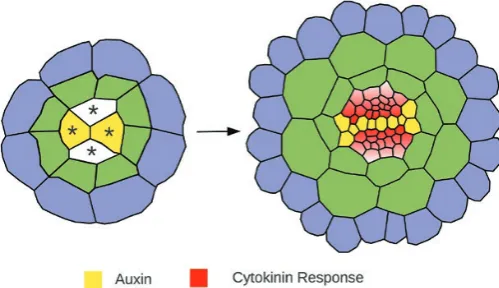
Over the last few years there has been considerable insight into the molecular mechanisms controlling the specification of the root vascular pattern. In Arabidopsis embryos, the
vascular cylinder forms from a group of four provascular ini-tial cells (Scheres et al., 1994) (Fig. 1). As the embryo devel-ops, these provascular initials proliferate through a sequence
Copyedited by: OUP
2.5
2.10
2.15
2.20
2.25
2.30
2.35
2.40
2.45
2.50
2.55
2.58
2.60
2.65
2.70
2.75
2.80
2.85
2.90
2.95
2.100
2.105
2.110
2.115 2.116 Page 2 of 12 | Mellor et al.
of highly regulated cell divisions to produce a vascular cylin-der of ~40 cells by the time the seed germinates. In addition to cell proliferation, cell specification is critical to establish the xylem and phloem cell lineages. These tissues go on to form the main conduits for long-distance transport of water, nutrients, and signaling molecules within the plant. As the xylem and phloem initials differentiate, a bisymmetric pat-tern becomes apparent, and this is defined by a central axis of xylem cells flanked by two domains of pluripotent procam-bial cells and two phloem poles.
Experimental studies have shown that the two hormones, auxin and cytokinin, are essential in mediating both the cell proliferation and specification processes. The auxin response factor MONOPTEROS (MP/ARF5) is a central regulator of vascular formation, and mutants lacking this gene show defects in the formative divisions that create the vascular cyl-inder (Hardtke and Berleth, 1998). Amongst other targets, MP promotes the expression of a basic helix–loop–helix tran-scription factor, TARGET OF MONOPTEROS 5 (TMO5) (Schlereth et al., 2010). Together with its homologs, TMO5 forms heterodimers with the LONESOME HIGHWAY (LHW) group of helix–loop–helix transcription factors to determine the frequency and orientation of cell divisions within the vascular cylinder (De Rybel et al., 2013; Katayama
et al., 2016).
Auxin and cytokinin also play a crucial role in regulat-ing patternregulat-ing, and the bisymmetric vascular pattern is the outcome of an initial bisymmetry in the signaling domains of these two hormones. Auxin response is highest in a cen-tral line of cells that will go on to become the xylem axis, while cytokinin signaling peaks in the two domains flank-ing this axis (Mähönen et al., 2006; Bishopp et al., 2011a) (Fig. 1). Mutants severely impaired in either auxin or cyto-kinin response lack bisymmetry and display a radially sym-metric vascular pattern (Mähönen et al., 2006; Bishopp et al., 2011a).
These distinct boundaries in the domains of hormo-nal sighormo-naling are maintained by two key interactions. High auxin response directly promotes transcription of the cytokinin inhibitor ARABIDOPSIS HISTINE
PHOSPHOTRANSFERASE 6 (AHP6) (Bishopp et al.,
2011a). In contrast, cytokinin signaling modulates the activity of a group of auxin transfer proteins known as PINFORMED proteins (PINs) (Ruzicka et al., 2009; Bishopp et al., 2011a; Pernisova et al., 2016), although the molecular mechanisms governing the control of PINs by cytokinin are not com-pletely understood. Cytokinin indirectly regulates PIN7 tran-scription, and modulates both the expression (Ruzicka et al., 2009) and the subcellular localization of PIN1 (Bishopp
et al., 2011a; Marhavý et al., 2011, 2014).
A second patterning process controls the disposition of the two cell types which make up the xylem axis. Protoxylem forms first at the marginal positions of the axis. It is charac-terized by the helical deposition of lignin that allows the cells to elongate as the root grows. Once the cells have completed elongation, larger metaxylem cells with a pitted secondary wall structure form in the center of the axis. This patterning of the axis is controlled by an additional group of transcrip-tion factors. The transcriptranscrip-tion factor SHORT ROOT (SHR) is expressed within the stele and moves to the endodermis where it forms a complex with SCARECROW (SCR) (Cui
et al., 2007). The SHR:SCR complex induces the expression of miRNA165/6, which moves into the vascular cylinder and targets the class III HD-ZIP transcription factors, including
PHABULOSA (PHB), for degradation (Carlsbecker et al.,
2010). Collectively, these HD-ZIP genes determine proto- versus metaxylem identity in a dose-dependent manner, and also interfere with the hormonal patterning mechanism by restricting AHP6 expression (Carlsbecker et al., 2010).
Why model biological systems?
Molecular research has traditionally focused on individual gene products. However, these products often undergo a complex series of interactions, often in non-linear pathways with multiple feedbacks occurring at both the cellular and tissue scales. Mathematical modeling provides a framework to formalize these interactions and understand how they can generate pattern in both time and space. While mathemati-cal models can serve to ‘document’ molecular processes and test the plausibility of interactive networks by recapitulating observed patterns of expression, they have a more powerful role in challenging experimental assumptions and identifying gaps in our knowledge to direct future theoretical and experi-mental work.
Previous models of hormone action in the
root tip
[image:5.612.47.297.78.222.2]There have been models of auxin transport for several decades (Mitchison, 1980), but only more recently has auxin trans-port been considered in multicellular models at the organ scale. In order to explore the transport dynamics of auxin Fig. 1. Schematic diagram showing cross-sections taken through an
3.5 3.10 3.15 3.20 3.25 3.30 3.35 3.40 3.45 3.50 3.55 3.58 3.60 3.65 3.70 3.75 3.80 3.85 3.90 3.95 3.100 3.105 3.110 3.115 3.116
Modeling vascular pattern | Page 3 of 12
within the root, a number of independent models of auxin transport have been generated based on structured grids of rectangular cells (Swarup et al., 2005; Grieneisen et al., 2007; Mironova et al., 2010). In Grieneisen et al. (2007), multiple auxin transporter types are placed within the cells based on experimental observations, while in Mironova et al. (2010), a single PIN type is modeled with its synthesis and degradation controlled by auxin. Both these models are able to generate an auxin maximum correctly at the root quiescent center. More recent auxin transport models have used realistic root geom-etries (Band et al., 2014) and new sensor lines (Brunoud et al., 2012) to incorporate a more detailed understanding of where auxin is localized within the root. While PIN levels change in response to perturbations in the mature root, PIN polarity seems to be fixed; therefore, most models of auxin action in the root are not concerned with the establishment and regula-tion of PIN polarity. These studies have shown that, in addi-tion to the PIN proteins controlling auxin efflux, a group of auxin importers (AUX/LAX) is also required to recreate the pattern of auxin seen at the root tip. In general, these models have all focused on the longitudinal flow of auxin; while there have been some models considering the radial flow of auxin in outer tissues (Swarup et al., 2005; Laskowski et al., 2008; Péret et al., 2013), these have not studied radial auxin flow through the vascular tissues. Other studies have considered radial pattering of the shoot using models formulated with a one-dimensional ring of cells (Ibañes et al., 2009; Fàbregas
et al., 2015). There have also been models which consider the crosstalk between auxin and cytokinin, initially within the context of a single cell, but later in a one-dimensional line of cells (Muraro et al., 2011, 2013).
Modeling root vascular patterning
In the last 2 years, there have been three independent pub-lications modeling root vascular patterning in Arabidopsis. At first glance, these models might seem redundant, but each model asks different questions and provides novel insights into the system. In this paper, we explore the commonali-ties between these models, as well as investigating their dif-ferences. We also run new simulations to test whether the findings of specific models are supported by the different modeling approaches. Finally, we discuss specific areas where there is as yet no clear consensus and highlight areas where future experimental programs may provide new insights.
The first of the three publications considered here (Muraro
et al., 2014) uses both a two-cell template and a multicellu-lar geometry to identify a minimal gene regulatory network involved in establishing and maintaining vascular pattern. The second (De Rybel et al., 2014) builds upon this pattern-ing mechanism to explore how the root vascular pattern is established and develops during embryogenesis. Importantly, it considers both cell growth and division and provides new data showing how auxin and cytokinin interact. The final publication (el-Showk et al., 2015) focuses on auxin transport in a spatially realistic model incorporating hormonal regula-tion of the auxin transporters. Hereafter, the three models are
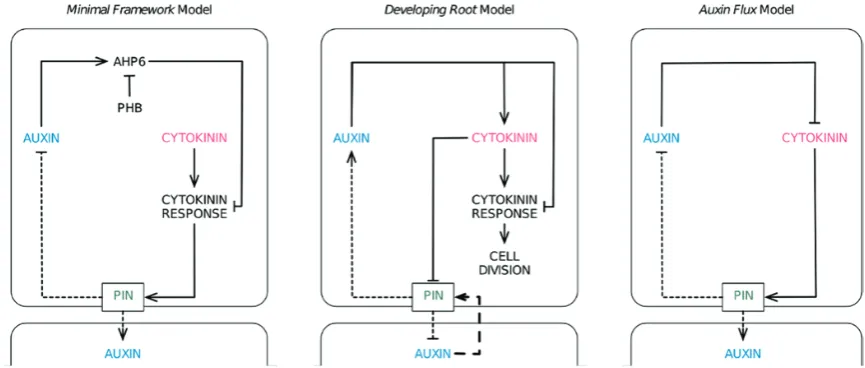
referred to as the Minimal Framework model (Muraro et al., 2014), the Growing Root model (De Rybel et al. 2014), and the Auxin Flux model (el-Showk et al. 2015). A summary of the network configurations in the different models is given in Fig. 2.
Model construction
Mathematical models will always be an abstraction of com-plex biological systems. There is never a clear answer as to how detailed to make them, and different teams will always take different approaches regarding how much informa-tion to include, depending on the quesinforma-tion being addressed. A summary of the different network configurations is given in Fig. 2.
The Minimal Framework model seeks to understand the interaction between molecular components and how these interact as a network to control pattern. To this end, it includes each key class of molecule modeled explicitly. This has the advantage of providing greater insight into the molec-ular circuitry, and indeed this model has led to new insights into the patterning of AHP6 by a PHB–miR165/6 module that has not been considered in the other models.
In contrast, the Auxin Flux model addresses a differ-ent question, asking how the hormonal activation of auxin transport is regulated in a spatial context. In terms of spatial structure, this is the most comprehensive of the three models, as it offers the most detail in terms of compartmentalization of cells by considering the apoplast as a separate compart-ment. In contrast, the molecular network in this model is designed using the most conservative approach, making the most parsimonious model of vascular development possible. Essentially, the model uses as few molecular components as possible while maintaining the ability to address the biologi-cal question. In this model, no distinction is made between hormone levels and hormone signaling output. Some key components, such as AHP6, are not modeled as discrete com-ponents; instead AHP6 is handled via a generic repression of cytokinin in response to auxin.
The Growing Root model asks how hormones control tis-sue development, and as such it is essential to use a growing template to investigate the effects on both cell growth/prolifer-ation and cell patterning. Since a new role for auxin-mediated cytokinin biosynthesis is an essential finding of this study, the authors investigate this by modeling cytokinin levels and cytokinin response separately, enabling the two quantities to be differentiated. However, they take a more parsimonious approach to some elements of their model where modeling would be unlikely to provide critical insights, applying a generic repression of cytokinin signaling by auxin (similar to the Auxin Flux model) and simplifying chains of interaction.
The three models used different modeling platforms: the Minimal Framework model was built using OpenAlea (Pradal
Copyedited by: OUP
4.5
4.10
4.15
4.20
4.25
4.30
4.35
4.40
4.45
4.50
4.55
4.58
4.60
4.65
4.70
4.75
4.80
4.85
4.90
4.95
4.100
4.105
4.110
4.115 4.116 Page 4 of 12 | Mellor et al.
are comprised of polygons, with each polygon representing a distinct cell. A set of ordinary differential equations (ODEs) determines the molecular processes occurring within each cell, and components can move between cells based on a set of terms in the ODEs governing the fluxes across membranes. Cellular Potts models differ in that cells are comprised of a number of pixels or voxels arranged in a grid and thus have internal space; in addition, the apoplast is explicitly included in the model. While the vertex-based approaches simulate movement of molecules purely as permeability across mem-branes, the Cellular Potts model, in contrast, also allows the investigation of diffusion within a cell and in the apoplast. Simulating the diffusion within cells has previously been shown to be important in templates with larger cells, such as those considering root bending or lateral root initiation (Laskowski et al., 2008). In general, vertex-based and Cellular Potts models have similar capabilities in a static setting; the major differences emerge when they are used to model cel-lular growth. This discussion is not within the scope of this paper and is covered in detail elsewhere (Prusinkiewicz and Runions, 2012; Liedekerke et al., 2015).
A minimal molecular framework for
vascular patterning
The Minimal Framework model (Muraro et al., 2014) inves-tigated the feasibility of an auxin–cytokinin mechanism as a control of tissue-specific patterning, first in a two-cell sys-tem but later in a multicellular sys-template. The two-cell syssys-tem was based on a pair of identical cells with a shared interface through which auxin and cytokinin could diffuse or, in the case of auxin, be transported through polar auxin transport. Within each cell, a series of equations calculated how the var-ious components (auxin, cytokinin, AHP6, and PIN7) inter-acted to determine the steady-state solutions for each cell. For simulations run with extremely high/low levels of either auxin or cytokinin, only one possible steady-state solution existed,
both cells having similar outputs. For example, extremely high auxin levels resulted in both cells expressing high levels of AHP6 and having a negligible cytokinin response. However, for a large subset of intermediate conditions, multiple steady-state solutions existed in which one cell had high AHP6 and the other high PIN7. The presence of multiple steady-state solutions suggests that the system can act in a ‘switch-like’ manner to determine discrete domains of gene activity, and reinforces the concept that the auxin–cytokinin interaction can act as a patterning mechanism. However, this two-cell approach does not address how these patterns would look in a realistic tissue.
[image:7.612.91.524.77.261.2]To introduce this model into a multicellular template, a series of simulations were run in which the expression/locali-zation of PINs were fixed based on experimental observa-tions and were not regulated by the model in order to explore genetic redundancy between the PINs. In an effort to sim-plify an otherwise complex network, the model later included only a single PIN type whose activity was based upon that of PIN7. During this work, the authors assumed a flat field of both cytokinin and auxin production, although subse-quent work has shown this not to be the case. In order to direct pattern formation, an initial asymmetry was required; this was supplied through an initial pre-pattern in PIN7 expression. An experimentally defined network of auxin and cytokinin regulation, including the regulation of AHP6 by PHB (Carlsbecker et al., 2010), was not sufficient to recre-ate the stable domains of gene expression as seen in roots. However, the authors were able to reproduce the observed patterns of gene expression stably by making two changes to this network configuration. The first involved altering the way in which PHB and miRNA165/6 interact. The introduc-tion of a mutual degradaintroduc-tion between these two components was required in order to produce stable gradients that could restrict AHP6 sufficiently to the marginal positions. The second change involved the incorporation of an additional, as yet unidentified, inhibitor of cytokinin (termed CKIN). Fig. 2. Schematic diagrams showing the network configurations of the three vascular patterning models. These have been re-arranged from the original figures to aid comparison between models. Activation or repression is shown with solid lines. Dashed lines indicate transport of auxin into and out of the cell, with the arrowhead indicating whether it promotes or inhibits auxin accumulation within that cell. The long dashed lines indicate a mechanism by which PIN proteins are polarized within a cell in a manner dependent on the concentration of auxin within neighboring cells (see text). Although only two cells are shown, these models are all embedded within multicellular templates.
5.5 5.10 5.15 5.20 5.25 5.30 5.35 5.40 5.45 5.50 5.55 5.58 5.60 5.65 5.70 5.75 5.80 5.85 5.90 5.95 5.100 5.105 5.110 5.115 5.116
Modeling vascular pattern | Page 5 of 12
This was required alongside AHP6 to restrict cytokinin response, and therefore PIN7, in the central parts of the xylem axis. At the time, the authors proposed that this com-ponent could target either cytokinin biosynthesis or signal-ing; however, subsequent studies suggest that the former is unlikely.
The revised network could reproduce a stable vascular pat-tern, but it required an initial asymmetry in PIN7. To test the robustness of this system, the output from a previous simulation was used as a set of initial conditions that closely resembled the pattern of gene expression seen in wild-type roots. Simulations were then run to steady state in a system in which every cell had the potential to express PIN7. These simulations revealed that the initial vascular pattern was maintained, suggesting that the network provides a robust mechanism for maintaining pattern around an initial asym-metry, even though it does not generate the initial asymmetry or address its possible causes.
Early events specifying the xylem axis
This question was addressed in the Growing Root model (De Rybel et al. 2014) by investigating how the xylem axis was specified during embryogenesis. The model incorporated both growth and patterning within a dynamic array of cells. This study identified a crucial new interaction through which auxin promotes the transcription of the LONELY GUY 4 (LOG4) gene via the TMO5/LHW dimer (De Rybel et al., 2014). LOG4 is a crucial enzyme involved in the final stages of the cytokinin biosynthesis pathway and is believed to be the rate-limiting step controlling cytokinin homeostasis (Kuroha et al., 2009). LOG4 is expressed in all four of the vascular initials, but in the growing root it is expressed throughout the xylem axis, suggesting that the xylem axis acts as a source of cytokinin (De Rybel et al., 2014). Although in this model both growth and patterning are regulated, it is likely that these activities are achieved through two independent cytokinin responses: while cytokinin signaling promotes periclinal cell division, PIN localization is regulated via cytokinin directly, in what the model assumes to be an independent cytokinin response path-way. The PIN dynamics differ from those used in the Minimal Framework and Auxin Flux models; cytokinin mediates the inhibition of PIN1 localization, as observed during lateral root formation (Marhavý et al., 2011), rather than inducing expression of PIN7 (Bishopp et al., 2011a). Furthermore, PIN1 is also polarized in response to auxin gradients, as in some other simulations in different developmental contexts (Jönsson et al., 2006). In this model, xylem cells are capable of producing cytokinin via TMO5/LHW-dependent activa-tion of LOG4. As a result of the mutual interacactiva-tion between cell growth and the reaction network, the production of cyto-kinin is constrained to the developing xylem axis in model simulations. As a result of high auxin levels in the xylem axis, cytokinin signaling is inhibited in these cells, which results in suppression of periclinal cell division.
The model itself comprised a combination of two inter-connected feed-forward loops that controlled both growth
and patterning (de Rybel et al., 2014). The first loop con-sidered cytokinin rather than cytokinin response to control PIN regulation and cell growth. The second incorporated the interaction between auxin and cytokinin response to control periclinal cell divisions. When applied to a template consisting of four provascular initial cells, these intercon-nected loops were sufficient to recapitulate both the growth and patterning processes necessary to create an axis of high auxin response in a growing template (de Rybel et al., 2014). However, this required two additional inputs within the ini-tial four-cell template (de Rybel et al., 2014). The first was a bias in which auxin was elevated in two source cells represent-ing the convergence points of the cotyledons. Secondly, the two source cells had to be connected by a small bridge (see Fig. 1), an assumption that subsequent experimental analy-ses have shown to be valid. Not only can these simulations recreate experimental observations based on limited prior information, but the simulations also showed gradients in both cytokinin and cytokinin response, with the highest cyto-kinin response in the cells adjacent to the xylem axis. While some cytokinin markers are not sensitive enough to reflect this gradient, re-analysis of others has shown such a gradient (de Rybel et al., 2014).
A parsimonious model of auxin fluxes
The Auxin Flux model (el-Showk et al. 2015) delves much deeper into the concentration and flux patterns of auxin. While the previous models only considered a single PIN protein, this model included PIN1, 3, and 7, together with a combined role for PIN2 and the PGPs. It also incorporated a generic auxin importer to account for AUX1, LAX1, and LAX2. The Auxin Flux model incorporates cytokinin-medi-ated up-regulation of PIN7 in a similar way to the Minimal Framework model. However, the role of AHP6 is simplified in this model; instead of explicitly modeling AHP6, the linear chain between auxin, AHP6, and the repression of cytokinin-mediated PIN activation is simplified to a generic repression of PIN7 and PIN1 by auxin.
Copyedited by: OUP
6.5
6.10
6.15
6.20
6.25
6.30
6.35
6.40
6.45
6.50
6.55
6.58
6.60
6.65
6.70
6.75
6.80
6.85
6.90
6.95
6.100
6.105
6.110
6.115 6.116 Page 6 of 12 | Mellor et al.
confirmed experimentally, as aux1 lax1 lax2 triple mutant plants were found to have unstable pattern formation.
Although the Auxin Flux model focused on vascular pat-terning in the root tip, it provided unexpected insights into the process of lateral root priming. Lateral roots originate from the pericycle cells flanking the xylem poles (Lavenus
et al., 2016). In the model, certain subcellular arrangements of PIN1 generate an auxin flux circuit that not only allows the xylem pole pericycle cells to accumulate auxin at the expense of the xylem axis, but also ensures that two the poles compete against each other for auxin. While this dynamic provides a potential mechanism to prime lateral roots, future work is needed to assess experimentally the subcellular status of PIN1 and to evaluate the auxin flux circuit in simulations of a growing, three-dimensional root.
What initial conditions are required to set
vascular pattern?
All three models require an initial asymmetry in order to establish the vascular pattern, but each addressed the asym-metry differently. In both the Minimal Framework and Auxin Flux models, this initial asymmetry was generated by an initial pre-placement of PINs. Furthermore, in the case of the Minimal Framework model, this initial asymmetry is required only transiently; once established, the system is able to maintain a stable pattern, even after the asymmetry is removed. The Growing Root model, in contrast, uses a per-sistent asymmetry in auxin input to drive pattern formation. Two of the four vascular cells continuously receive higher auxin input than other cells in the vascular cylinder, based on the observation that symmetry breakage first occurs in the apical part of the embryo and leads to an asymmetric pro-duction and transport of auxin at the incipient cotyledon, as
cells immediately subtending the cotyledons have been shown to have higher auxin response (De Rybel et al., 2014), while mutants with altered numbers of cotyledons have been shown to generate roots with irregular numbers of xylem poles (Help
et al., 2011). A key question, which we now investigate with a new set of simulations, is whether a transient asymmetry in auxin input can be used to drive patterning in the Minimal Framework model.
[image:9.612.127.490.507.723.2]To test this, we reran the Minimal Framework model using the original parameters and allowing each component (including PIN7) to be expressed in any cell, but with an ini-tial condition of high auxin at both protoxylem cells and all four xylem-pole pericycle cells, where high AHP6 expression has been observed. The production rate of auxin is uniform throughout the tissue. We made one additional change to the model (see Supplementary Model S1 at JXB online). In the original model, we had a hypothetical component termed ‘CKIN’ that acted redundantly to AHP6 to inhibit cytokinin in the metaxylem. We changed this from a repressor of cyto-kinin levels to a repressor of cytocyto-kinin response, in keeping with the subsequent discovery of the xylem as a source of cytokinin. With these changes, the resulting steady-state pat-tern of AHP6 expression closely resembles the initial condi-tions, with only a minor shift in the position of the xylem axis (Fig. 3). These simulations support the idea that an asymme-try in auxin drives pattern formation in the root but suggest that such an asymmetry is required only transiently. While in the embryo and primary root of Arabidopsis the continuous transport of auxin from the cotyledons/pre-existing vascular is likely to provide a continuous asymmetry in auxin input, as incorporated within the Growing Root model, this is not necessarily the case in newly formed roots (such as lateral or crown roots) or during pattern specification in other plant species with three or more vascular poles. To investigate the
7.5
7.10
7.15
7.20
7.25
7.30
7.35
7.40
7.45
7.50
7.55
7.58
7.60
7.65
7.70
7.75
7.80
7.85
7.90
7.95
7.100
7.105
7.110
7.115 7.116
Modeling vascular pattern | Page 7 of 12
importance of a continuous asymmetric auxin input for the functionality of the Growing Root model, we ran model sim-ulations using a transient asymmetric auxin input; the model does not produce the correct pattern of auxin and cytokinin. However, when run in a static template (Fig. 4A), the same simulation produces the correct cytokinin and auxin patterns. The results from the Growing Root model suggest that con-tinuous asymmetric auxin input is necessary during establish-ment of the vasculature, while this input is not required in later stages and to maintain auxin and cytokinin signaling domains in mature roots.
We believe that the observation that transient changes in auxin can be propagated as stable changes in vascular pattern will allow future models to address the patterning process in other species.
The xylem axis as a source of cytokinin
Although there are multiple markers for observing cytokinin response at a cellular/tissue scale, there are no methods for imaging the location of cytokinin itself at this resolution. Although the Minimal Framework and Auxin Flux models could produce stable patterning with a homogenous field of cytokinin production in each cell, experimental results pub-lished with the Growing Root model showed that the xylem
serves as the major source of cytokinin. One output of the Growing Root model is that it is possible to create a gradient of cytokinin by driving cytokinin synthesis in the xylem axis only. Does such a gradient exist in plants and is it required for patterning? Analyses of independent cytokinin-responsive marker genes suggest that a gradient in cytokinin response does occur, but current technology does not allow us to know whether this is mirrored by a gradient in cytokinin itself. The presence of a cytokinin gradient in these tissues is an issue where there are different viewpoints between the authors.
The Growing Root model investigates both cell division and patterning through regulation of the PINs. Although cyto-kinin regulates both of these processes, it is likely that it does so using different downstream regulatory components; cyto-kinin-mediated patterning and cell proliferation are therefore handled separately in the model. As little is known about the mechanism through which cytokinin regulates PIN1, this was modeled as a direct interaction between cytokinin and PIN1 based on data from lateral root organogenesis (Marhavý
[image:10.612.136.481.394.705.2]et al., 2011). This simplification was introduced in order to reduce unnecessary parameters, although it has subsequently been shown that this interaction is dependent on cytokinin signaling rather than being a direct activity modulated by the hormone itself (Marhavý et al., 2014). Within the Growing Root model, a gradient of cytokinin promotes increased
Copyedited by: OUP 8.5 8.10 8.15 8.20 8.25 8.30 8.35 8.40 8.45 8.50 8.55 8.58 8.60 8.65 8.70 8.75 8.80 8.85 8.90 8.95 8.100 8.105 8.110 8.115 8.116 Page 8 of 12 | Mellor et al.
polarization of PINs away from the xylem axis. Here we test whether a gradient of cytokinin is an absolute requirement for this model.
By running simulations in the Growing Root model with increased cytokinin diffusion, we were able to observe that the desired model output can be achieved with much shal-lower gradients than were previously published. In the most extreme simulations, cytokinin diffusion could be increased up to 40-fold with near-homogeneous levels and maintain correct patterning. Nevertheless, to specify pattern correctly, there is a requirement for cytokinin levels to be higher in the xylem than in adjacent cambial cells. The idea that only a shallow gradient of cytokinin is required is appealing, as other sources of cytokinin exist, such as phloem (Bishopp
et al., 2011b), although the majority of LOG activity is within the xylem axis (De Rybel et al., 2014). In order to explore whether it is the regulation of PINs that is responsible for the requirement for a cytokinin gradient, we ran new simulations using the Growing Root model, in which PIN localization is promoted by cytokinin (Supplementary Model S2). These simulations resulted in correct patterning even when the rate of cytokinin diffusion was raised so high that it produced a homogeneous field of cytokinin (Fig. 4B).
In reality, multiple modes of cytokinin-mediated PIN activity probably co-exist in plants, and further experimen-tal analyses documenting the exact interaction between cyto-kinin and individual PINs in the given developmental context is needed. Collectively, these simulations show that a shallow gradient of cytokinin is required in simulations incorporating PIN1-like regulation, as this appears to be the most important PIN during embryonic root formation (Friml et al., 2003); it is possible that there are different requirements for cytokinin gradients during the formation of embryonic and mature roots. These studies highlight the need for detailed analyses analyzing exactly how each PIN responds to cytokinin in spe-cific tissue types. Whilst this is feasible for the growing root, investigating this process during embryogenesis would repre-sent a significant technical challenge.
A gradient of any molecule across a multicellular tissue is possible providing it is synthesized (either exclusively or at higher levels) in a group of source cells and that it moves between cells (e.g. via diffusion). The slope of the gradi-ent results exclusively from the balance between the rates of diffusion and degradation. A molecule that diffuses very quickly or degrades very slowly will form a shallow gradient or become homogenously distributed throughout the tissue; a slower rate of diffusion or a faster rate of degradation will form a steeper gradient. The identification of the xylem axis as a key source for cytokinin provides a group of source cells, but what kind of gradient forms around this source?
Unfortunately, as we are unable to visualize individual molecules, the degradation rate and diffusion coefficient of cytokinin are both unknown. In the Auxin Flux paper, the authors argue that the parameters required to generate an informative gradient of cytokinin in tissues the size of an Arabidopsis root are unrealistic. Since all three models only include passive movement of cytokinin via diffusion, the choice of these parameters is critical for determining
the shape of the resulting gradient; however, problems arise because these parameters are simply unknown.
Can a gradient of cytokinin exist?
In the Minimal Framework and Growing Root models, move-ment of individual molecules is governed only by permeability across a membrane. In the Auxin Flux model, movement also occurs via diffusion within cells and in the apoplast, arguably giving a more realistic modeling of diffusion. This is of par-ticular importance for hormonal signaling since, at least over a short range, signaling molecules are thought to propagate faster apoplasticaly than symplasticaly (Robert and Friml, 2009). The Auxin Flux model uses the same diffusion coef-ficient for cytokinin and auxin based on the rationale that they are similar sized molecules. Although the parameter for auxin diffusion has been used in other computational models and is based on experimental values, these were not meas-ured in plants but were generated using a polar membrane created between egg lecithin and decane (Gutknecht and Walter, 1980). Within this system, diffusion was dependent on pH, and the rate of auxin flux across the membrane was increased through conversion of auxin to an ionized form at the membrane surface. Whilst these may represent the ‘best estimates’ of cytokinin diffusion, they are open to debate, and only direct measurements will be able to provide irrefutable parameters.
Since an informative gradient would require that the cyto-kinin diffusion or degradation rates differ by several orders of magnitude from those used in the Auxin Flux model, the authors carried out an experiment to establish a lower limit on the diffusion rate of cytokinin in plants. They treated roots with exogenous cytokinin and measured the change in cytokinin response within the root; cytokinin response increased in both the outer and inner layers of the root within 6 h, suggesting that cytokinin can traverse the radius of the Arabidopsis root in a matter of hours at most (el-Showk et al., 2015). This is a rate of cytokinin movement which is incom-patible with the formation of an informative cytokinin gra-dient via diffusion unless the degradation rate is also orders of magnitude higher; however, such rapid degradation would also place limits on how far cytokinin could travel and would affect the time scales of regulatory networks. The authors therefore suggest that other mechanisms, such as active cyto-kinin transport or uneven expression of the cytocyto-kinin per-ception machinery, might be responsible for the observed cytokinin signaling patterns.
Although the models use a different system for dealing with diffusion, the Auxin Flux model was able to reproduce the gradient formed in the Growing Root model with an appro-priate choice of parameters. We therefore endeavored to eval-uate the questions of whether the xylem axis can act as a sole source of cytokinin and whether an informative cytokinin gradient can exist using the Minimal Framework model.
9.5
9.10
9.15
9.20
9.25
9.30
9.35
9.40
9.45
9.50
9.55
9.58
9.60
9.65
9.70
9.75
9.80
9.85
9.90
9.95
9.100
9.105
9.110
9.115 9.116
Modeling vascular pattern | Page 9 of 12
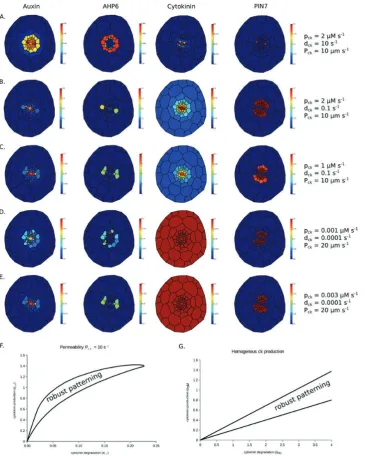
an initial asymmetry in PIN localization and then test the robustness of any pattern by removing this restriction on PIN placement. Using the original model parameters produces a sharp gradient of cytokinin away from the xylem axis, result-ing in very low PIN7 expression except in the protoxylem, and AHP6 expression spreads throughout the pericycle and adjacent cells in the stele (Fig. 5A).
As discussed above, while the Auxin Flux model uses a dif-fusion coefficient in a Cellular Potts model to simulate cyto-kinin movement within and between cells, the other models only simulate movement from cell to cell using a permeability parameter. Based on an approximate average width of the cell layers in the cross-section of 30 μm, we estimate that, for a
given cytokinin production and degradation rate, the cyto-kinin distribution in the Auxin Flux model with Dck=600 μm2 s–1 can be reproduced in the other two models with P
[image:12.612.126.513.247.704.2]ck=20μm s-1. Similarly, we predict the results with permeability Pck=10 μm s–1 used in the Minimal Framework model can be roughly reproduced in the Auxin Flux with a diffusion coef-ficient of 300 μm2 s–1. Though this diffusion coefficient is of the same order of magnitude to that used in the Auxin Flux model, cytokinin degradation (dck), the other key param-eter in dparam-etermining the sharpness of any cytokinin gradi-ent, is much higher in the Minimal Framework model than in both the Growing Root and Auxin Flux models, resulting in the sharp gradient in (Fig. 5A). Although the exact data
Copyedited by: OUP 10.5 10.10 10.15 10.20 10.25 10.30 10.35 10.40 10.45 10.50 10.55 10.58 10.60 10.65 10.70 10.75 10.80 10.85 10.90 10.95 10.100 10.105 10.110 10.115 10.116 Page 10 of 12 | Mellor et al.
regarding the turnover of cytokinin are not available, reduc-ing this in line with the other models (from 10 s–1 to 0.1 s–1), so that it degrades over the time scale of minutes rather than seconds, results in improved patterning of the xylem axis (Fig. 5B). A further adjustment, so that the level of cytokinin in the procambium regions is closer to that in the published model, results in the correct patterning of the root vascular cylinder and the formation of a gradient of cytokinin peak-ing within the xylem axis (Fig. 5C).
Using the parameters from the Auxin Flux model (cyto-kinin production pck=0.001 arbitrary units s–1, degradation dck=0.0001 s–1, estimated permeability Pck=20 μm s–1) in the Minimal Framework model with cytokinin production restricted to the xylem results in little or no gradient in cyto-kinin, but also no regular pattern due to insufficient over-all levels of cytokinin (Fig. 5D). However, when raising the level of cytokinin production to pck=0.003 arbitrary units s–1, while there is still no cytokinin gradient, the overall level of cytokinin is raised sufficiently so that the correct pattern is generated and maintained robustly (Fig. 5E).
As these new simulations required a change of parameters, we tested the sensitivity of the three key parameters relating to cytokinin activity: cytokinin production (pck), cytokinin degra-dation (dck), and cytokinin permeability (Pck). Using the value of Pck from the Minimal Framework model (Pck=10 μm s–1) as a starting point, we found ranges of the other two parameters, pck and dck, for which the desired pattern is maintained robustly. To assess whether the pattern is formed correctly, we use k-means clustering to categorize cells into two clusters based on the level of AHP6 protein. If and only if the cluster of cells with the highest AHP6 level is exactly equal to the set of six cells com-prising the two protoxylem and four xylem pole pericycle cells do we conclude that the model has patterned the tissue correctly.
Plotting the region of two-parameter space for which the desired pattern is formed (Fig. 5F) shows that with the perme-ability Pck=10 μm s–1 no pattern can be formed for degrada-tion rates above around dck=0.22 s–1. Below this degradation rate, there always exists a range for which patterning occurs. This shows that while the model is able to form a pattern with very shallow or intermediate cytokinin gradients, when the cytokinin degradation rate is too high, the cytokinin gradi-ent away from the xylem axis becomes too steep to be able to maintain a stable pattern. We also note that for a given degra-dation rate below the threshold value, there is only a relatively narrow range of cytokinin production that will support a pat-tern. This parameter does not affect the cytokinin gradient, but instead determines the overall level of cytokinin in the tissue. Since PIN7 is sensitive to the level of cytokinin, if pro-duction is too high, PIN7 expression dominates throughout the stele, while if it is too low AHP6 expression dominates instead. The narrow range of viability to form the correct pattern further illustrates the importance of the regulation of cytokinin production as shown by De Rybel et al. (2014). Similarly shaped regions of parameter spaces, but with differ-ent numerical ranges, are produced when repeating the exer-cise using the permeability from the Growing Root model (Pck=0.1 μm min–1) and the estimated representative perme-ability from the Auxin Flux model (Pck=20 μm s–1).
If cytokinin is produced evenly throughout the tissue, rather than just in the xylem axis, it is still possible to produce the desired pattern of AHP6 expression. Plotting the region of two-parameter space for which the desired pattern occurs as before, we see that while for a given cytokinin degradation rate there is a corresponding range of production rates for which there is the correct, stable pattern, there is no upper limit on the degradation rate, as is the case when production is limited to the xylem (Fig. 5G).
Together these simulations suggest that, while restricting cytokinin production to the xylem axis is a plausible method for vascular patterning, at least in the Minimal Framework and Growing Root models, it is not an absolute requirement for the patterning process. Furthermore, these results suggest that the overall cytokinin level as well as cytokinin distribu-tion can act as the driving force for this patterning process.
Conclusion
The organization of root vascular tissues provides a fascinat-ing model to investigate how patterns form in multicellular structures, and the surge in research in vascular development is testament to this. Experimental studies have identified the key components involved in this process and determined how they interact. More recently, theoretical studies have investi-gated these non-linear interactions and feedback mechanisms and revealed how they collectively determine patterning out-put. While each of the models incorporates known values where possible, all models by necessity rely on parameter estimation. To address the uncertainty of these parameters, each of the models has made an exploration of the parameter space that allows patterning.
11.5 11.10 11.15 11.20 11.25 11.30 11.35 11.40 11.45 11.50 11.55 11.58 11.60 11.65 11.70 11.75 11.80 11.85 11.90 11.95 11.100 11.105 11.110 11.115 11.116
Modeling vascular pattern | Page 11 of 12
relevant to cytokinin diffusion are in the range to form an informative gradient or demonstrate that cytokinin is pat-terned by an alternative mechanism. We have also highlighted several other key areas that need further research, namely fur-ther insight into how cytokinin regulates PIN activity and the factor(s) that operate alongside AHP6 to limit auxin response in the xylem axis.
The three independent modeling approaches together offer considerable insight into the patterning process. All of the models support each other in some aspects; but at the same time each model provides new insights into the network that are unique to that model. In a few areas, they find disagree-ments. By comparing the three modeling approaches, we are able to focus future experimentation on aspects where the modeling has indicated that there is additional complexity. We believe that the approach of integrating multiple independent models of root vascular patterning serves as an exemplar for understanding other developmental processes in plants.
Supplementary data
Supplementary data are available at JXB online.
Supplementary Model S1. Minimal framework model/ Supplementary Model S2. Growing root model. Table S1. Parameters used for the simulation in Fig. 4.
Acknowledgements
NM was funded by the Biotechnology and Biological Sciences Research Council (BB/L023555/1). BDR was funded by the Netherlands Organisation for Scientific Research (NWO; VIDI 864.13.001) and by The Research Foundation - Flanders (FWO; Odysseus II G0D0515N and Post-doc grant 12D1815N). APM was funded by Academy of Finland (grant nos 266431, 273130, ad 271832). AB was funded by a Royal Society University Research Fellowship.
References
Band LR, Wells DM, Fozard Ja, et al. 2014. Systems analysis of auxin transport in the Arabidopsis root apex. The Plant Cell 26, 862–875. Bishopp A, Help H, El-Showk S, Weijers D, Scheres B, Friml J, Benková E, Mähönen AP, Helariutta Y. 2011a. A mutually inhibitory interaction between auxin and cytokinin specifies vascular pattern in roots. Current Biology 21, 917–926.
Bishopp A, Lehesranta S, Vatén A, Help H, El-Showk S, Scheres B, Helariutta K, Mähönen AP, Sakakibara H, Helariutta Y. 2011b. Phloem-transported cytokinin regulates polar auxin transport and maintains vascular pattern in the root meristem. Current Biology 21, 926–932.
Brunoud G, Wells DM, Oliva M, et al. 2012. A novel sensor to map auxin response and distribution at high spatio-temporal resolution. Nature 482, 103–106.
Carlsbecker A, Lee J-Y, Roberts CJ, et al. 2010. Cell signalling by microRNA165/6 directs gene dose-dependent root cell fate. Nature 465, 316–321.
Cui H, Levesque MP, Vernoux T, Jung JW, Paquette AJ, Gallagher KL, Wang JY, Blilou I, Scheres B, Benfey PN. 2007. An evolutionarily conserved mechanism delimiting SHR movement defines a single layer of endodermis in plants. Science 316, 421–425.
De Rybel B, Adibi M, Breda AS, et al. 2014. Integration of growth and patterning during vascular tissue formation in Arabidopsis. Science 345, 1255215–1255215.
De Rybel B, Möller B, Yoshida S, Grabowicz I, Barbier de Reuille P, Boeren S, Smith RS, Borst JW, Weijers D. 2013. A bHLH complex controls embryonic vascular tissue establishment and indeterminate growth in Arabidopsis. Developmental Cell 24, 426–437.
el-Showk S, Help-Rinta-Rahko H, Blomster T, Siligato R, Marée AFM, Mähönen AP, Grieneisen VA. 2015. Parsimonious model of vascular patterning links transverse hormone fluxes to lateral root initiation: auxin leads the way, while cytokinin levels out. PLoS Computational Biology 11, 1–40.
Fàbregas N, Formosa-Jordan P, Confraria A, Siligato R, Alonso JM, Swarup R, Bennett MJ, Mähönen AP, Caño-Delgado AI, Ibañes M. 2015. Auxin influx carriers control vascular patterning and xylem differentiation in Arabidopsis thaliana. PLoS Genetics 11, e1005183. Friml J, Vieten A, Sauer M, Weijers D, Schwarz H, Hamann T, Offringa R, Jürgens G. 2003. Efflux-dependent auxin gradients establish the apical–basal axis of Arabidopsis. Nature 426, 147–153.
Grieneisen VA, Xu J, Marée AFM, Hogeweg P, Scheres B. 2007. Auxin transport is sufficient to generate a maximum and gradient guiding root growth. Nature 449, 1008–1013.
Gutknecht J, Walter A. 1980. Transport of auxin (indoleacetic acid) through lipid bilayer membranes. Journal of Membrane Biology 56, 65–72. Hardtke CS, Berleth T. 1998. The Arabidopsis gene MONOPTEROS encodes a transcription factor mediating embryo axis formation and vascular development. EMBO Journal 17, 1405–1411.
Help H, Mähönen AP, Helariutta Y, Bishopp A. 2011. Bisymmetry in the embryonic root is dependent on cotyledon number and position. Plant Signaling and Behavior 6, 1837–1840.
Ibañes M, Fàbregas N, Chory J, Caño-Delgado AI. 2009.
Brassinosteroid signaling and auxin transport are required to establish the periodic pattern of Arabidopsis shoot vascular bundles. Proceedings of the National Academy of Sciences, USA 106, 13630–13635.
Jönsson H, Heisler MG, Shapiro BE, Meyerowitz EM, Mjolsness E. 2006. An auxin-driven polarized transport model for phyllotaxis. Proceedings of the National Academy of Sciences, USA 103, 1633–1638. Katayama H, Iwamoto K, Kariya Y, Asakawa T, Kan T, Fukuda H, Ohashi-Ito K. 2016. A negative feedback loop controlling bHLH complexes is involved in vascular cell division and differentiation in the root apical meristem. Current Biology 25, 3144–3150.
Kuroha T, Tokunaga H, Kojima M, Ueda N, Ishida T, Nagawa S, Fukuda H, Sugimoto K, Sakakibara H. 2009. Functional analyses of LONELY GUY cytokinin-activating enzymes reveal the importance of the direct activation pathway in Arabidopsis. The Plant Cell 21, 3152–3169. Laskowski M, Grieneisen VA, Hofhuis H, Ten Hove CA, Hogeweg P, Marée AFM, Scheres B. 2008. Root system architecture from coupling cell shape to auxin transport. PLoS Biology 6, 2721–2735.
Lavenus J, Goh T, Roberts I, Guyomarc’h S, Lucas M, De Smet I, Fukaki H, Beeckman T, Bennett M, Laplaze L. 2016. Lateral root development in Arabidopsis: fifty shades of auxin. Trends in Plant Science 18, 450–458.
Liedekerke P Van, Palm M, Jagiella N, Drasdo D, Liedekerke P Van, Palm M, Jagiella N, Simulating DD. 2015. Simulating tissue mechanics with agent based models: concepts and perspectives. Computational Particle Mechanics 2, 401–444.
Mähönen AP, Bishopp A, Higuchi M, Nieminen KM, Kinoshita K, Törmäkangas K, Ikeda Y, Oka A, Kakimoto T, Helariutta Y. 2006. Cytokinin signaling and its inhibitor AHP6 regulate cell fate during vascular development. Science 311, 94–98.
Mähönen AP, Bonke M, Kauppinen L, Riikonen M, Benfey PN, Helariutta Y. 2000. A novel two-component hybrid molecule regulates vascular morphogenesis of the Arabidopsis root. Genes and Development 14, 2938–2943.
Marhavý P, Bielach A, Abas L, et al. 2011. Cytokinin modulates endocytic trafficking of PIN1 auxin efflux carrier to control plant organogenesis. Developmental Cell 21, 796–804.
Marhavý P, Duclercq J, Weller B, Feraru E, Bielach A, Offringa R, Friml J, Schwechheimer C, Murphy A, Benková E. 2014. Cytokinin controls polarity of PIN1-dependent auxin transport during lateral root organogenesis. Current Biology 24, 1031–1037.
Copyedited by: OUP
12.5
12.10
12.15
12.20
12.25
12.30
12.35
12.40
12.45
12.50
12.55
12.58
12.60
12.65
12.70
12.75
12.80
12.85
12.90
12.95
12.100
12.105
12.110
12.115 12.116 Page 12 of 12 | Mellor et al.
Merks RM, Guravage M, Inzé D, Beemster GT. 2011. VirtualLeaf: an open-source framework for cell-based modeling of plant tissue growth and development. Plant Physiology 155, 656–666.
Mironova VV, Omelyanchuk Na, Yosiphon G, Fadeev SI, Kolchanov Na, Mjolsness E, Likhoshvai Va. 2010. A plausible mechanism for auxin patterning along the developing root. BMC Systems Biology 4, 98. Mitchison GJ. 1980. The dynamics of auxin transport. Proceedings of the Royal Society B: Biological Sciences 209, 489–511.
Muraro D, Byrne H, King J, Bennett M. 2013. The role of auxin and cytokinin signalling in specifying the root architecture of Arabidopsis thaliana. Journal of Theoretical Biology 317, 71–86.
Muraro D, Byrne H, King J, Voß U, Kieber J, Bennett M. 2011. The influence of cytokinin–auxin cross-regulation on cell-fate determination in Arabidopsis thaliana root development. Journal of Theoretical Biology 283, 152–167.
Muraro D, Mellor N, Pound MP, et al. 2014. Integration of hormonal signaling networks and mobile microRNAs is required for vascular patterning in Arabidopsis roots. Proceedings of the National Academy of Sciences, USA 111, 857–862.
Péret B, Middleton AM, French AP, et al. 2013. Sequential induction of auxin efflux and influx carriers regulates lateral root emergence. Molecular Systems Biology 9, 699.
Pernisova M, Prat T, Grones P, Harustiakova D, Matonohova M, Spichal L, Nodzynski T, Friml J, Hejatko J. 2016. Cytokinins influence
root gravitropism via differential regulation of auxin transporter expression and localization in Arabidopsis. New Phytologist 212, 497–509.
Pradal C, Dufour-Kowalski S, Boudon F, Fournier C, Godin C. 2008. OpenAlea: a visual programming and component-based software platform for plant modeling. Functional Plant Biology 35, 751–760.
Prusinkiewicz P, Runions A. 2012. Computational models of plant development and form. New Phytologist 193, 549–569.
Robert HS, Friml J. 2009. Auxin and other signals on the move in plants. Nature Chemical Biology 5, 325–332.
Ruzicka K, Simásková M, Duclercq J, Petrásek J, Zazímalová E, Simon S, Friml J, Van Montagu MCE, Benková E. 2009. Cytokinin regulates root meristem activity via modulation of the polar auxin transport. Proceedings of the National Academy of Sciences, USA 106, 4284–4289.
Scheres B, Wolkenfelt H, Willemsen V, Terlouw M, Lawson E, Dean C, Weisbeek P. 1994. Embryonic origin of the Arabidopsis primary root and root meristem initials. Development 2487, 2475–2487.
Schlereth A, Möller B, Liu W, Kientz M, Flipse J, Rademacher EH, Schmid M, Jürgens G, Weijers D. 2010. MONOPTEROS controls embryonic root initiation by regulating a mobile transcription factor. Nature 464, 913–916.