www.impactjournals.com/oncotarget/ Oncotarget, Vol. 7, No. 12
Role of erythropoietin in the angiogenic activity of bone marrow
endothelial cells of MGUS and multiple myeloma patients
Aurelia Lamanuzzi1,*, Ilaria Saltarella1,*, Arianna Ferrucci1, Roberto Ria1, Simona
Ruggieri2, Vito Racanelli1, Luigia Rao1, Tiziana Annese2, Beatrice Nico2, Angelo
Vacca1, Domenico Ribatti2,3
1Department of Internal Medicine and Clinical Oncology, University of Bari Medical School, Bari, Italy
2Department of Basic Medical Sciences, Neurosciences and Sensory Organs, University of Bari Medical School, Bari, Italy 3National Cancer Institute “Giovanni Paolo II”, Bari, Italy
*These authors contributed equally to this work
Correspondence to: Domenico Ribatti, e-mail: domenico.ribatti@uniba.it
Keywords: angiogenesis, erythropoietin, endothelial cells, monoclonal gammopathy of undetermined significance, multiple myeloma
Received: September 16, 2015 Accepted: January 31, 2016 Published: February 22, 2016
ABSTRACT
Increasing evidences suggest several biological roles for erythropoietin and its receptor (Epo and EpoR), unrelated to erythropoiesis, including angiogenesis. Here, we detected the expression of EpoR in bone marrow-derived endothelial cells from monoclonal gammopathy of undetermined significance (MGUS) and multiple myeloma (MM) patients (MGECs and MMECs, respectively) and assessed whether Epo plays a role in MGECs- and MMECs-mediated angiogenesis. We show that EpoR is expressed by both MGECs and MMECs even though at a higher level in the first ones. Both EC types respond to rHuEpo in terms of cell proliferation, whereas other responses, including activation of JAK2/STAT5 and PI3K/Akt pathways, cell migration and capillarogenesis are enhanced by Epo in MGECs, but not in MMECs. In addition, the conditioned media of both Epo-treated cells induce a strong angiogenic response in vivo in the chorioallantoic membrane assay, comparable to that of vascular endothelial growth factor (VEGF). Overall, these data highlight the effect of Epo on MGECs- and MMECs-mediated angiogenesis: MGECs are more responsive to Epo treatment than MMECs, probably because over-angiogenic phenotype of MMECs is already activated by their autocrine/paracrine loops occurring in the “angiogenic switch” from MGUS.
INTRODUCTION
Endothelial cells (ECs) isolated from the bone marrow of multiple myeloma patients (MMECs) express EC markers, including Tie2, vascular endothelial growth
factor receptor-2 (VEGFR-2), fibroblast growth factor
receptor-2 (FGFR-2), CD105-endoglin and vascular endothelial (VE)-cadherin. Furthemore, MMEC in vitro and in vivo angiogenic activity is enhanced by an increase in matrix metalloproteinase-2 and -9 (MMP-2 and MMP-9) secretion and by the up-regulation of angiogenic-related genes [1].
Erythropoietin (Epo) is a pleiotropic cytokine that exerts different biological effects, and angiogenesis is one of its extra-hematopoietic functions [2]. Epo and
Epo receptor (EpoR) are expressed in the vasculature during embryogenesis, and their deletion in null embryos leads to angiogenic defects [3]. In post-natal life Epo stimulates both proliferation and migration of human and bovine ECs in vitro as well as in the rat aortic ring model [4–8]. Moreover, Epo induces endothelin-1 (ET-1) expression in EC cultures [6, 9], and recombinant human Epo (rHuEpo) induces an increased proliferation, MMP-2 expression and differentiation into vascular tubes of human ECs in vitro [7]. EpoR mRNA is expressed in different ECs [4, 8].
This study was designed to determine the effects of Epo on ECs from monoclonal gammopathy of
undetermined significance [MGUS, (MGECs)] and
RESULTS
RHuEpo proliferation of BMECs expressing EpoR
To assess the mitogenic ability of rHuEpo, MGECs
and MMECs were treated with 15 U/ml, 30 U/ml and 60 U/ml rHuEpo for 24, 48 and 72 h. The addition of rHuEpo significantly increased cell proliferation in a time
and dose manner, and the highest proliferative rate was obtained for both MGECs and MMECs at a concentration
of 30 U/ml for 48 h and 72 h, (Figure 1). In order to
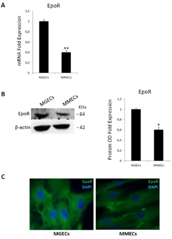
evaluate EpoR expression in MGECs and in MMECs, we performed a Real-Time RT PCR and a Western blotting analysis. Real-Time RT PCR demonstrated that MGECs had a higher expression of EpoR than
MMECs (Figure 2A). Western blotting analysis as well
as immunofluorescence staining confirmed these data
(Figure 2B and 2C).
RhuEpo regulates secretion of pro-angiogenic factors by BMECs
To evaluate if rHuEpo could modify the angiogenic
cytokine secretion, we carried out a multiplex ELISA testing CM from MGECs and MMECs treated with 30U/ml of rHuEpo for 24, 48 and 72 h. ELISA revealed that
ANG-2 secretion gradually decreased in MGECs, but it increased in MMECs. HGF was not secreted by MGECs,
[image:2.612.142.478.259.692.2]while its level significantly increased in MMECs in a time-dependent manner. Similarly, IL-8 and VEGF levels were not modified by rHuEpo treatment in MGECs but
Figure 1: RHuEpo promotes bone-marrow endothelial cells proliferation. Cell proliferation assay using CellTiter-Glo®
in MMECs IL-8 release increased in a time-dependent manner and VEGF secretion raised at 24 h as well as at 48 and 72 h (Figure 3A).
Real-Time RT PCR experiments were performed. In MGECs an inhibition of ANG-2 gene expression occurred; IL-8 mRNA levels decreased at 24 h and increased at 48 and 72 h, as well as HGF and VEGF (Figure 3B). On the contrary, in MMECs Real-Time RT-PCR experiments showed a decrease in gene expression of all the pro-angiogenic genes (Figure 3B).
Stimulation of EpoR elicits activation of JAK2/ STAT5 signaling pathway in BMECs
To evaluate time correlation of pathways activated by rHuEpo, we have studied the phosphorylation of Epo
downstream effectors JAK2, STAT5 and Akt in both
MGECs and MMECs (Figure 4). In MGECs rHuEpo
triggered an increase of Akt phosphorylation on Ser473 after 30 min of treatment and of STAT5 phosphorylation
[image:3.612.139.477.179.648.2]after 5 min, returning to basal levels after activation.
Figure 2: Bone marrow endothelial cells express Epo receptor. (A) mRNA levels are analyzed by Real Time RT PCR and
normalized to GAPDH; fold increase of mRNA in MGECs versus MMECs as mean ± SD of 6 MGUS and 8 MM patients.(B) Western blot
of representative MGECs and MMECs (left); fold increase of optical density (OD) in MGECs versus MMECs as mean ± SD of 8 MGUS and 12 MM patients (right). Significances *P < 0.03 and **P < 0.003 by Wilcoxon signed-rank test.(C)Immunofluorescence for Epo-R (green signal) and nuclei (blue signal) in ECs from representative MGUS and MM patients. Merge show more cell surface expression of
Epo-R in MGECs versus MMECs. Left panel: merged picture of Epo-R and nuclei in MGECs; right panel: merged picture of Epo-R and
Figure 3: RHuEpo regulates secretion of pro angiogenic factors by bone marrow endothelial cells. (A) Secreted ANG-2, HGF, IL-8 and VEGF levels quantified by multiplex ELISA using Q-Plex™ Array Human Angiogenesis Antigen in conditioned media (CM)
of 7 MGECs and 5 MMECs treated with rHuEPO for 24, 48 and 72 h; histograms are expressed in pg/ml.(B) mRNA levels of the same
while No change in JAK2 phosphorylation was observed (Figure 4A). Instead, a 48 h treatment led to an increase of JAK2 phosphorylation and Akt
activation, but not of STAT5 (Figure 4C). On the other
hand, in MMECs RHuEpo did not induce significative
modulation of JAK2, STAT5 and Akt phosphorylation
[image:5.612.81.534.75.678.2]at treatments neither short nor long-term (Figure 4B and 4D).
Figure 4: Stimulation of EpoR elicits activation of JAK2/STAT5 signaling pathway in bone marrow endothelial cells.
Western blot of representative MGECs and MMECs rHuEPO-treated for 5ʹ, 15ʹ, 30ʹ and 60ʹ (A and B) and for 24, 48 and 72 h (C and D).
RHuEpo treatment enhances BMECs motility
in vitro
To test chemo-induced migration of MGECs and
MMECs treated with 30 U/ml of rHuEpo for 24, 48
and 72 h, Boyden chamber assay was used (Figure 5). Pre-treatment with rHuEpo did not change MGEC and MMEC migratory ability, but the addition of rHuEpo,
as chemoattractant factor, at 24 and 48 h significantly
enhanced MGEC induced migration more than of VEGF and FGF-2 agents. Conversely, MMECs did not response to chemoattractant activity of rHuEpo, except for not pre-treated cells (Figure 5A).
We also studied spontaneous cell migration through a scratch wound healing assay. MGECs closed more rapidly the wound when they were treated for 48 with
30 U/ml of rHuEpo. On the contrary, MMECs were not
responsive to rHuEpo treatment (Figure 5B).
RHuEpo stimulates angiogenesis in vitro and
in vivo
[image:6.612.140.474.221.668.2]To verify if rHuEpo treatment enhances BMECs angiogenic ability, we performed in vitro and in vivo assays. As previously demonstrated (1, 15), MGECs are not able to form new capillary tubes in vitro on Matrigel®,
Figure 5: RHuEpo treatment enhances bone marrow endothelial cells motility in vitro. (A) Boyden micro-chamber assay
of MGECs (left) and MMECs (right) rHuEPO-treated for 24 h, 48 h and 72 h, fold increase of migrated cells in MGUS versus MM as mean ± SD of 8 MGUS and 6 MM patients; negative control arbitrary set as 100.(B) Wound healing assay of MGECs and MMECs
but after pre-treatment with rHuEpo for 48 h, they acquired a strong angiogenic activity, forming branching, anastomosing tubes with multicentric junctions, and originating a meshwork of capillary-like structures (Figure 6). Otherwise, MMECs had intrinsic angiogenic ability in vitro. Pre-treatment with rHuEpo for 24–48 h did not lead to an increase in forming capillary-like structures, while pre-treatment for 72 h induced MMECs to form a higher number of vascular tubes assessed by number of branching points analysis (Figure 6).
We have previously demonstrated that MMECs and rHuEpo alone exerts a strong angiogenic activity in the CAM in vivo assay [1, 7]. Macroscopic observation of the CAMs showed that CM of MGECs and MMECs treated for 72 h with rHuEpo induced a strong angiogenic response (mean number of vessels: 24 ± 3 and 27 ± 4 respectively), characterized by newly formed capillaries spreading radially towards the sponges as compared to medium alone (mean number of vessels: 7 ± 2) (Figure 7).
Figure 6: RHuEpo stimulates angiogenesis in vitro. (A) Angiogenesis in vitro on Matrigel©. Images are representative of one
MGUS and one MM patient out of total 8 MGUS and 6 MM analyzed by EVOS “Micron” image software. Significances *P < 0.03 and
DISCUSSION
Anagnostou et al. demonstrated that Epo enhances
the proliferation and migration of Human Umbilical Cord Vein ECs (HUVECs) as well as a strong positive
EpoR staining of the in vivo vascular endothelium [10].
These evidences have been confirmed in Bovine Adrenal
Capillary ECs (BACECs) [6, 11]. In 1999, we observed that rHuEpo induced an increased cell proliferation, the MMP-2 expression, and the differentiation into vascular tubes of the human EA.hy926 EC line in vitro [7]. Moreover, in the CAM assay, rHuEpo exerted an angiogenic activity comparable to that of FGF-2, and CAM’s ECs expressed both EpoR and factor VIII-related antigen [7].
In this study, we show that EpoR is expressed by both MGECs and MMECs even though at a higher level in
the first ones. Both EC types respond to rHuEpo in terms
of cell proliferation, whereas other responses, including
activation of JAK2/STAT5 and PI3K/Akt pathways, cell
migration and capillarogenesis, are enhanced by Epo in MGECs, but not in MMECs. In addition, the CM of both Epo-treated cells induce a strong angiogenic response in vivo in the CAM assay, comparable to that of VEGF. In this context, Epo may be considered a key
player in the “angiogenic switch” -occurring in the BM
microenvironment during MM progression [12].
Epo is an endogenous stimulator of vessel growth
during tumor progression through an autocrine/paracrine
loops [2]. Tumor cells release increasing amounts of VEGF and placental growth factor (PlGF) in response to Epo [13].
In cerebral EC cultures, rHuEpo enhanced angiogenesis by increasing VEGF levels which were inhibited by anti-Epo antibody [14]. In the experimental model of femoral artery
legation, blood flow recovery, activation of VEGF/VEGFR
system, and mobilization of endothelial precursor cells (EPCs) were impaired in EpoR-null mice as compared to wild-type mice [15]. In the Lewis lung carcinoma xenograft model, subcutaneous administration of Epo promoted tumor growth through enhancement of angiogenesis [16]. In the dorsal skin-fold window chambers, the co-injection of Epo with mammary carcinoma cells stimulated tumor
neovascularization and growth [17]. Finally, Epo/EpoR
levels correlated with angiogenesis and progression in different human tumors [18–22].
Our results have also clinical implications. RHuEpo
and Epo-stimulating agents (ESA), used to treat or
to prevent anemia in oncological patients receiving chemotherapy including MM patients, negatively affected patient survival [23, 24]. Increased EpoR expression was
identified as negative prognostic factor for overall survival
and progression free survival [25, 26]. Moreover, Epo administration to patients with MM and myelodysplastic syndrome induced BM angiogenesis and further malignant transformation in plasma cell leukemia and
acute monoblastic leukemia, respectively [27, 28]. ESA
treatment of non-cancer related disease, including end-stage renal disease, could stimulate dormant tumor growth by mechanisms of promoting angiogenesis and tumor
growth [29]. ESA may also facilitate tumor invasion and
[image:8.612.101.514.431.662.2]metastasis through angiogenesis, tumor cell mobilization, and upregulation of MMPs.
Figure 7: RHuEpo stimulates angiogenesis in vivo. Macroscopic pictures of gelatin sponges soaked with serum free medium
(SFM) alone (A) or supplemented with VEGF (B), or with 30 U/ml rHuEpo (C) ,or with the CM of MGECs and MMECs pre-treated with 30 U/ml rHuEpo (C, D), implanted on the chick embryo chorioallantoic membrane. Note numerous allantoic vessels developing radially
towards the implants with the exception of the sponge adsorbed with SFM alone. Original magnification × 50. Significance P < 0.001 for
Administration of anti-Epo-antibody, soluble EpoR or an inhibitor of JAK2 resulted in a delay in tumor growth in a experimental model with rat mammary
adenocarcinoma cells [30]. Similarly, injection of an
anti-Epo antibody or the soluble form of EpoR into xenograft mice of uterine and ovarian cancer models reduced capillaries leading to tumor destruction [31]. Administration of putative anti-angiogenic agents targeting
Epo/EpoR may be limited by development of anemia due
to the inhibition of erythropoiesis. Otherwise, alleviation of anemia by systemic rHuEpo treatment can decrease hypoxia, enhances proliferation or survival of cancer cells, radiosensitivity of vessels and tumor perfusion by oxygen and chemotherapeutic agents, favouring their delivery [32]. Overall, our data suggest that Epo is involved in the regulation of angiogenic response occurring in MM through a direct effect on ECs, as well as on other cells of tumor microenvironment, including macrophages, as we have previously demonstrated [33]. MGECs are more responsive to Epo treatment than MMECs, probably because over-angiogenic phenotype of MMECs is already
activated by their autocrine/paracrine loops occurring in the “angiogenic switch” from MGUS.
MATERIALS AND METHODS
Patients
Fifty patients fulfilling the International Myeloma
Working Group diagnostic criteria for active MM (n = 27)
and MGUS (n = 23) were studied. The study was approved
by the local Ethics Committee of the University of Bari Medical School, and all patients provided their informed
consent in accordance with the Declaration of Helsinki.
Reagents
Recombinant Human Epo (rHuEpo, Eprex®, epoietin
alfa)was provided by Janssen-Cilag (Sauderton, UK). Dulbecco’s modified Eagle’s medium (DMEM), heat-inactivated fetal bovine serum (FBS), antibiotic/ antimycotic, tripsyn/EDTA and PBS without Ca2+ and
Mg2+ were from Sigma-Aldrich (St Louis, MO).
Isolation and characterization of MGECs and MMECs
Bone marrow aspirates were centrifuged on
Ficoll-Hypaque (Pharmacia Biotech, Uppsala, Sweden) gradient
centrifugation, and the separated mononuclear cells were left to adhere to 25-cm2 polystyrene flasks in complete
medium (RPMI-1640 medium supplemented with 10%
fetal calf serum [FCS] and 1% glutamine) for 2 h in culture
conditions. Adherent cells were stromal cells, including ECs. To isolate ECs, stromal cells were harvested in
trypsin/ethylendiaminotetraacetate (EDTA) solution [0.05/0.02% in phosphate-buffered saline (PBS)], washed
twice with PBS, suspended in FCS-free medium (SFM),
and immunodepleted of macrophages and possible residual
plasma cells by a 30ʹ incubation in CD14 (a
monocyte-macrophage marker) plus CD38 (a plasma cell and hematopoietic cell marker) monoclonal antibody (MoAb)–
coated flasks (Immunotech, Coulter, Marseilles, France).
Residual cells were suspended at 0.25 to 1 × 10⁶/mL
in SFM and incubated for 30ʹ at 37°C with magnetic
microbeads (Dynal, Oslo, Norway) at 0.15 to 0.5 × 10⁶/mL,
respectively, coated with Ulex europaeus agglutinin-1 (UEA-1; Sigma Chemical, St Louis, MO), a lectin binding a specific receptor highly expressed by and restricted to
ECs, in rotation. Microbeads with bound cells were recovered using a side-pool magnetic separation unit,
transferred to 12-well plates in 3 mL complete medium/
well, and left to migrate to the plate surface and grow. Fifteen to 20 days were needed to obtain 1 to 2 × 10⁶ cells
per patient. The MGEC and MMEC population contained more than 95% factor VIII–related antigen (FVIII-RA)+
and CD31+cells, as assessed by fluorescence activated
cell sorting (FACS; FACS Calibur; Becton Dickinson, San Jose, CA). Contamination by macrophages and plasma cells was either ruled out or very insignificant, as evaluated by FACS with the CD14 and CD38 MoAbs,
respectively, and by RT-PCR and Western blot for CD38. The trypan blue viability was more than 90%. To obtain their conditioned medium (CM), MGECs and MMECs
at 90% confluence were cultured in SFM (approximately 1 × 10⁶/mL) for 24 h. CM were collected, sequentially
centrifuged at 1200 and 12 000 rpm for 10ʹ, respectively, filtered through sterilized 0.22 μm pore-size filters (Costar, Cambridge, MA), and stored at –80°C. FACS analysis
for FVIII-RA, CD14, and CD38 was followed by RT-PCR and Western blot for FVIII-RA and CD38. RT-RT-PCR
was performed on 2 μg total RNA extracted with Trizol
reagent (Invitrogen, Life Technologies, Carlsbad, CA) and reverse transcribed by Moloney murine leukemia virus reverse transcriptase (MMLV-RT; Invitrogen). Then,
1 μg cDNA was amplified by 22 to 35 cycles using human FVIII-RA primers (5ʹ-GTTCGTCCTGGAAGGATCGG- 3ʹ-and 5ʹ-CACTGACACCTGAGTGAGAC-3), human CD38 primers (5ʹ-ACCCCGCCTGGAGCCCTATG-3ʹ and 5ʹ-GCTAAAACAACCACAGCGACTGG-3ʹ), and
glyceraldehyde-3-phosphate dehydrogenase (GAPDH)
control primers (5-CCCTCCAAAATCAAGTGGGG-3ʹ and 5ʹCGCCACAGTTTCCCGGAGGG-3ʹ) (Invitrogen).
Cell preparations with more than 95% FVIII-RA+ cells
and no (or very insignificant) CD14+ and CD38+ cells were
admitted to the sequence of tests.
Real-time reverse transcription-polymerase chain reaction (real-time RT-PCR)
Total RNA was isolated using the RNeasy Mini kit
were carried out using the “StepOne Real-Time RT-PCR System” (Applied Biosystems) and Taqman® Real Time
PCR technology (Applied Biosystems) [34]. The relative gene expression (fold change) was measured with the comparative threshold cycle (Ct) method using GAPDH as endogenous control and the 2–ΔΔCt formula [35].
Western blotting
Total protein lysate (35 µg) from MGECs and MMECs were separated on 4–12% NuPAGE® gels
(Invitrogen), electro-transferred to a polyvinylidene
difluoride membrane (PerkinElmer Life Science Inc.,
Boston, MA) and immunoblotted with anti-Epo-R (M20,
Santa Cruz Biotechnology Inc., Santa Cruz, CA),
anti-pJAK2(Tyr1007/1008), anti-JAK2, anti-pSTAT5(Tyr694),
anti-STAT5, anti-Akt, anti-pAkt(Ser473) (Cell Signaling Technology
Inc., Danvers, MA) and β-actin antibodies (Sigma-Aldrich).
Then, the membrane was incubated with mouse and rabbit horseradish peroxidase–conjugated IgG (Bio-Rad,
Hercules, California, U.S.). Immunoreactive bands were
visualized by enhanced chemiluminescence (LiteAblot extend substrate, Euroclone) and the Gel Logic 1,500
Imaging System (Eastman Kodak Co., Rochester, NY), quantified with the Kodak Molecular Imaging Software,
and the expression bands were quantify as arbitrary optical density units (OD).
Immunofluorescence
Five × 103 MMECs and MGECs were cultured
on fibronectin-coated chamber slides (LabTek, Nalge Nunc International, Naperville, IL, USA), fixed
(paraformaldehyde) and incubated with an anti-Epo-R
rabbit polyclonal Ab (Santa Cruz Biotechnology Inc.), then with the anti-rabbit IgG-FITC Ab (Sigma-Aldrich). Nuclei were counterstained with 4ʹ,6-diamidino-2-phenylindole
(DAPI), and mounting medium was used (Vectashield®,
Vector, Burlingame, CA, USA). Pictures were acquired by
an Axioplan-2 microscope (Carl Zeiss, Jena, Germany),
and analyzed by the Leica Application Suite Advanced
Fluorescence software (Leica Microsystems, Wetzlar, Germany).
Cytokine measurement
Conditioned Media (CM) were obtained by seeding
2 × 10⁵ MGECs and MMECs and treating or not the cells
with 30U/ml rHuEpo for 24 h, 48 h and 72 h. CM were collected and centrifuged at 1500 rpm at 4°C for 5ʹ to
eliminate cell debris. Cytokines were measured by using
Q-PlexTM Array Human Angiogenesis Antigen (Quansys
Biosciences, Logan, Utah) allowing simultaneous quantification of the following cytokines in simple
samples: angiopoietin-2 (ANG-2), FGF-2, hepatocyte growth factor (HGF), interleukin-8 (IL-8), platelet derived growth factor-BB (PDGF-BB), tissue inhibitor of matrix
metalloproteinase-1 and -2 (TIMP-1, TIMP-2), tumor
necrosis factor alpha (TNF-α) and VEGF according to the manufacturer’s instructions. Secreted levels of cytokines were quantified through Q-View Software (Quansys Biosciences, Logan, Utah).
Proliferation assay
MGECs and MMECs were seeded at 250 cells per well in 96-well plates in completed medium. After 24 h, medium was removed and the cells were washed twice
with PBS 1X. Then, the cells were cultured in DMEM supplemented with 5% FBS and were treated with 15 U/ml, 30 U/ml and 60 U/ml rHuEpo for 24 h, 48 h and 72 h.
The cell proliferation was evaluated with CellTiter-Glo®
Luminescent Cell Viability Assay (Promega Corporation,
Madison, WI, USA) according to the manufacturer’s
instructions. The results are expressed as relative luminescence and this latter is directly proportional to cell number. Experiments were performed in quadruplicate.
Chemotaxis assay
To evaluate MGECs and MMECs chemotaxis
ability induced by rHuEpo treatment, cells (5 × 10⁴) were
pre-treated and not pre-treated with 30 U/ml rHuEpo
for 24, 48 and 72 h. The cells were tested in a Boyden microchamber on a polycarbonate membrane (Neuro
Probe, Inc., Warwickshire, UK) pre-coated with 10 µg/ml fibronectin (Sigma-Aldrich) using SFM (as negative control), DMEM with 1.5% FBS added VEGF and FGF-2 (both 10 ng/ml; Miltenyi Biotec, Bergisch Gladbach,
Germany) as chemoattractants (as positive control) and
DMEM with 30U/ml rHuEpo. After 16 h at 37°C, the migrated cells were fixed, stained (Hema-Fast Kit, Exaxol Italia, Genova, Italy) and counted in five randomly chosen fields/well under a digital inverted light microscope EVOS (EuroClone) at 40 X.
Wound healing assay
Eightx104 MGECs and MMECs/well were seeded
on fibronectin-coated (10 μg/ml; Sigma-Aldrich) 24-well plates and treated with 30 U/ml rHuEpo for 24, 48 and 72 h. FBS 20% was used as positive control and SFM alone as negative one. Cells were scraped as a “wound”
with a pipette tip, and left to move into the wound for
16 h, then fixed and counted in at least three randomly chosen 10X wound fields on the EVOS microscope.
In vitro capillarogenesis assay on matrigel
MGECs and MMECs treated and not
pre-treated with 30 U/ml rHuEpo for 24, 48 and 72 h were plated (2 × 10⁴) on 48-well plates coated with
was followed by measurement of mesh areas and vessel
length in three randomly chosen fields with the EVOS inverted microscope (EuroClone) at 40 × magnification.
In vivo CAM angiogenesis assay
Fertilized chicken eggs were incubated at 37°C at
constant humidity. On day 8, sterilized gelatin sponges
adsorbed with SFM alone or supplemented with VEGF (100 ng/embryo) or with CM of MGECs and MMECs pre-treated for 24 h with 30 U/ml rHuEpo, were implanted on
the top of the CAM, as previously described [36]. CAMs were examined daily until day 12 and photographed in ovo with a stereomicroscope. Blood vessels entering the sponges within the focal plane of the CAMs were counted by two observers in a double blind fashion at
50 × magnification.
ACKNOWLEDGMENTS AND FUNDING
This work was supported by European Union Seventh Framework Programme (FP7/2007–2013) undergrant agreement n.278570 to DR and n.278706 to AV, and Associazione Italiana per la Ricerca sul Cancro (AIRC), Investigator Grant (n. 14095 to AV).
CONFLICTS OF INTEREST
All authors declare no conflicts of interest.
REFERENCES
1. Vacca A, Ria R, Semeraro F, Merchionne F, Coluccia M, Boccarelli A, Scavelli C, Nico B, Gernone A, Battelli F,
Tabilio A, Guidolin D, Petrucci MT, et al. Endothelial cells in the bone marrow of patients with multiple myeloma. Blood. 2003; 102:3340–3348.
2. Ribatti D. Angiogenic effects of erythropoietin. Int Rev Cell Mol Biol. 2012; 299:199–234.
3. Kertesz N, Wu J, Chen THP, Sucov HM, Wu H. The role of
erythropoietin in regulating angiogenesis. Dev Biol. 2004; 276:101–110.
4. Anagnostou A, Lee ES, Kessimian N, Levinson R, Steiner M.
Erythropoietin has a mitogenic and positive chemotactic
effect on endothelial cells. Proc Natl Acad Sci. 1990;
87:5978–5982.
5. Ashley RA, Dubuque SH, Dvorak B, Woodward SS, Williams SK, Kling PJ. Erythropoietin stimulates
vasculogenesis in neonatal rat mesenteric microvascular endothelial cells. Pediatr Res. 2002; 51:472–478.
6. Carlini RG, Reyes AA, Rothstein M. Recombinant human erythropoietin stimulates angiogenesis in vitro. Kidney International. 1995; 47:740–745.
7. Ribatti D, Presta M, Vacca A, Ria R, Giuliani R, Dell’Era P, Nico B, Roncali L, Dammacco F. Human erythropoietin
induces a pro-angiogenic phenotype in cultured endothelial cells and stimulates neovascularization in vivo. Blood. 1999; 93:2627–36.
8. Yamaji R, Okada T, Moriya M, Naito M, Tsuruo T, Miyatake K, Nakano Y. Brain capillary endothelial cells
express two forms of erythropoietin receptor mRNA. Eur J Biochem. 1996; 239:494–500.
9. Vogel V. Effects of erythropoietin on endothelin-1 synthesis and the cellular calcium messenger system in vascular endothelial cells. Am J Hypertens. 1997; 10:289–296.
10. Anagnostou A, Liu Z, Steiner M, Chin K, Lee ES,
Kessimian N, Noguchi CT. Erythropoietin receptor mRNA
expression in human endothelial cells. Proc Natl Acad Sci.
1994; 91:3974–3978.
11. Carlini RG, Dusso AS, Obialo CI, Alvarez UM, Rothstein M.
Recombinant human erythropoietin (rHuEPO) increases endothelin-1 release by endothelial cells. Kidney International. 1993; 43:1010–1014.
12. Ribatti D, Nico B, Vacca A. Importance of the bone marrow microenvironment in inducing the angiogenic response in multiple myeloma. Oncogene. 2006; 25:4257–4266. 13. Perelman N. Placenta growth factor activates monocytes
and correlates with sickle cell disease severity. Blood. 2003; 102:1506–1514.
14. Wang L, Zhang Z, Wang Y, Zhang R, Chopp M. Treatment
of stroke with erythropoietin enhances neurogenesis and angiogenesis and improves neurological function in rats.
Stroke. 2004; 35:1732–1737.
15. Nakano M, Satoh K, Fukumoto Y, Ito Y, Kagaya Y, Ishii N, Sugamura K, Shimokawa H. Important role of
erythropoietin receptor to promote VEGF expression and angiogenesis in peripheral ischemia in mice. Circ Res. 2007; 100:662–669.
16. Okazaki T, Ebihara S, Asada M, Yamanda S, Niu K, Arai H.
Erythropoietin promotes the growth of tumors lacking its receptor and decreases survival of tumor-bearing mice by enhancing angiogenesis. Neoplasia. 2008; 10:932–939.
17. Hardee ME, Cao Y, Fu P, Jiang X, Zhao Y, Rabbani ZN,
Vujaskovic Z, Dewhirst MW, Arcasoy MO. Erythropoietin blockade inhibits the induction of tumor angiogenesis and
progression. PLoS ONE. 2007; 2:e549.
18. Li H-G, Li J-S, Chen W-L, Wang L, Wu D-H, Lin Z-Y. Prognostic significance of erythropoietin and erythropoietin
receptor in tongue squamous cell carcinoma. Br J Oral
Maxillofac Surg. 2009; 47:470–475.
19. Nico B, Annese T, Guidolin D, Finato N, Crivellato E, Ribatti D. Epo is involved in angiogenesis in human glioma. J Neuro-Oncol. 2010; 102:51–58.
20. Ribatti D, Marzullo A, Gentile A, Longo V, Nico B, Vacca A,
Dammacco F. Erythropoietin/erythropoietin-receptor
system is involved in angiogenesis in human hepatocellular carcinoma. Histopathol. 2007; 50:591–596.
21. Ribatti D, Poliani PL, Longo V, Mangieri D, Nico B,
is involved in angiogenesis in human neuroblastoma. Histopathol. 2007; 50:636–641.
22. Wang L. Prognostic significance of erythropoietin and
erythropoietin receptor in gastric adenocarcinoma. WJG. 2011; 17:3933.
23. Leyland-Jones B. Breast cancer trial with erythropoietin terminated unexpectedly. Lancet Oncol. 2003; 4:459–460.
24. Powles T, Shamash J, Liu W. Erythropoietin to treat
anaemia in patients with head and neck cancer. Lancet. 2004; 363:82.
25. Lin Y-T, Chuang H-C, Chen C-H, Armas G, Chen H-K, Fang F-M, Huang C-C, Chien C-Y. Clinical significance
of erythropoietin receptor expression in oral squamous cell carcinoma. BMC Cancer. 2012; 12:194.
26. Volgger B, Kurz K, Zoschg K, Theurl I, Ciresa-Konig A, Marth C, Weiss G. Importance of erythropoietin receptor expression in tumour tissue for the clinical course of breast cancer. Anticancer Res. 2010; 30:3721–3726.
27. Olujohungbe A, Handa S, Holmes J. Does erythropoietin
accelerate malignant transformation in multiple myeloma? Postgrad Med J. 1997; 73:163–164.
28. Bunworasate U. Erythropoietin-dependent transformation of
myelodysplastic syndrome to acute monoblastic leukemia. Blood. 2001; 98:3492–3494.
29. Cao Y. Erythropoietin in cancer: a dilemma in risk therapy.
Trends Endocrinol Metab. 2013; 24:190–199.
30. Arcasoy MO, Amin K, Karayal AF, Chou S-C, Raleigh JA, Varia MA, Haroon ZA. Functional significance of
erythropoietin receptor expression in breast cancer. Lab Invest. 2002; 82:911–918.
31. Yasuda Y, Fujita Y, Masuda S, Musha T, Ueda K, Tanaka H, Fujita H, Matsuo T, Nagao M, Sasaki R, Nakamura Y.
Erythropoietin is involved in growth and angiogenesis in malignant tumours of female reproductive organs. Carcinogenesis. 2002; 23:1797–1805.
32. Tovari J, Gilly R, Raso E, Paku S, Bereczky B, Varga N,
Vago A, Tímar J. Recombinant human erythropoietin alpha targets intratumoral blood vessels, improving chemotherapy in human xenograft models. Cancer Res. 2005; 65: 7186–7193.
33. De Luisi A, Binetti L, Ria R, Ruggieri S, Berardi S,
Catacchio I, Racanelli V, Pavone V, Rossini B, Vacca A, Ribatti D. Erythropoietin is involved in the angiogenic potential of bone marrow macrophages in multiple myeloma. Angiogenesis. 2013; 16:963–973.
34. Ria R, Todoerti K, Berardi S, Coluccia AML, De Luisi A,
Mattioli M, Ronchetti D, Morabito F, Guarini A, Petrucci MT, Dammacco F, Ribatti D, Neri A, et al. Gene expression
profiling of bone marrow endothelial cells in patients with
multiple myeloma. Clin Cancer Res. 2009; 15:5369–5378.
35. Livak KJ, Schmittgen TD. Analysis of relative gene expression data using Real-Time Quantitative PCR and the 2−ΔΔCT Method. Methods. 2001; 25:402–408.