Original Article
Effect of p38 mitogen-activate protein
kinase on MUC5AC protein expression of bile
duct epithelial cells in hepatolithiasis patients
Ping Wang1, Xiaodong Ma2, Yu He3, Beiwang Sun1, Canhua Zhu1, Rujin Zhao1, Shaoling Zhang1, Xianxian Huang1, Yanmin Liu1
1Department of Hepatobiliary Surgery, The First Affiliated Hospital of Guangzhou Medical University, Guangzhou,
Guangdong, P.R. China; 2Institute of Materia Medica, South China Normal University, Guangzhou, Guangdong, P.R.
China; 3Department of Hepatobiliary Surgery, Southwest Hospital, Third Military University, Chongqing, P.R. China
Received August 26, 2015; Accepted September 28, 2015; Epub October 1, 2015; Published October 15, 2015
Abstract: Primary hepatolithiasis is a common bile duct disease with benign nature but complicated mechanisms.
Current studies have revealed its correlation with cytokine release by chronic inflammation, which also increased
mucin (MUC) synthesis. This study investigated the role of p38 mitogen-activated protein kinase (MAPK) in regu-lating cytokine release and mucin synthesis, in an attempt to elucidate the role of p38 signaling molecule in the pathogenesis of hepatolithiasis. In human intrahepatic bile duct endothelial cells (HIBECs), lipoprotein (LPS) was used to induce the high expression of MUC. Small interference RNA (siRNA) was then used to silencing p38 gene
expression. Cytokines including interleukin (IL)-1β and tumor necrosis factor (TNF)-α were measured, along with MUC5AC protein and mRNA expression assay. The interference of p38 gene expression inhibited the release of IL-1β and TNF-α in cultured cells. It also depressed both mRNA and protein levels of MUC5A. P38 MAPK signal pathway
may be involved in the formation and progression of hepatolithiasis. This study provides potential new strategy for treating hepatolithiasis using p38 MAPK signal pathway as the drug target.
Keywords: Hepatolithiasis, lipoprotein, p38 MAPK, mucin
Introduction
Primary hepatolithiasis is a common bile duct
disease in China and Far East regions [1]. It
has complicated pathogenesis mechanism and may lead to severe consequences such as liver failure at the terminal stage. Pathological
stud-ies have confirmed that dilated intrahepatic
bile duct calculus was accompanied with
chron-ic hyper-proliferative inflammation, fibrosis of
bile duct wall and hyperplasia of peripheral glands of the bile duct, but without obstruction
of extrahepatic bile duct [2]. The abundantly
secretion of mucus is also one pathological fea-ture of bile ducts in hepatolithiasis patients, suggesting the role of mucin (MUC), which is the major component of mucus, in the
forma-tion of calculi [3]. One possible mechanism
involves the secretion of MUC under the stimu-lation on bile duct epithelial cells for cytokine
and inflammatory factor activation [4]. MUC is
aggregated on the wall and cavity of bile duct,
cholestasis, both of which facilitate the
forma-tion of calculi and aggravate local inflammatory
response, for further potentiation of MUC pro- duction.
As one kind of intracellular signal molecule, p38 mitogen-activated protein kinase (MAPK) can be activated by a cascade of stress re-
sponse including osmotic challenge, inflamma -tory cytokine, lipoprotein (LPS), ultraviolet and growth hormone shock, to exert pluripotent
bio-logical functions via downstream signals [5].
The activation of p38 has been detected in various hepatocyte injuries complicated with
inflammation, such as the chemical liver injury [6], alcoholic or non-alcoholic fatty liver disease [7]. In general, the activation of p38 can aggra
-vate inflammation. The production of mucin in
intrahepatic bile duct calculi is also caused by
inflammatory response. The effect of interfer
howev-This study interfered synthesis of p38 by small interference RNA (siRNA) approach. The release
level of IL-1β and TNF-α from cultured cells
was examined, along with mRNA and protein levels of MUC5AC, in order to elucidate the role of p38 signal molecule in pathogenesis of hepatolithiasis.
Materials and methods
Cell culture and transfection
Human intrahepatic bile duct epithelial cell (HIBEC) was provided by Mingshan Biotech
(Guangzhou, China) and was cultured in DMEF-F12 medium containing fetal bovine serum (FBS, Hyclone, UT, US). Cells with confluence between 30%~50% were seeded into 24-well
plate. Synthesized siRNA (Table 1 for sequenc-es, produced by Sigma, Hong Kong) was
pre-pared as previously documented [8] and was
mixed in Opti-MEM medium (Invitrogen, CA, US), along with medium containing Lipofecta- mine 2000 (Invitrogen, CA, US). After 20-min incubation, the mixture was added into the
plate. The transfection efficiency was quanti
-fied under a fluorescent microscope. 24 hours
after transfection, all cells were given normal or LPS (100 mM, Sigma, Hong Kong) containing
medium for induction of inflammation.
Western blotting
Cultured cells were collected and homogenized with RIPA lysis buffer (1 mM) for iced incubation
(15 min). After centrifugation (14 000 rpm for
15 min), supernatants were collected for
pro-tein quantification using BCA method. After
washed by TBST, followed by rabbit anti-mouse IgG (H+L) (Proteintech, Wuhan, China) incuba-tion for 2 hours. The membrane was then devel-oped by ECL method.
Enzyme-linked immunosorbent assay (ELISA)
Using ELISA kits for human IL-1β and TNF-α
(RayBio, GA, US), we measured cytokine levels from transfected HIBEC cells. In brief, stan-dards and diluted samples were added into 96-well plate. After incubation for 2.5 hours, the supernatants were discarded, followed by washing and addition of biotin-labelled anti-body (0.1 mL each well). The plate was rinsed after 1 hour, and was added with
HRP-Strptavidin reagents for 45-min incubation. The
chromogenic substrate (TMB) was added for dark incubation. The reaction was quenched by stopping buffer after 30 min. The plate was loaded onto a microplate reader for
absor-bance value at 450 nm.
Real-time quantitative PCR
Total RNA was extracted from collected cells in 1 mL Trizol reagents (Invitrogen, CA, US). After complete lysis of tissues, 0.2 mL chloroform was added and mixed for extracting RNA. After 15-min incubation and 10 000 g centrifuga-tion, the supernatant was saved and tated in 0.5 mL isopropanol. RNA was precipi-tated under room temperature and collected by centrifugation (10 000 g, 10 min), follow- ed by washing in 75% ethanol and air drying.
RNA was re-suspended in 15 μL DEPC-treated
water.
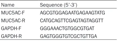
Using specific primers for MUC5AC as previ
-ously reported [9] (Table 2, Sigma, Hong Kong),
[image:2.612.91.290.180.249.2]PCR amplification was carried out using the fol -lowing conditions: 50°C for 30 min and 95°C pre-denature for 5 min, followed by 30 cycles each containing 95°C denature for 30 sec, 55°C annealing for 30 sec and 72°C elongation for 50 sec. The reaction ended with 72°C elon-gation for 10 min. The relative expression of the Table 1. siRNA sequence
Name Sequence (5’-3’) Bp
p38 siRNA sense UGUGUAUCUGGUGACCCAUCUTdT 21 p38 siRNA anti-sense AGAUGGGUCACCAGAUACACATdT 21 control siRNA sense UUCUCCGAACGUGUCACGUUU 21 control siRNA non-sense ACGUGACACGUUCGGAGAAUU 21
denature in boiled water for 5 min, pro-teins were separated by SDS-PAGE, and
were transferred to PVDF membrane under an electrical field. Primary anti -bodies, including anti-Mucin 5AC and anti-p38 (1:1 000, Abcam, Hong Kong) were added for overnight incubation. On the next day, the membrane was Table 2. Primer sequence for RT-PCR
Name Sequence (5’-3’)
MUC5AC-F AGCGTGGAGAATGAGAAGTATG MUC5AC-R CATGCAGTTCGAGTAGTAGGTT
target genes was determined by 2-ΔΔCt method
using GAPDH as the reference.
Statistical analysis
SPSS 10.0 software was used to collect all data, which were presented as mean ± stan-dard deviation (SD). Analysis of variance (ANOVA) was used to perform comparisons among all groups, followed by SNK-Q test to compare means between two groups.
Results
Cytokine release from HIBECs
The stimulus of LPS can activate the synthesis
and release of IL-1β and TNF-α from HIBECs
(Figure 1). The introduction of siRNA significant -ly depressed the release of both cytokines when compared to siRNA control and LPS groups (P<0.05, Figure 1).
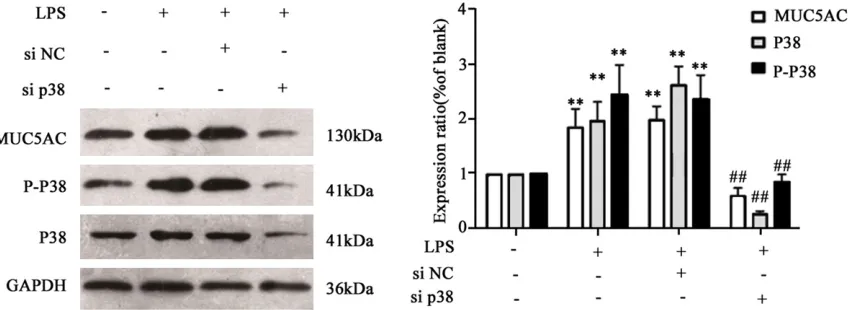
MUC5AC and phosphorylated p38 expression
Multiple inflammatory related signal pathways
can induce the elevated synthesis of MUC5AC
[10]. Meanwhile the activation of p38 can
induce further downstream signal pathways
[image:3.612.96.521.73.252.2]such as NF-κB [11]. To investigate the correla -tion between p38 expression and MUC5AC syn-thesis, we found that LPS treatment can acti-vate the phosphorylation of p38, accompanied with elevated MUC5AC expression (Figure 2). The interference of p38 caused depressed MUC5AC level (Figure 2, P<0.05). These results Figure 1. IL-1β (left) and TNF-α (right) levels in cultured medium of HIBECs. **P<0.05 compared to normal control
group; ##P<0.05 compared to siRNA control (si NC) group.
Figure 2. P38 phosphorylation (left) and MUC5AC expression (right) of cells. **P<0.05 compared to normal control
[image:3.612.96.523.305.460.2]suggested the potential involvement of p38 signal pathway in LPS-induced MUC5AC over- expression.
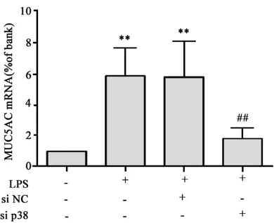
MUC5AC mRNA level
Consistent results have been obtained from mRNA of MUC5AC, as quantitative RT-PCR
showed significantly suppressed MUC5AC
mRNA level after the application of p38 siRNA (P<0.05, Figure 3).
Discussion
As a common surgical complication, intrahe-patic bile duct calculi, or hepatolithiasis, have unclear pathogenesis yet. Mucin is a type of high-molecular weight glycoprotein to coat or
protect epithelial cells [12]. Past studies have
revealed mucin as one important factor in bile duct calculi, as the intrahepatic bile duct disease is often accompanied with abnormal secretion of mucin. It has now widely accepted the over-secretion of MUC played an important role in the formation of intrahepatic bile duct
calculi [13]. Among various subtypes of MUC,
the elevation of MUC2 and MUC5AC in
hepato-lithiasis was evident [14].
The mechanism of endothelial hyperplasia and MUC over-secretion, however, still remain un- clear. More and more evidences supported the
participation of inflammation in those process
-es [15]. Related pathological proc-ess-es may
involve the aggregation of abundant MUC in the inner wall or the cavity of bile duct, causing mechanical bile duct obstruction and
cholesta-sis, both of which may further aggravate the
condition of inflammatory response. Therefore, the inhibition of inflammatory factor and cyto -kine release and related MUC production, has the potency to become the new drug target. Early clinical observations have found the
occurrence of bile duct inflammation accompa
-nied with calculi [16]. Other scholars also dem
-onstrated the correlation between inflamma -tion-induced hyperplasia of bile duct endotheli-al cells and the pathogenesis of intrahepatic
bile duct calculi. In such chronic inflammation
of bile duct, the re-formation of bile duct epithe-lial cells increases, along with over-secretion of MUC into the cavity. Similar process occurs in
the bronchial epithelial cells, whose IL-1β induc -es MUC5AC secretion via cAMP-PKA signal
pathway [17]. The over-expression of MUC5AC
has also been suggested to be related with
MAPK signal pathway [18]. Early studies have
demonstrated the activation of MUC2 and MUC5AC expression by LPS via up-regulating
TNF-α and protein kinase C signal pathway in mouse gall bladder epithelial cells [19]. In bac -terial infection, LPS, as the major component of bacterial cell wall, can stimulate the release of
multiple pro-inflammatory cytokines and fac -tors via stimulating HIBECs, which were also
induced to secrete abundant MUC [20].
As one important member of MAPK family, p38 is expressed in various cells and exerts certain
pro-inflammatory roles [21, 22]. Its role in regu -lating MUC5AC in intrahepatic bile duct calculi, however, remained unclear. This study thus investigated if the inhibition of p38 may regu-late MUC5AC secretion, for further treating intrahepatic bile duct calculi.
HIBEC can express abundant of MUC and thus mimics the formation of intrahepatic bile duct
calculi [23]. We thus selected HIBEC as the drug target. As previously reported [24], LPS can induce the occurrence of inflammation,
which is accompanied with MUC over-expres-sion in HIBEC, therefore mimicking the in vivo formation of intrahepatic bile duct calculi.
siRNA, with 20~24 nt length, can specifically
bind with target mRNA by complementary base paring, and induce the post-transcriptional gene silencing under the direction of Dicer enzyme, thus down-regulating the level of tar-get protein and further downstream signal
[image:4.612.92.287.70.229.2]pathways and activity of actors [25]. This study
Figure 3. MUC5AC mRNA level. **P<0.05 compared
utilized siRNA to inhibit the expression of p38, thus observing the role of p38 in calculi forma-tion. We used HIBEC with the help of LPS induc-tion to elevate MUC5AC expression. Our results found that LPS induced the over-expression of MUC5AC via p38 signal pathway in HIBECs.
The transfection by p38-siRNA can significantly decrease the level of IL-1β and TNF-α in culture
medium, plus the expression of MUC5AC. Our results collectively suggest the participation of p38 MAPK signal pathway in the formation and progression of intrahepatic bile duct calculi, and provide potential novel strategy for treating patients using p38-MAPK as the drug target. Acknowledgements
Research supported by the Medical Scientific Research Foundation of Guangdong Province, China (Grant No. A2013254).
Disclosure of conflict of interest
None.
Address correspondence to: Dr. Ping Wang, Depart-
ment of Hepatobiliary Surgery, The First Affiliated
Hospital of Guangzhou Medical University, 151 Yanjiang West Road, Yuexiu District, Guangzhou 510120, Guangdong, China. Tel: +86-137111769-
09; Fax: +86-13711176909; E-mail: wagnping006@
163.com
References
[1] Anninga JK, Gelderblom H, Fiocco M, Kroep
JR, Taminiau AH, Hogendoorn PC, Egeler RM. Chemotherapeutic adjuvant treatment for os-teosarcoma: where do we stand? Eur J Cancer 2011; 47: 2431-45.
[2] Tsui WM, Lam PW, Lee WK, Chan YK. Primary hepatolithiasis, recurrent pyogenic cholangitis, and oriental cholangiohepatitis: a tale of 3 countries. Adv Anat Pathol 2011; 18: 318-28.
[3] Feng X, Zheng S, Xia F, Ma K, Wang S, Bie P, Dong J. Classification and management of
hepatolithiasis: A high-volume, single-center’s experience. Intractable Rare Dis Res 2012; 1: 151-6.
[4] Sasaki M, Ikeda H, Ohira S, Ishikawa A, Nakanuma Y. Expression of trefoil factor family 1, 2, and 3 peptide is augmented in
hepatoli-thiasis. Peptides 2004; 25: 763-70.
[5] Feng YJ and Li YY. The role of p38 mitogen- activated protein kinase in the pathogenesis of
inflammatory bowel disease. J Dig Dis 2011; 12: 327-32.
[6] Liu CM, Ma JQ, Xie WR, Liu SS, Feng ZJ, Zheng
GH, Wang AM. Quercetin protects mouse liver against nickel-induced DNA methylation and
inflammation associated with the Nrf2/HO-1 and p38/STAT1/NF-kappaB pathway. Food
Chem Toxicol 2015; 82: 19-26.
[7] Geng S, Zhu W, Xie C, Li X, Wu J, Liang Z, Xie W, Zhu J, Huang C, Zhu M, Wu R, Zhong C. Medium-chain triglyceride ameliorates insulin
resistance and inflammation in high fat
diet-induced obese mice. Eur J Nutr 2015; [Epub ahead of print].
[8] Tormos AM, Taléns-Visconti R, Nebreda AR, Sastre J. p38 MAPK: a dual role in hepatocyte proliferation through reactive oxygen species.
Free Radic Res 2013; 47: 905-16.
[9] Dogru M, Okada N, Asano-Kato N, Tanaka M,
Igarashi A, Takano Y, Fukagawa K, Shimazaki J, Tsubota K, Fujishima H. Atopic ocular surface
disease: implications on tear function and ocu-lar surface mucins. Cornea 2005; 24 Suppl 8: S18-s23.
[10] Seshadri S, Lu X, Purkey MR, Homma T, Choi AW, Carter R, Suh L, Norton J, Harris KE, Conley DB, Kato A, Avila PC, Czarnocka B, Kopp PA,Peters AT, Grammer LC, Chandra RK, Tan BK, Liu Z, Kern RC, Schleimer RP. Increased expression of the epithelial anion transporter
pendrin/SLC26A4 in nasal polyps of patients
with chronic rhinosinusitis. J Allergy Clin Immunol 2015; [Epub ahead of print].
[11] Menden H, Welak S, Cossette S, Ramchan- dran R, Sampath V. Lipopolysaccharide (LPS)-mediated angiopoietin-2-dependent autocrine angiogenesis is regulated by NADPH oxidase 2 (Nox2) in human pulmonary microvascular en-dothelial cells. J Biol Chem 2015; 290:
5449-61.
[12] Yang L, Junmin S, Hong Y, Shuodong W. PGE(2) induces MUC2 and MUC5AC expression in hu-man intrahepatic biliary epithelial cells via
EP4/p38MAPK activation. Ann Hepatol 2013;
12: 479-86.
[13] Liu Z, Tian F, Feng X, He Y, Jiang P, Li J, Guo F,
Zhao X, Chang H, Wang S. LPS increases
MUC5AC by TACE/TGF-alpha/EGFR pathway
in human intrahepatic biliary epithelial cell. Biomed Res Int 2013; 2013: 165715.
[14] Sasaki M, Nakanuma Y and Kim YS. Expression of apomucins in the intrahepatic biliary tree in hepatolithiasis differs from that in normal liver and extrahepatic biliary obstruction. Hepato- logy 1998; 27: 54-61.
[15] Vilkin A, Nudelman I, Morgenstern S, Geller A, Bar Dayan Y, Levi Z, Rodionov G, Hardy B,
[16] Gatto M and Alvaro D. Cholangiocarcinoma: risk factors and clinical presentation. Eur Rev Med Pharmacol Sci 2010; 14: 363-7.
[17] Gray T, Nettesheim P, Loftin C, Koo JS, Bonner J, Peddada S, Langenbach R. Interleukin-1beta-induced mucin production in human air-way epithelium is mediated by cyclooxygen-ase-2, prostaglandin E2 receptors, and cyclic AMP-protein kinase A signaling. Mol Pharmacol
2004; 66: 337-46.
[18] Lee J, Komatsu K, Lee BC, Lim JH, Jono H, Xu H, Kai H, Zhang ZJ, Yan C, Li JD. Phosphodie-
sterase 4B mediates extracellular signal-regu -lated kinase-dependent up-regulation of mu-cin MUC5AC protein by Streptococcus pneu-moniae by inhibiting cAMP-protein kinase A- dependent MKP-1 phosphatase pathway. J Biol Chem 2012; 287: 22799-811.
[19] Zen Y, Harada K, Sasaki M, Tsuneyama K, Katayanagi K, Yamamoto Y, Nakanuma Y. Lipopolysaccharide induces overexpression of MUC2 and MUC5AC in cultured biliary epithe-lial cells: possible key phenomenon of hepato-lithiasis. Am J Pathol 2002; 161: 1475-84. [20] Van Seuningen I, Pigny P, Perrais M, Porchet N,
Aubert JP. Transcriptional regulation of the 11p15 mucin genes. Towards new biological
tools in human therapy, in inflammatory dis
-eases and cancer? Front Biosci 2001; 6:
D1216-34.
[21] Thouverey C and Caverzasio J. Focus on the
p38 MAPK signaling pathway in bone develop-ment and maintenance. Bonekey Rep 2015; 4:
711.
[22] Kuroyanagi G, Otsuka T, Yamamoto N, Matsu- shima-Nishiwaki R, Kozawa O, Tokuda H. Re-
sveratrol suppresses TGF-beta-induced VEGF synthesis in osteoblasts: Inhibition of the p44/ p42 MAPKs and SAPK/JNK pathways. Exp
Ther Med 2015; 9: 2303-2310.
[23] Lleo A, Zhang W, McDonald WH, Seeley EH, Leung PS, Coppel RL, Ansari AA, Adams DH, Afford S, Invernizzi P, Gershwin ME. Shotgun
proteomics: identification of unique protein profiles of apoptotic bodies from biliary epithe
-lial cells. Hepatology 2014; 60: 1314-23. [24] Tojima I, Shimizu S, Ogawa T, Kouzaki H,
Omura S, Sunazuka T, Shimizu T.
Anti-inflam-matory effects of a novel non-antibiotic macro-lide, EM900, on mucus secretion of airway epithelium. Auris Nasus Larynx 2015; 42:
332-6.
[25] Kaur IP, Chopra K, Rishi P, Puri S, Sharma G.
Small RNAs: the qualified candidates for gene
manipulation in diverse clinical pathologies.