and review of literatures
Feng Zhou1, Bingjian Lv1, Lifeng Dong2, Fang Wan2, Jiale Qin3, Lili Huang4
Departments of 1Pathology, 2Surgery, 3Ultrasound, 4Obstetrics and Gynecology, Women’s Hospital, School of Medicine, Zhejiang University, Hangzhou, Zhejiang Province, China
Received May 22, 2014; Accepted July 2, 2014; Epub June 15, 2014; Published July 1, 2014
Abstract: We report a very rare case of Peutz-Jeghers syndrome (PJS) composed of multiple genital tract tumors and mucinous adenocarcinoma. A 46-year-old woman presented to our hospital with lower abdominal pain result-ing from PJS involves sex cord tumor with annular tubules (SCTAT), ovarian mucinous tumor, ovarian serous tumor, mucinous adenocarcinoma of colon. The CEA concentration is high before surgery, and decreases after the surgery and subsequent chemoradiotherapy. This case demonstrates a classic clinical presentation of a patient with PJS. PJS patients have increased risk of malignancy and early detection and regular surveillance of the high-risk patients with PJS is crucial. Surgery may be required for obstructive gastrointestinal lesions as well as those exhibiting ma-lignant degeneration.
Keywords: PJS, SCTAT, ovarian mucinous cystadenoma, ovarian serous cystadenoma, mucinous adenocarcinoma of colon
Introduction
Peutz-Jeghers syndrome (PJS) is an inherited cancer syndrome characterized by mucocuta-neous melanin pigmentation and hamartoma-tous. Gastrointestinal polyps can result in chronic bleeding and anemia and also cause recurrent obstruction and intussusception requiring repeated laparotomy and bowel resection. Mucocutaneous hyperpigmentation presents as dark blue to dark brown macules around the mouth, eyes, and nostrils, and on
the fingers [1].
The incidence of PJS is estimated to be between 1 in 50,000 to 1 in 200,000 live births, and predisposition to benign and malignant tumors of the stomach, small intestine, pancreas, cer-vix, breast and ovaries. Polyps are the most common in the small intestine, but may occur anywhere in and outside the gastrointestinal
tract [1-3]. We present the unusual case of a
46-year-old woman with PJS who had a sex
cord tumor with annular tubules, ovarian nous tumor, ovarian serous tumor and muci-nous adenocarcinoma of colon.
Case report
The patient was 46-year-old, she had com-plained of lower abdominal pain for 2 weeks, and visited our hospital for a close evaluation. Since early childhood, she was noted to have hyperpigmented lesions over the perioral region (Figure 1A) and fingers (Figure 1B), and a his-tory of colon intussusception due to hamarto-matous polyps that was treated by partial col-ectomy for twice when she was 15 and 29 years old. Since his partial colectomy surgery, the patient had been on a three-year colonoscopic surveillance schedule for his remaining colon. He was treated by hysterectomy for leiomyoma-ta at 36 years old. The patient’s father also has perioral pigmentation, and died for intestinal intussusception at years old. Lower endoscopy
found no mass in the lumen of colon. The Pap smear showed normal cells. Pelvic ultrasound showed a cyst 15.1 × 13.8 × 12.6 cm at the left adnexal region, a cyst 4.7 × 4.4 × 4.1 cm at the right adnexal region and a mass 5.9 × 5.0 × 6.2 at the pelvic. The CEA levels were 53.8 ng/ml. A subsequent bilateral salpingo-oophorectomy was performed and a 25-cm large intestine with the mass and numerous 0.2-2.0 cm polyps removed surgically Macroscopically, the left and right ovary were cystic, 9.0 × 9.0 × 8.0 cm
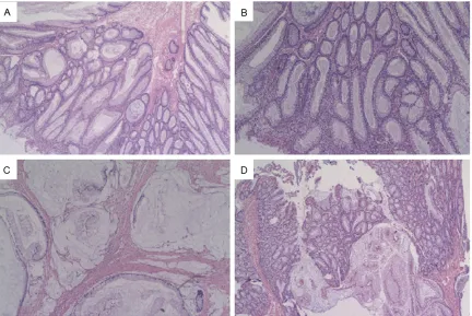
and 4.0 × 3.0 × 3.0 cm, respectively. Both ovi-ducts were unremarkable on gross examina-tion. There was numerous 0.2-1.5 cm polyps in the lumen and a pale yellow-tan and slimy mass under the polyps measuring 6 × 5 × 4 cm in the wall of the colon (Figure 1C).
[image:2.612.90.523.73.169.2]Histologically, the left and right ovary showed benign mucinous cystadenoma (Figure 2A) and benign serouscystadenoma (Figure 2B), re- spectively. Additionally, there were scattered Figure 1. Pigmentation around her lips (A) and fingers (B). Surgical specimen from colon resection with the lumen
opened displaying the numerous polyps and mass under the mucosa (C).
Figure 2. The cyst’s walls are lined with a one-layered mucin producing cubic to cylindrical epithelium, which is similar to an endocervical epithelium (A, 50×). Prominence of the ciliated cell, producing small fan-like projections
(B, 50×). Scattered small and calcified nests of sex cord tumors with annular tubules, and the sex cord element
[image:2.612.92.524.220.509.2]small nests of sex cord tumors with annular
tubules, some of which were calcified, in the
cortex and septa of the cysts from the both the ovaries (Figure 2C, 2D). Polyps from the colon
revealed mild acute inflammation superim -posed on architectural disorganization without dysplasia, suggestive of P-J polyps (Figure 3A), there are some adenomas with mild dysplasia in the colon (Figure 3B). The mass of the colon
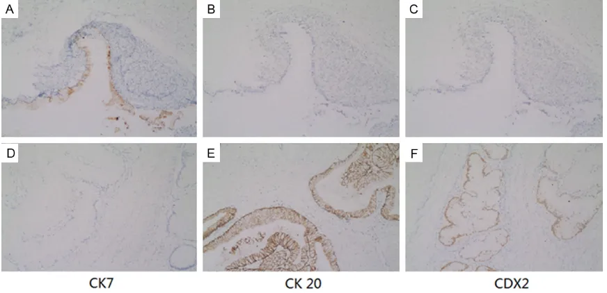
showed a deeply infiltrative, but well differenti -ated mucinous adenocarcinoma, which have penetrated through to the outer wall of the colon. The epithelial cells that lined the muci-nous glands were 2 to 3 times taller than con-ventional mucinous cells and the cytoplasm appeared pale and homogenous without dis-tinct goblet cell vacuolation (Figure 3C, 3D). By immunohistochemical methods, the tumor cells of mucinous tumor were strongly positive for cytokeratin 7 (CK7); negative for CK20 and CDX2 (Figure 4A-C). While the tumor cells of
the colon were strongly positive for CK20 and CDX2; negative for CK7 (Figure 4D-F). The CK7+/CK20- and CK7-/CK20+ pattern is typi-cal of epithelial ovarian tumors and intestinal tumors, respectively. CDX2, a critical nuclear transcription factor for intestinal development, is expressed in intestinal epithelium and ade-nocarcinomas. The pattern of this case indicat-ed that both the mucinous tumor of left ovary and mucinous adenocarcinoma of the colon are primary tumors. The CEA concentration is high (53.8 ng/ml) before surgery, and decreas-es after the surgery and subsequent chem- oradiotherapy.
Discussion
PJS is an autosomal dominant disorder charac-terized by the development of hamartomatous polyposis in the gastrointestinal tract from the stomach to the large intestine and melanin-pigmented macules on the skin mucosa, includ-Figure 3. Histologically, hamartomatous polyps showing arborizing musculature and hyperplastic glands without
dysplasia (A, 25×). Tubular adenomas shows a slightly modified mucosal architccture,glands are lined by enlarged
[image:3.612.90.522.72.374.2]ing the oral mucosa, lips, nasal wings and inter-digits. The diagnostic criteria for PJS include the presence of small bowel hamartomatous polyps, characteristic mucocutaneous pigmen-tation, and family history. Two of these criteria must be met in order to make a clinical
diagno-sis of PJS [1].
The responsible gene is a tumor suppressor, STK11/LKB1, on chromosome 19p13.3. PJS complicates with benign and malignant tumors in various organs.
A significantly increased risk of both gastroin -testinal and nongastroin-testinal malignancies has been demonstrated for patients with PJS. A report of 133 Dutch PJS patients from 54
fami-lies [2], a meta-analysis has been performed by Giardiello et al. [3], assessing 210 patients from six studies and Hearle et al. [4] examined
the incidence of cancer in 419 individuals with PJS, 297 of which had documented STK11 mutations. These three articles offer the most comprehensive data for cancer risk, demon-strate that PJS patients carry a markedly ele-vated cancer risk in PJS patients, and higher in females than in males, but independent of
fam-ily history and STK11 mutation status [2-4].
The incidence of malignant gastrointestinal tumors is highest in the large intestine, fol-lowed by the stomach, small intestine, duode-num and pancreas, and the incidence of
malig-nant tumors in other organs is highest in the uterine cervix, followed by the ovary and lung
[2-4]. In this case, the mucinous adenocarcino -ma is well differentiated, and the glands lined by tall columnar cells in comparison with con-ventional mucinous glands with goblet cells. So, we can not exclude the possibility of metas-tasis, such as cervical malignancy minimal deviation adenocarcinoma (MDA) and pancre-atic cancer. But the CK7-/CK20+ pattern and
expression of the CDX2 are highly specific of
colorectal origin.
The exact mechanism of carcinogenesis in PJS remains to be established. Two possible modes of cancer development have been proposed in PJS: de novo carcinogenesis and a
hamartoma-adenoma-carcinoma sequence [2]. In this case,
P-J polyps had hyperplastic glands and the
epi-thelial misplacement was florid and extended
into the serosa. Chains or irregular cell clusters
floating freely in mucinous lakes. Thus, the car -cinomas may occur in contiguity with p-j polyps. There are some tubular adenomas with dyspla-sia in the colon, so the colon cancer might have developed through hamartoma-adenoma-car-cinoma sequence.
[image:4.612.89.525.72.283.2]The clinical manifestations of SCTAT differ between patients with and without PJS. Young
[6] et al. conducted a comparative study in 21
SCTAT patients with PJS and 47 SCTAT patients without PJS, and found that SCTAT complicated with PJS is commonly multifocal, bilateral, small
(detected microscopically), and calcified in
>50% of cases, and has a good prognosis. In contrast, SCTAT without PJS is unilateral, large
(palpable), calcified in 12% of cases, and has a poor prognosis in 20%. Song [7] et al. described
the case of a 41-year-old woman with PJS who had multiple genital tract tumors and breast cancer. In our case, the woman had SCTAT, epi-thelial ovarian tumors and colon cancer with PJS.
Mucinous and serous epithelial ovarian tumors are also seen with increased frequency in patients with PJS and benign lesions that devel-op into tumors with mucinous to serous ratios
of 8:1 [8]. Serous cystadenoma of one ovarian
and mucinous cystadenoma of the other one with PJS in a patient is very rare and interest-ing. Three cases of serous tumor of the ovary
associated with PJS have been published [7, 9, 10]. Mucinous ovarian tumors with PJS can be benign, borderline, malignant [11-15], one of
them was diagnosed as ovarian mixed serous and mucinous borderline tumor, three ovarian mucinous adenocarcinoma were metastatic from the cervix, and in three cases the ovaries contained both primary and metastatic tumors. In our case, the mucinous tumor of right ovary is primary tumor for the CK7+/CK20- pattern. Conclusion
The case with the multiple genital tract tumors and mucinous adenocarcinoma of colon in a person with PJS has not been reported to date. This case demonstrates a classic clinical pre-sentation of a patient with PJS. PJS patients have increased risk of malignancy and early detection and regular surveillance of the high-risk patients with PJS is crucial. Surgery may be required for obstructive gastrointestinal lesions
partment of Obstetrics and Gynecology, Women’s Hospital, School of Medicine, Zhejiang University, 1 Xueshi Road, Hangzhou, Zhejiang Province, 310006, People’s Republic of China. Tel: 0086-571-87061501-2022; Fax: 0086-571-87061878; E-mail: 29402335@qq.com
References
[1] Westerman AM, Entius MM, de Baar E, Boor PP, Koole R, van Velthuysen ML, Offerhaus GJ, Lindhout D, de Rooij FW, Wilson JH. Peutz-Jeghers syndrome: 78-year follow-up of the original family. Lancet 1999; 353: 1211-5.
[2] van Lier MG, Westerman AM, Wagner A, Looman CW, Wilson JH, de Rooij FW, Lemmens VE, Kuipers EJ, Mathus-Vliegen EM, van Leerdam ME. High cancer risk and increased mortality in patients with Peutz-Jeghers syn-drome. Gut 2011; 60: 141-147.
[3] Giardiello FM, Brensinger JD, Tersmette AC, Goodman SN, Petersen GM, Booker SV, Cruz-Correa M, Offerhaus JA. Very high risk of can-cer in familial PeutzeJeghers syndrome. Gastroenterology 2000; 119: 1447-53.
[4] Hearle N, Schumacher V, Menko FH, Olschwang S, Boardman LA, Gille JJ, Keller JJ, Westerman AM, Scott RJ, Lim W, Trimbath JD, Giardiello FM, Gruber SB, Offerhaus GJ, de Rooij FW, Wilson JH, Hansmann A, Möslein G, Royer-Pokora B, Vogel T, Phillips RK, Spigelman AD, Houlston RS. Frequency and spectrum of can-cers in the Peutz Jeghers syndrome. Clin Cancer Res 2006; 12: 3209-15.
[5] Scully RE. Sex cord tumor with annular tu-bules: a distinctive ovarian tumor of the Peutz-Jeghers syndrome. Cancer 1970; 25: 1107-21.
[6] Young RH, Welch WR, Dickersin GR, Scully RE. Ovarian sex cord tumor with annular tubules: review of 74 cases including 27 with PeutzeJeghers syndrome and four with adeno-ma adeno-malignum of the cervix. Cancer 1982; 50: 1384-402.
[7] Song SH, Lee JK, Saw HS, Choi SY, Koo BH, Kim A, Yeom BW, Kim I. PeutzeJeghers Syndrome with multiple genital tract tumors and breast cancer: a case report with a review of literatures. J Korean Med Sci 2006; 21: 752-7.
tumors in patients with Peutz-Jeghers syn-drome (PJS). Open J Genetics 2011; 1: 65-9.
[9] Srivatsa PJ, Keeney GL, Podratz KC. Disseminated cervical adenoma malignum and bilateral ovarian sex cord tumors with an-nular tubules associated with Peutz-Jeghers Syndrome. Gynecol Oncol 1994; 53: 256-64.
[10] Dozois RR, Judd ES, Dahlin DC, Bartholomew LG. The Peutz-Jeghers syndrome: Is there a pre-disposition to the development of intesti-nal malignancy? Arch Surg 1969; 98: 509-17.
[11] Brichard B, Chantrain C, Wese F, Gosseye S, Vermylen C. Peutz-Jeghers syndrome and bilat-eral ovarian tumors in a 14-year-old girl. J Pediatr Hematol Oncol 2005; 27: 621-3.
[12] Mangili G, Taccagni G, Garavaglia E, Carnelli M, Montoli S. An unusual admixture of neoplastic and metaplastic lesions of the female genital tract in the Peutz-Jeghers syndrome. Gynecol Oncol 2004; 92: 337-42.
[13] Matseoane S, Moscovic E, Williams S, Huang JC. Mucinous neoplasm in the cervix associat-ed with a mucinous neoplasm in the ovary and concurrent bilateral sex cord tumors with an-nular tubules: im-munohistochemical study. Gynecol Oncol 1991; 43: 300-4.
[14] Lucidarme D, Dridba M, Khoury S, Vandermolen P, Foutrein P, Vandevenne P, Leduc M, Creusy C, Filoche B. Ovarian tumor and Peutz-Jeghers syndrome: A case report. Gastroenterol Clin Biol 1990; 14: 1015-8.