RESEARCH ARTICLE
The presence and role of interstitial cells of Cajal in the proximal
intestine of shorthorn sculpin (
Myoxocephalus scorpius
)
Jeroen Brijs1,*, Grant W. Hennig2, Anna-Maria Kellermann3, Michael Axelsson1and Catharina Olsson1
ABSTRACT
Rhythmic contractions of the mammalian gastrointestinal tract can occur in the absence of neuronal or hormonal stimulation owing to the generation of spontaneous electrical activity by interstitial cells of Cajal (ICC) that are electrically coupled to smooth muscle cells. The myogenically driven component of gastrointestinal motility patterns in fish probably also involves ICC; however, little is known of their presence, distribution and function in any fish species. In the present study, we combined immunohistochemistry andin vivorecordings of intestinal motility to investigate the involvement of ICC in the motility of the proximal intestine in adult shorthorn sculpin (Myoxocephalus scorpius).Antibodies against anoctamin 1 (Ano1, a Ca2+-activated Cl− channel), revealed a dense network of multipolar, repeatedly branching cells in the myenteric region of the proximal intestine, similar in many regards to the mammalian ICC-MY network. The addition of benzbromarone, a potent blocker of Ano1, altered the motility patterns seen in vivoafter neural blockade with TTX. The results indicate that ICC are integral for the generation and propagation of the majority of rhythmic contractile patterns in fish, although their frequency and amplitude can be modulated via neural activity.
KEY WORDS: Anoctamin 1, Enteric nervous system, Fish, Gut, Motility, Myogenic
INTRODUCTION
Complex gastrointestinal motility patterns occur throughout the gastrointestinal tract to accomplish crucial functions such as food digestion, nutrient absorption and removal of waste products (Kunze and Furness, 1999; Sanders et al., 2006). Recently, we
described a diverse array of in vivo motility patterns in adult
shorthorn sculpin (Myoxocephalus scorpius) for the first time (Brijs
et al., 2014). Many of the motility patterns were reminiscent of, and/ or may have similar functions to motility patterns documented in other vertebrates (e.g. peristalsis, migrating motor complexes,
‘myogenic ripples’; Clench and Mathias, 1992; D’Antona et al.,
2001; Hennig et al., 1999; Wingate, 1981).
Gastrointestinal motility patterns are achieved through
coordinated contractions and relaxations of smooth muscle in the gut wall which are controlled by a number of intrinsic and extrinsic mechanisms including: (1) myogenic factors such as slow waves, which are generated and propagated in interstitial cells of Cajal
(ICC) networks, (2) neurogenic factors such as intrinsic enteric reflexes modulated by extrinsic pathways, and (3) humoral factors such as hormones and paracrine factors (see Kunze and Furness, 1999; Olsson and Holmgren, 2001; Sanders et al., 2006). Nerves may also act directly on smooth muscle cells or via intermediaries such as intramuscular ICC (ICC-IM) and platelet-derived growth
factor receptor cells (i.e. PDGFRα; Heldin et al., 1998; Sanders
et al., 2012a; Sanders et al., 2006). To date, studies examining gastrointestinal motility in fish, including the effects of various neuropeptides and other signalling substances, have primarily used
in vitromodels (isolated segments of gut; Gräns and Olsson, 2011; Olsson and Holmgren, 2001). Only a few studies have examined the
underlyingin vivocontrol mechanisms (Brijs et al., 2014; Holmberg
et al., 2007, 2006; Rich et al., 2013). Findings from some of these studies have revealed that although blockade of neural activity (using tetrodotoxin, TTX) abolishes some motility patterns, other patterns persist, indicating that they are myogenic in origin (Brijs et al., 2014; Holmberg et al., 2007). The myogenically driven component of gastrointestinal motility patterns in fish has repeatedly been suggested to involve ICC (Brijs et al., 2014; Holmberg et al., 2007; Rich et al., 2013, 2007); however, currently our knowledge regarding the presence, distribution and function of these cells in fish is limited.
In mammals, ICC act as pacemakers (myenteric ICC/ICC-MY) or modulators of neurotransmission (ICC-IM and deep muscular plexus ICC/ICC-DMP) (Sanders et al., 2006). ICC-MY are essential for the generation and propagation of electrical slow waves (the rhythmic oscillations of smooth muscle membrane potentials that form the basis of smooth muscle contraction). Loss or disruption of ICC networks has been linked with many gastrointestinal disorders (Ward and Sanders, 2001). Much is known about the network behaviour of mammalian ICC as a result of direct investigations
utilizingc-kit(the proto-oncogene encoding the receptor tyrosine
kinase, Kit; Huizinga et al., 1995; Ward et al., 1994) and Ca2+
indicators (Hennig et al., 2004); however, indirect studies of ICC behaviour via the analysis of myogenically mediated motor
behaviours have also significantly contributed to our
understanding of the role of ICC (Bercik et al., 2000; D’Antona
et al., 2001; Ferens et al., 2005; Hennig et al., 2010a; Kobayashi et al., 1996; Yoneda et al., 2002).
ICC were originally described over a century ago, as small fusiform or stellate cells with prominent varicose processes that formed networks in gastrointestinal tissues (Cajal, 1911). Antibodies against Kit have been used extensively to label ICC throughout the gastrointestinal tract in a wide range of vertebrates (Christensen, 1992), but success in labelling of ICC using Kit antibodies in teleosts has been variable (Ball et al., 2012; Mellgren and Johnson, 2005; Parichy et al., 1999; Rich et al., 2007; Wallace
et al., 2005). Recently, anoctamin 1 (Ano1), a Ca2+-activated Cl−
channel found to be expressed exclusively in ICC has been used as a more specific marker for ICC (Espinosa et al., 2008; Gomez-Pinilla
Received 23 June 2016; Accepted 3 November 2016
1Department of Biological and Environmental Sciences, University of Gothenburg,
SE-405 30 Göteborg, Sweden.2Department of Physiology and Cell Biology,
University of Reno, Nevada, NV 89557, USA.3Department of Nature and
Engineering, Bremen University of Applied Sciences, Bremen 28199, Germany.
*Author for correspondence ( jeroen.brijs@bioenv.gu.se)
J.B., 0000-0002-3671-5038
Journal
of
Experimental
et al., 2009; Hwang et al., 2009). It has been shown that this channel plays a fundamental role in the generation of slow waves in the gastrointestinal tract in a range of mammals, as animals lacking functional Ano1 fail to develop slow waves in the gastrointestinal tract (Hwang et al., 2009). High-throughput screening for Ano1 inhibitors found that benzbromarone was a specific and potent blocker of this channel, thereby providing specific pharmacological tools to determine the role of Ano1 in the pacemaker activity of ICC (Bernstein et al., 2014; Huang et al., 2012).
The presence of Ano1-positive cells has been demonstrated in the zebrafish intestine from 3 days post-fertilization (dpf ) (Uyttebroek et al., 2013). The appearance of these cells corresponds well with the onset of propagating contractions observed in zebrafish larvae (Holmberg et al., 2003), supporting the idea that these Ano1-positive cells may be pacemaker ICC. Furthermore, support for the involvement of ICC in the generation of gastrointestinal motility in fish lies in the fact that shallow
propagating contractions (‘ripples’) observed in shorthorn sculpin
share many characteristics with myogenic contractions in
mammals (Brijs et al., 2014; D’Antona et al., 2001). However,
the majority of the evidence is circumstantial and therefore direct manipulation of ICC activity is required to determine the role of ICC in gastrointestinal motility of fish.
The objectives of the present study were to investigate whether Ano1 is a suitable marker for identifying ICC in shorthorn sculpin, and if so, to determine their presence in the myenteric region of the proximal intestine. Furthermore, the functional involvement of ICC in intestinal motor behaviour was investigated using a pharmacological antagonist of Ano1 channels and a previously
testedin vivomethod for qualitatively and quantitatively measuring
motility patterns in teleost fish (Brijs et al., 2014).
MATERIALS AND METHODS
Experimental animals and holding conditions
Shorthorn sculpin (Myoxocephalus scorpiusLinnaeus 1758) are a
benthic marine species described as opportunistic ambush predators (e.g. intermittent feeding strategy and carnivorous diet; Dick et al., 2009; Moore and Moore, 1974; Scott and Scott, 1988) that possess a
gastrointestinal system consisting of aU-shaped sac-like stomach,
pyloric caeca and a relatively short intestine (Olsson, 2011b; Seth and Axelsson, 2009). Individuals of both sexes, ranging between 100 and 400 g were captured off the west coast of Sweden. Fish were transported to the University of Gothenburg and kept in a fiberglass tank containing 1000 litres of re-circulated, aerated
seawater (30–33 parts per thousand, ppt) with shelter (clay plant
pots that have been split in half ) and substrate (stones) at 10°C. Fish were acclimated for a minimum period of 6 weeks with a 12 h light:12 h dark photoperiod and fed once a week with commercial
white fish corresponding to∼5–15% of body mass.
The fish were fasted for 3 weeks to ensure no food remained in the stomach or intestine, as potential postprandial motility patterns would confound our interpretations (Brijs et al., 2014). The fasting period was based on (1) a previous study on shorthorn sculpin,
which demonstrated that even 72 h post-feeding∼20% of the meal
still remained in the stomach (Seth and Axelsson, 2009) and (2) our own documentation of food contents still remaining in individuals after 2 weeks of fasting.
Animal care and all physiological experimental procedures were performed in accordance with guidelines and regulations approved by the ethical committee of Gothenburg, Sweden (ethical permit 167-2013). Unless otherwise stated, all reagents used in the study were purchased from Sigma-Aldrich (St Louis, MO, USA).
Immunohistochemistry
Shorthorn sculpin (N=3) were killed by a sharp blow to the head. A
ventral incision was made and the entire gastrointestinal tract was removed. The proximal intestine was cut open, pinned flat on to
dental wax and fixed in 4% paraformaldehyde (in 0.1 mol l−1
phosphate buffer, pH 7.3) at 4°C for 2–4 h. The preparations were
washed (3×10 min) with 0.1 mol l−1 phosphate buffered saline
(PBS; 0.9% NaCl, pH 7.2) and stored in 0.1 mol l−1PBS including
0.2% sodium azide.
Whole-mount preparations exposing the myenteric region were obtained by gently peeling off the mucosa, submucosa and most of the circular muscle layer, leaving a thin layer of circular muscle over the myenteric region (to minimize or prevent the removal of enteric
neurons and ICC from the region). The preparations (∼5×5 mm)
were incubated overnight with either a primary antibody against anoctamin 1 alone or in combination with one of two neuronal markers, anti-Hu C/D or anti-acetylated tubulin (see Table S1). After washing (3×10 min) with PBS, the preparations were incubated with the appropriate secondary antibody or antibodies
(see Table S1) for 1–2 h, then washed with PBS (3×10 min) and
subsequently mounted on slides with carbonate-buffered glycerol. All incubations took place in a humid chamber at room temperature.
The antibodies were diluted using 0.1 mol l−1PBS, containing 2%
NaCl, 0.1% bovine serum albumin and 0.2% NaN3.
The preparations were observed using a Nikon Eclipse E1000 digital fluorescence microscope equipped with a Nikon Digital Camera DXM1200 and the Nikon software, ACT-1 (Nikon, Melville, NY, USA). Adjustment of colour, contrast and brightness of pictures were made in Adobe Photoshop CS5
(Adobe Systems, New York, USA). Density and shape
measurements were performed in VolumetryG8d (a custom-made program designed and developed by G.W.H.).
Recording and analysis of gastrointestinal motility patterns
Individual shorthorn sculpin were anaesthetized in water containing
75 mg l−1MS222 (ethyl-3-aminobenzoate methanesulphonic acid)
buffered with 150 mg l−1 NaHCO
3. Length and mass of the fish
were recorded prior to the fish being placed ventral side up on soft, water-saturated foam on the surgical table. To maintain anaesthesia, gills were continuously flushed with aerated water containing
50 mg l−1MS222 buffered with 100 mg l−1NaHCO
3at 10°C.
Relative cardiac output and heart rate were monitored throughout the experiment to ensure fish were sufficiently anaesthetized by using a 20 MHz Doppler flow crystal (Iowa Doppler products, Iowa
City, IA, USA) embedded in 1.6–2.3 mm cuffs (depending on size
of fish) on the ventral aorta adjacent to the bulbus arteriosus. The lead from the Doppler flow probe was connected to a directional-pulsed Doppler flowmeter (model 545C-4, Iowa Doppler products), which was in turn connected to a PowerLab 8/30 system (ADInstruments, Castle Hill, Australia). Data was collected on a PC using ADInstruments acquisition software Chart 7 Pro v.7.2.5, at
a sampling rate of 10 Hz. ‘Pre-operative’ heart rate and relative
blood flow of anaesthetized fish was measured for 15–20 min prior
to accessing the proximal intestine. If heart rate or relative blood
flow values decreased to less than 70% of‘pre-operative’values
during the experimental period then the fish was excluded (one individual was excluded because of a deterioration in cardiac performance).
The proximal intestine was accessed and prepared for video-recording using methods described in detail in Brijs et al. (2014). Briefly, a mid-ventral incision was made to access the abdominal
cavity, whereupon the proximal intestine was gently extracted from
Journal
of
Experimental
the cavity and placed in a modified Petri dish (total volume 125 ml). The proximal intestine was submerged with re-circulating shorthorn
sculpin Ringer’s solution (206 mmol l−1NaCl, 81 mmol l−1KCl,
2 mmol l−1 CaCl
2·2H2O, 1 mmol l−1 MgCl2·6H2O, 1 mmol l−1
NaH2PO4·2H2O, Na2HPO4·2H2O, pH 7.45) bubbled with air at
10°C. The intestine was held in place by guiding it around a large
pin positioned in the area where the intestine forms aU-bend. Care
was taken not to twist, restrict or damage any blood vessels or nerves during this process. A piece of black cotton was placed over the mesentery and middle intestine, with fiber-optic lights strategically placed to ensure optimum contrast between the background and proximal intestine. The exposed abdominal cavity was covered with
a chamois saturated in Ringer’s solution and the fish was left
undisturbed for 1 h before video recording commenced. The recovery time was deemed necessary from our previous study (Brijs et al., 2014), as rhythmic and repeatable intestinal motility
patterns returned 30–60 min after the completion of the surgical
procedure.
A calibration bar (25 mm) was placed in the field of view as a spatial reference. Images were captured using a DMK31AF03 monochrome, FireWire camera (The Imaging Source, Putzbrunn, Germany). Resolution was 1024×768 pixels; corresponding to a field of view at the magnification used of 54×39 mm. Images were captured at 3.75 frames per second for 30 min for three successive videos. A recording period of 30 min was selected to ensure that a representative sample of motility patterns was recorded, as our previous study demonstrated periods of inactivity and low contraction frequency in shorthorn sculpin (Brijs et al., 2014). The sequence of video-recordings consisted of the following:
control period (only Ringer’s solution), neuronal blockade (TTX,
Alomone Labs, Jerusalem, Israel) and blockade of ICC
(benzbromarone). Fish were left undisturbed for 15 min between each period to allow the blockers sufficient time to affect the proximal intestine prior to the subsequent recording. During our previous study, we observed that preparations remained viable for long periods of time (>5 h) (Brijs et al., 2014), but to ensure that the section of proximal tissue was still functional at the end of the
experiment, carbachol was added to the Ringer’s solution to induce
contractions. All fish responded to carbachol and were hence included in the final analysis. At the end of the experimental period, fish were killed with an overdose of anaesthetic.
All drugs were administered by adding 125 µl of stock solution
(0.001 mol l−1 TTX, 0.1 mol l−1 benzbromarone, 0.001 mol l−1
carbachol) to the modified Petri dish to achieve final bath
concentrations of 1 µmol l−1 (TTX and carbachol) and
100 µmol l−1(benzbromarone). The final concentrations selected
for TTX and carbachol (1 µmol l−1) have previously been
demonstrated to diminish/abolish or induce gastrointestinal motility in fish, respectively (Jensen and Holmgren, 1985; Karila and Holmgren, 1995; Kitazawa et al., 2012). The final bath
concentration selected for benzbromarone (100 µmol l−1) was
selected after initial testing of both 10 µmol l−1and 100 µmol l−1,
which were concentrations demonstrated to successfully block Ano1 in mammalian studies (Bernstein et al., 2014; Huang et al., 2012). TTX and carbachol were dissolved in 0.9% saline, whereas benzbromarone was dissolved in dimethyl sulfoxide (DMSO). To minimize any potential effects of the solvent, we ensured that the final concentration of DMSO in the bath did not exceed 0.1% (v/v), as this concentration has been previously shown to have no effects
on gastrointestinal motility in a range ofin vitroandin vivostudies
on fish (Jensen and Conlon, 1997; Shahbazi et al., 1998; Zhou et al., 2014).
Video recordings were analyzed by importing files into Volumetry G8d to construct spatiotemporal maps (ST maps). ST maps can be used to visualize and quantitatively describe complex motility patterns, as well as their spatial (regional distribution) and temporal (frequency) characteristics, as previously described (Brijs et al., 2014; Hennig et al., 1999). Briefly, images were thresholded to define the outer edges of the proximal intestine, which indented as contractile waves passed. The distance between the thresholded edges of the intestine was calculated for each pixel along the intestine and colour coded as a percentage change compared with
the average outer diameter (ΔODavg) at that pixel calculated over the
entire recording period (30 min). Diameters less than the average were colour-coded red (i.e. maximal contraction coded as fully saturated red), diameters equal to the average were colour-coded white, and diameters greater than the average were colour-coded blue (maximal dilation coded as fully saturated blue). Each video frame produced a single row of colour-coded pixels, corresponding to the relative diameters of the intestine at each point along the preparation. All the video frames in the recording (30 min: 6750 frames) were used to construct an ST map. Different types of contractions and their respective motion characteristics (i.e. frequency, velocity, direction) were quantified in VolumetryG8d (see below). Cumulative histograms of the prevalence of the range of relative diameters in different conditions during the recording period were used to assess overall contraction amplitudes.
Particle analysis
Particle analysis was used to more accurately assess the nature and prevalence of specific types of motor behaviours. Briefly, contraction peaks were enhanced using an inflexion detector
(Δt=75–187.5 s), smoothed (Gauss 7×7 pixel kernel, s.d.=1.5),
thresholded (inflexion sum≥50%) and flood-filled. The space–time
coordinates of each contraction peak were stored and the overall object ( particle) numbered. The leading edge coordinates of each particle were extracted and used to calculate the instantaneous velocity at each point along the particle (using linear regression over a span±0.3 mm), as well as the interval between each contraction peak. Results were plotted as histograms. Quantitative descriptions of contractions were limited after benzbromarone due to the lack of resolvable contractions (see Results and Discussion).
Parameters of wall motion
Three main parameters of wall motion were compared before and after drugs in this study. The frequency of phasic contractions was measured as the interval between successive events and was
calculated automatically for‘ripples’and manually for slow
anally-propagating contractions. Longer intervals equate to a slower frequency of events. The amplitude of contractions/relaxations is commonly used to measure neural motor output to the muscle, but can also reflect excitation-coupling between ICC (slow waves) and muscle. The velocity and direction of propagating contractions is a measure of the stability of initiation sites, and the coupling between cells in the network in which activity is propagating through (e.g. slow waves in ICC-MY or migrating motor complexes).
Statistical analysis
Data were assessed for outliers and normality (Shapiro–Wilk>0.05).
To meet these assumptions, the frequency of resolvable contractions
data were transformed using a log10 transformation. A one-way
repeated measures ANOVA and Scheffepost hoctest were used to
determine whether significant differences in overall contraction
amplitude occurred between the different treatments (i.e. control,
Journal
of
Experimental
TTX and benzbromarone). The use of normalized contraction values to better identify active contractions prevented comparisons between groups using the mean (normalized to 0). Instead, the prevalence of contractions that reduced the diameter by 10% or more was chosen as a comparison point as it was representative of
the overall shift between the curves and the strength of these contractions were likely to be of biological importance.
Paired-samplet-tests were used to determine whether TTX significantly
affected any parameters of the contractions (i.e. frequency of resolvable contractions and amplitude of specific motility patterns).
A B
Ano1 Ano1D
C E
Ano1 Hu C/D Ano1/Hu C/DG
F H
J
I K
M
L N
Ano1 AcT Ano1/AcT
Ano1 AcT Ano1/AcT
Ano1 AcT Ano1/AcT
*
*
[image:4.612.55.560.134.657.2]*
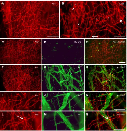
Fig. 1. Ano1 immunoreactivity in the proximal intestine of shorthorn sculpin (Myoxocephalus scorpius).(A) A dense network of Ano1-immunoreactive cells between the circular and longitudinal muscle layers. (B) Multipolar Ano1-immunoreactive cells (asterisks), with branching (arrowhead) interconnected processes shown at higher magnification. Arrows indicate broad, flattened processes. (C-N) Double labelling with Ano1 and the neuronal markers Hu C/D (C-E) or acetylated tubulin (AcT) (F-N) shows the relationship between the ICC network and the myenteric nervous plexus. (C-E) There is no overlap between the Ano1-immuoreactive network (C) and Hu C/D-immunoreactive nerve cell bodies (D), as shown by the merged image in E. (F-N) Neither is there any overlap between Ano1-immuoreactive processes (F,I,L) and the AcT-immunoreactive nerve fibres (G,J,M) as shown by the merged images (H,K,N). I-N show increased magnification of different regions of F-H; here, it is clearly visible that some immunoreactive processes run alongside but separate from individual nerve fibres (I-K). It also shows that Ano1-immunoreactive cells (arrow) can be observed close to the thicker nerve strands (L-N). Scale bars: 50 µm (B,I-N), 100 µm (A,C-H).
Journal
of
Experimental
A
B
C
D
E
F
G
H
I
Control TTX Benz
Control TTX Benz
Control TTX Benz
5 mm 5 mm 5 mm
5 mm
15 mm 10 mm
5 mm 5 mm 5 mm 5 mm
300 s 300 s
300 s
–2.5 %
+2.5 %
ΔODavg
–5.0 %
+5.0 %
ΔODavg
–5.0 %
+5.0 %
ΔODavg
>200
0
s
[image:5.612.107.499.56.587.2]* *
* *
Fig. 2. The effect of neuronal blockade and blockade of ICC onin vivomotility patterns in the proximal intestine of shorthorn sculpin.Spatiotemporal (ST) maps portray changes in the diameter along the exteriorized section of the proximal intestine (ΔODavg,fully contracted=red, no contractile activity=white, fully dilated=blue). The horizontal axis represents the distance from the oral (left) to mid (right) section of the proximal intestine, whereas the vertical axis represents the recording period (0–30 min). Displayed below each ST map are the overall outlines of the exteriorized intestines throughout the 30 min experimental periods, which demonstrate on a 16-bit grayscale, the amount of the time that the edges of the intestine spent at a particular position (from black to white=from 0 s to >200 s). (A,D,G) The predominant rhythmic motility patterns in the proximal intestine of three individual shorthorn sculpin under control conditions are slow anally-propagating (see dashed arrows that either overlay the contraction or the distention located anally to the contraction) and orally anally-propagating ripples (sloping to the left, asterisks in D). Slow anally propagating contractions are prolonged, circular muscle contractions that slowly propagate in an anal direction (i.e. from left to right on the ST map) over a large proportion of the proximal intestine. Ripples are the rhythmic, shallow circular muscle contractions primarily propagating in an oral direction (i.e. from right to left on the ST map) over relatively smaller distances when compared with the slow anally propagating contractions. (B,E,H) Neurogenic blockade (1 µmol l−1TTX) did not abolish the slow anally propagating contractions or ripples, but altered their frequency and amplitude. (C,F,I) Blockade of interstitial cells of Cajal (ICC) with benzbromarone (100 µmol l−1) significantly reduced or abolished ripples and slow anally propagating contractions. Shallow, low-frequency contractile activity was seen in some fish, as well as a few random and minor non-propagating contractions.
Journal
of
Experimental
P<0.05 was regarded as statistically significant. All statistical
analyses were performed with IBM SPSS Statistics 21.
Immunohistochemical data are presented as means±s.d.; unless otherwise stated, the remaining data are presented as means±s.e.m.
RESULTS
Presence of ICC in proximal intestine
Ano1 immunoreactivity revealed a dense network of Ano1-positive cells in the myenteric region (i.e. between the circular and longitudinal muscle layers) of the shorthorn sculpin proximal intestine (Fig. 1A). The network consisted of interconnected cells with repeatedly branching processes (Fig. 1B). The processes seemed to form anastomoses, giving the appearance of a mesh (Fig. 1A,B). Individual cell bodies were multipolar, with a comparatively large ovoid nucleus (nucleus major axis=8.8±
1.1 µm, minor axis=4.2±1.0 µm, mean±s.d., N=12 cells from 4
photos, Fig. 1B). Owing to the complex, overlapping structure of cell processes in the network, it was difficult to precisely determine cell density in terms of cells per unit area; however, the area labelled by Ano1 immunoreactivity (cell bodies and processes) in relation to
the total field of view was 25±2% (N=4).
Double-labelling with the neuronal markers acetylated tubulin (AcT) or Hu C/D showed that the Ano1-immunoreactive network was clearly distinct from the neuronal myenteric plexus (Fig. 1C-N). As previously shown (Olsson, 2011a), Hu C/D-immunoreactive enteric nerve cell bodies were found scattered throughout the plexus, whereas AcT-immunoreactive nerve fibres composed a dense network that included both single fibres and thicker nerve strands (see Fig. 1G). Although some Ano1-immunoreactive processes run very close to AcT-immunoreactive nerve fibres, merged images showed no overlap between the different structures (see Fig. 1H,K, N). In addition, most of the Ano1-immunoreactive network seemed to be located in a slightly different focal plane than the nerve plexus. These results suggest that the sculpin has a developed ICC network at the level of the myenteric plexus (ICC-MY).
Spontaneous gastrointestinal motility patterns
The motility patterns observed in the proximal intestine were similar to those observed in our previous study (Brijs et al., 2014). The most
prevalent patterns consisted of ‘ripples’ and slow
anally-propagating contractions (i.e. observed in all fish, N=8, see
examples in Fig. 2A,D,G and Movie 1). The range (95% confidence intervals) of overall contraction amplitudes quantified
during control conditions was−18 to +18% of the average outer
diameter of the intestine (ODavg; Fig. 3). Under control conditions,
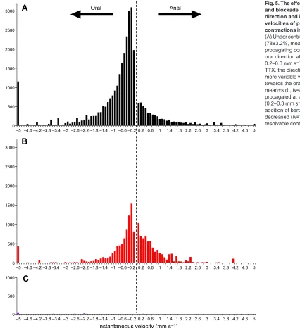
‘ripples’propagated primarily in an oral direction (77±3%, mean±
s.d.,N=8) at a modal frequency of 2.8 contractions per minute (cpm;
Fig. 4) and modal velocity of 0.20–0.30 mm s−1 (Fig. 5). Slow
anally propagating contractions occurred at an average frequency of
0.33±0.02 cpm and had an average velocity of 0.10±0.01 mm s−1.
These slowly propagating contractions had graded effects on the higher-frequency contractions of the proximal intestine ranging
from: (1) little effect on the pattern of underlying ‘ripples’but a
slight enhancement of their amplitude (Fig. 2A), (2) noticeable
enhancement of‘ripples’often with a sustained or nearly sustained
contraction at the leading edge of the propagating contraction (Fig. 2D) and (3) sustained, high-amplitude contractions during
which underlying‘ripples’were no longer apparent (Fig. 2G, top).
In many cases, this motor behaviour resulted in a significant displacement of contents in the anal direction, as evidenced by the slow-propagating dilations ahead of these types of contractions (blue streaks in Fig. 2A,B,G; see overlaid yellow dashed lines).
Myogenic gastrointestinal motility patterns
Blockade of neural activity with tetrodotoxin (TTX: 1 µmol l−1) did
not abolish‘ripples’or slow anally propagating contractions, but
instead altered the characteristics of these motility patterns (Fig. 2B, E,H and Movie 1). The range (95% confidence intervals) of overall
contraction amplitudes quantified during neural blockade (−17 to
+21% ODavg.; Fig. 3) was not significantly different than under
control conditions. However, further investigation into the amplitude of specific motility patterns revealed that neural
blockade significantly reduced the amplitude of ‘ripples’ (ΔOD
control=−11±2% versus ΔOD TTX=−8±1%; t=2.65, d.f.=7,
P=0.03) and slow anally propagating contractions (ΔOD control=
−41±5% versus ΔOD TTX=−24±3%; t=3.18, d.f.=7, P=0.02).
Furthermore, TTX altered the frequency distribution, increasing the prevalence of lower-frequency contractions (modal intervals of 70 and 130 s corresponding to 0.86 and 0.46 cpm, respectively; Fig. 4B). Although the modal instantaneous velocity was
unchanged compared with controls (0.20-0.30 mm s−1; Fig. 5B),
the strong bias of contractions propagating in an oral direction was
reduced (60±26%, mean±s.d., N=8) and there was an overall
reduction in resolvable contractions (down to 50% of that in
controls,t=2.54, d.f.=7,P=0.04).
Effect of blockade of Ano1 currents on gastrointestinal motor patterns
Subsequent addition of benzbromarone (100 µmol l−1) after TTX
(1 µmol l−1) had a substantial impact on the contractile activity of
the proximal intestine of shorthorn sculpin (Fig. 2C,F,I and
Movie 1). Benzbromarone not only abolished‘ripples’ and slow
anally propagating contractions but nearly all forms of contractile activity in the proximal intestine of shorthorn sculpin with the exception of a few random and minor non-propagating contractions (Fig. 2C,F,I and Movie 1). The range of overall contraction amplitudes following the addition of benzbromarone was reduced to
−6 to +7% ODavg(95% confidence intervals; Fig. 3). Resolvable
contractions were reduced to <2% of controls and detailed
Diameter (ΔODavg)
–50 –10 –40 10 –30 –20 20 30 40 50
ST
map area (%)
50
*
*
Control
TTX (1 μmol l–1)
Benzbromarone (100 μmol l–1)
[image:6.612.318.555.56.258.2]0
Fig. 3. The effect of neuronal blockade and blockade of ICC on overall contraction amplitudes in the proximal intestine.The prevalence of all diameters (ΔODavg) along the proximal intestine during the 30 min recording period in 8 fish is similar between controls (black) and after the addition of TTX (1 µmol l−1: red). Further addition of benzbromarone (100 µmol l−1) significantly reduces contraction amplitudes when compared with control and TTX [ANOVA at−10% ODavg(see shaded box):P<0.05,N=8].
Journal
of
Experimental
frequency/velocity information could not be quantified. However, in a few fish (Fig. 2C,F) very faint contractions were still observed at
a frequency of 0.40–0.46 cpm. All fish included in the analysis
responded with marked contractions of the intestine after the addition of carbachol, indicating that benzbromarone did not negatively impact cholinergic or contractile mechanisms.
DISCUSSION
Evidence for ICC in the gastrointestinal tract of shorthorn sculpin
Immunohistochemistry revealed a dense network of multipolar Ano1-positive cells in the myenteric region of the proximal intestine in adult shorthorn sculpin. The general appearance of the network (i.e. location, multipolar cells and branching interconnected processes) is similar to the ICC-MY described in mammals (Komuro, 2006), and is unlikely to be due to labelling of other cells types with branched morphology in this region (e.g. macrophages) that do not form an interconnected network (Sanders et al., 2006; Ward and Sanders, 2001). Therefore, based on the location and morphology of Ano1-positive cells, we suggest that these cells are similar to mammalian ICC (Gomez-Pinilla et al., 2009; Komuro, 2006) in this species and by extension, play a putative role as pacemakers for gastrointestinal motility.
The presence of ICC-like cells in the myenteric region was reported for some teleost species using Methylene Blue in the 1940s (Kirtisinghe, 1940). Since then, subsequent investigations using
Kit-antibodies to detect ICC in zebrafish (Danio rerio) have been
conflicting, with some studies failing to detect them (Mellgren and Johnson, 2005; Parichy et al., 1999; Uyttebroek et al., 2013), whereas others have suggested the presence of putative ICC in the myenteric region and circular muscle layer (Ball et al., 2012; Rich et al., 2007). Recently, a network of Ano1-positive cells has also been described in larval and adult zebrafish (Uyttebroek et al., 2013), which in combination with our observations strongly indicate that Ano1 is a more reliable marker for ICC than Kit in the gastrointestinal tract of fish. The few successful studies using Kit antibodies for identifying ICC in fish (Ball et al., 2012; Rich et al., 2007) described cells with similar characteristics as the Ano1-positive cells reported here; however, they were only able to detect these cells at 7 dpf. In contrast, Ano1-positive cells could be identified as early as 3 dpf (Uyttebroek et al., 2013). In mammals, Ano1 and Kit are simultaneously expressed by all ICC but Ano1 is considered to be a more specific marker as Kit immunoreactivity can
be weaker in some ICC (i.e.‘Kit-dim’ICC) and Kit has also been
shown to label mast cells (Gomez-Pinilla et al., 2009). The present study strongly indicates that Ano1 can be utilized as a specific
Interval (s)
A
B
C
Combined count
0 1000 2000 3000 4000 5000 6000
0 15 30 45 60 75 90 105 120 135 150 165 180 195 210 225 240 255 270 285 300
0 1000 2000 3000 4000 5000 6000
0 15 30 45 60 75 90 105 120 135 150 165 180 195 210 225 240 255 270 285 300
0 1000
0 15 30 45 60 75 90 105 120 135 150 165 180 195 210 225 240 255 270 285 300
0 500 1000 1500 2000
Interval (s) 0
500 1000 1500 2000
Interval (s)
[image:7.612.49.398.52.469.2]0 5 10 15 20 25 30 35 40 45 50 55 60 65 70 75 80 85 90 0 5 10 15 20 25 30 35 40 45 50 55 60 65 70 75 80 85 90
Fig. 4. The effect of neuronal blockade and blockade of ICC on the frequency of contractions in the proximal intestine. (A) Histogram of the prevalence of intervals between contractions throughout the section of proximal intestine. The dominant interval was 21–22 s, corresponding to a frequency of
∼2.8 cpm. Lower-frequency contractions (up to 3–4 min intervals, 0.33–0.25 cpm) were observed, but tapered off at greater intervals. Inset shows a detail of intervals between 0 and 90 s. (B) Trimodal distribution of intervals after the addition of TTX (see detailed 0–90 s inset). Dominant intervals were 17, 30 and 70 s, and as for controls, lower-frequency contractions were detected (up to 3–4 min intervals,
0.33–0.25 cpm). (C) After the addition of benzbromarone, very few contractions could be measured. The dashed vertical line provides a reference point at 90 s.
Journal
of
Experimental
marker for ICC in fish, which provides a platform for future investigations into the currently unknown distribution of ICC in the different regions and layers of the piscine gastrointestinal tract.
Role of ICC in the control of gastrointestinal motility in fish
Using a previously developed and tested method for examining spatial and temporal gastrointestinal contractile activity (Brijs et al., 2014), without severing the gut from its blood supply and extrinsic nervous control systems, we were able to investigate the
mechanisms that generate differentin vivomotility patterns in the
proximal intestine of shorthorn sculpin. Control gastrointestinal motility patterns in the proximal intestine of shorthorn sculpin were
qualitatively and quantitatively similar toin vivomotility patterns
described in our previous study (Brijs et al., 2014).
The most common motility patterns in the present study were the
shallow, rhythmic contractions (2.8 cpm) referred to as ‘ripples’
(Brijs et al., 2014), in analogy with the initial description of similar
patterns in the guinea pig proximal colon (D’Antona et al., 2001).
Ripples primarily propagated in an oral direction, which has been suggested to promote or optimize absorption (Chen et al., 2013; Dinning et al., 2012; Hennig et al., 2010a), as the contractions may aid in the mixing and circulation of intestinal contents over the mucosal surface of the gastrointestinal tract (Lee, 1983). Ripples persisted in all individuals following neuronal blockade, similar to
the‘myogenic ripples’described in numerous mammalian studies
(Benard et al., 1997; D’Antona et al., 2001; Dinning et al., 2012;
Hennig et al., 2010a; Huizinga et al., 2011). However, instead of the fairly constant frequency seen under control conditions, the prevalence of lower-frequency ripples was markedly increased. This suggests that some form of neural activity is required to stabilize high-frequency ripples under control conditions. It has been suggested that myogenic ripples in mammals are the phasic contractions in the gastrointestinal tract caused by underlying slow waves, which are generated and actively propagated by ICC (Bercik et al., 2000; Hennig et al., 2010a; Sanders et al., 2006). The
major initiating inward current in ICC is due to the opening of Ca2+
-0 500 1000 1500 2000 2500 3000
–5 –4.6 –4.2 –3.8 –3.4 –3 –2.6 –2.2 –1.8 –1.4 –1 –0.6 –0.2 0.2 0.6 1 1.4 1.8 2.2 2.6 3 3.4 3.8 4.2 4.6 5
0 500 1000 1500 2000 2500 3000
–5 –4.6 –4.2 –3.8 -3.4 –3 –2.6 –2.2 –1.8 –1.4 –1 –0.6 -0.2 0.2 0.6 1 1.4 1.8 2.2 2.6 3 3.4 3.8 4.2 4.6 5
0 500 1000
–5 –4.6 –4.2 –3.8 –3.4 –3 –2.6 –2.2 –1.8 –1.4 –1 –0.6 –0.2 0.2 0.6 1 1.4 1.8 2.2 2.6 3 3.4 3.8 4.2 4.6 5
Instantaneous velocity (mm s–1)
Anal Oral
A
B
C
[image:8.612.57.483.57.523.2]Combined count
Fig. 5. The effect of neuronal blockade and blockade of ICC on the overall direction and instantaneous velocities of propagating
contractions in the proximal intestine. (A) Under control conditions, the majority (78±3.2%, mean±s.d.,n=8) of propagating contractions travelled in the oral direction at a modal velocity of 0.2–0.3 mm s−1. (B) After the addition of TTX, the direction of propagation was more variable without the strong bias towards the oral direction (60±26%, mean±s.d.,N=8) but contractions propagated at a similar modal velocity (0.2–0.3 mm s−1). (C) The subsequent addition of benzbromarone dramatically decreased (N=3) or abolished (N=5) resolvable contractions.
Journal
of
Experimental
activated Cl− channels that are highly expressed in mammals (Gomez-Pinilla et al., 2009; Hwang et al., 2009). This channel also appears to be involved in the propagation of the slow wave front through the ICC-MY network in the mouse intestine (Singh et al., 2014). Blockade of this channel prevents the depolarization step that
opens other Ca2+channels and prevents the pacemaker transients
from being generated in ICC (Sanders et al., 2006, 2012b).
Although membrane potential and Ca2+ activity within the ICC
were not directly measured in this study, the blockade of these channels produced an effect consistent with blocking slow waves in ICC (i.e. the loss of rhythmic propagating contractions). Our findings strongly suggest that the generation and propagation of slow waves in the ICC in the proximal intestine of shorthorn sculpin
is fundamental for the initiation and propagation of in vivo
gastrointestinal motility patterns. Similar results have been observed in a range of mammals, as pharmacological blockade of Ano1 expressed in ICC prevented the generation of slow waves in the stomach and intestine with consequent adverse effects on gastrointestinal motility (Hwang et al., 2016, 2009; Zhu et al., 2009). Our findings concerning the function of ICC in the gastrointestinal tract of fish are also in line with observations made on the embryonic intestine of zebrafish, which show that the appearance of Ano1-positive cells (Uyttebroek et al., 2013) coincides with the onset of propagating contractions (Holmberg et al., 2003).
The other major gastrointestinal motility pattern observed in shorthorn sculpin during this study was the rhythmic pattern of slow
anally propagating contractions (∼a contraction every 3 min). This
motility pattern was documented in our previous study (Brijs et al., 2014) and was argued to resemble and have a similar role as the migrating motor complexes (MMCs) observed during the fasted state of most mammals with an intermittent food intake (Bueno and Ruckebusch, 1976; Ruckebusch and Fioramonti, 1975; Ruckebusch and Bueno, 1976; Szurszewski, 1969). However, the underlying mechanisms of these motility patterns differ as mammalian MMCs are neurally mediated (Brierley et al., 2001; Spencer et al., 2000), whereas slow anally propagating contractions persisted in all individuals following neuronal blockade by TTX, albeit with a reduction in amplitude. This motor pattern was also abolished following pharmacological blockade of Ano1 with benzbromarone
suggesting that the ICC can enable two very different
gastrointestinal motility patterns in fish. If so, it remains to be determined if they are both generated by the ICC-MY network, or whether the different patterns are generated by different ICC cell types. Although ICC-MY is the main cell type responsible for slow wave activity in mammals, both ICC-MY and ICC-IM have been reported to generate slow waves in mammals (e.g. guinea-pig; Hirst
et al., 2002). Similarly, in W/Wvmice in which ICC-MY fail to
develop in the intestine, the smooth muscle layers themselves are capable of generating rhythmic, yet somewhat disordered patterns of contractions (Hennig et al., 2010b). Whether the remaining
motility patterns observed in this study after TTX and
benzbromarone are intrinsic to smooth muscle, or are due to some form of mechanical, paracrine or endocrine activation of smooth muscle remains to be determined.
Although both‘ripples’and slow anally propagating contractions
can be modulated by neural activity, myogenic mechanisms seem to play a dominant role in the generation of propagating contractions in the proximal intestine of shorthorn sculpin. This is in agreement with results from zebrafish larvae, showing that neither enteric nor extrinsic innervation are required for the initiation and propagation of contractions, but instead play an important role as modulators of
intestinal activity later on in development (Holmberg et al., 2007).
However, future investigations concerningin vivogastrointestinal
motility patterns and their respective control mechanisms in different regions of the gastrointestinal tract in fish would be necessary to determine whether the patterns and control mechanisms observed in the present study are similar throughout the entire gastrointestinal tract.
Conclusions
We propose that the dense network of Ano1-labeled cells with repeatedly branched, overlapping processes found in the myenteric region of the proximal intestine of a large adult fish correspond to the ICC-MY network in mammals. Furthermore, the role of ICC in the shorthorn sculpin seems to be more encompassing than in mammals, as contractions still occurred after addition of TTX but were abolished when Ano1 channels were blocked, suggesting that ICC are integral for the generation and propagation of the majority
of rhythmicin vivocontractile patterns in this fish species. Finally,
the present study provides a platform for future studies to further investigate the distribution and functional roles of ICC in other regions of the gastrointestinal system in fish and other ectothermic species.
Acknowledgements
We gratefully acknowledge Linn Bengtén for her technical assistance in the laboratory.
Competing interests
The authors declare no competing or financial interests.
Author contributions
Conceived and designed the experiment: J.B., G.W.H., M.A., C.O. Performed the experiments: J.B., A.-M.K., C.O. Analyzed the data: J.B., A.-M.K., C.O., G.W.H. Wrote the paper: J.B., G.W.H., A.-M.K., M.A., C.O.
Funding
This research was supported by grants from the Swedish Research Council (Vetenskapsrådet) to M.A.; Stiftelsen Wilhelm och Martina Lundgrens
Vetenskapsfond to J.B. and C.O.; Herbert och Karin Jacobssons Stiftelse, Helge Axelsson Johnsson Stiftelse and Kungliga Vetenskaps- och Vitterhets-Samhället i Göteborg (KVVS) to J.B.; National Center for Research Resources (5 P20 RR018751-09) to G.W.H.; and the National Institutes of Health through the National Institute of General Medical Sciences (8 P20 GM103513-09) to G.W.H. Deposited in PMC for release after 12 months.
Supplementary information
Supplementary information available online at
http://jeb.biologists.org/lookup/doi/10.1242/jeb.141523.supplemental
References
Ball, E. R., Matsuda, M. M., Dye, L., Hoffmann, V., Zerfas, P. M., Szarek, E., Rich, A., Chitnis, A. B. and Stratakis, C. A.(2012). Ultra-structural identification of interstitial cells of Cajal in the zebrafishDanio rerio. Cell Tissue Res.349, 483-491.
Benard, T., Bouchoucha, M., Dupres, M. and Cugnenc, P.(1997).In vitroanalysis of rat intestinal wall movements at rest and during propagated contraction - a new method.Am. J. Physiol.36, 776-784.
Bercik, P., Bouley, L., Dutoit, P., Blum, A. L. and Kucera, P.(2000). Quantitative analysis of intestinal motor patterns: spatiotemporal organization of nonneural pacemaker sites in the rat ileum.Gastroenterology119, 386-394.
Bernstein, K., Vink, J. Y., Fu, X. W., Wakita, H., Danielsson, J., Wapner, R. and Gallos, G.(2014). Calcium-activated chloride channels anoctamin 1 and 2 promote murine uterine smooth muscle contractility.Am. J. Obstet. Gynecol.211, 688.e1-e10.
Brierley, S. M., Nichols, K., Grasby, D. J. and Waterman, S. A.(2001). Neural mechanisms underlying migrating motor complex formation in mouse isolated colon.Br. J. Pharmacol.132, 507-517.
Brijs, J., Hennig, G. W., Axelsson, M. and Olsson, C.(2014). Effects of feeding on in vivo motility patterns in the proximal intestine of shorthorn sculpin (Myoxocephalus scorpius).J. Exp. Biol.217, 3015-3027.
Journal
of
Experimental
Bueno, L. and Ruckebusch, M.(1976). Insulin and jejunal electrical activity in dogs and sheep.Am. J. Physiol.230, 1538-1544.
Cajal, S. R.(1911).Histologie du systeme nerveux de l’homme et des vertebres. Paris: Maloine.
Chen, J.-H., Zhang, Q., Yu, Y., Li, K., Liao, H., Jiang, L., Hong, L., Du, X., Hu, X., Chen, S. et al.(2013). Neurogenic and myogenic properties of pan-colonic motor patterns and their spatiotemporal organization in rats. PLoS ONE8, e60474.
Christensen, J. (1992). A commentary on the morphological identification of interstitial cells of Cajal in the gut.J. Autonomic Nervous Syst.37, 75-88.
Clench, M. H. and Mathias, J. R.(1992). A complex avian intestinal motility response to fasting.Am. J. Physiol.262, G498-G504.
D’Antona, G., Hennig, G. W., Costa, M., Humphreys, C. M. and Brookes, S. J. H.
(2001). Analysis of motor patterns in the isolated guinea-pig large intestine by spatio-temporal maps.Neurogastroenterol. Motil.13, 483-492.
Dick, T., Chambers, C. and Gallagher, C. P.(2009). Parasites, diet and stable isotopes of shorthorn sculpin (Myoxocephalus scorpius) from Frobisher Bay, Canada.Parasite J. Soc. Fr. Parasitol.16, 297-304.
Dinning, P. G., Costa, M., Brookes, S. J. and Spencer, N. J.(2012). Neurogenic and myogenic motor patterns of rabbit proximal, mid, and distal colon.
Am. J. Physiol. Gastrointest. Liver Physiol.303, G83-G92.
Espinosa, I., Lee, C.-H., Kim, M. K., Rouse, B.-T., Subramanian, S., Montgomery, K., Varma, S., Corless, C. L., Heinrich, M. C., Smith, K. S. et al. (2008). A novel monoclonal antibody against DOG1 is a sensitive and specific marker for gastrointestinal stromal tumors. Am. J. Surg. Pathol. 32, 210-218.
Ferens, D. M., Chang, E. C., Bogeski, G., Shafton, A. D., Kitchener, P. D. and Furness, J. B.(2005). Motor patterns and propulsion in the rat intestine in vivo recorded by spatio-temporal maps.Neurogastroenterol. Motil.17, 714-720.
Gomez-Pinilla, P. J., Gibbons, S. J., Bardsley, M. R., Lorincz, A., Pozo, M. J., Pasricha, P. J., Van de Rijn, M., West, R. B., Sarr, M. G., Kendrick, M. L. et al.
(2009). Ano1 is a selective marker of interstitial cells of Cajal in the human and mouse gastrointestinal tract.Am. J. Physiol. Gastrointest. Liver Physiol. 296, G1370-G1381.
Gräns, A. and Olsson, C.(2011). Gut motility. InEncyclopedia of Fish Physiology: From Genome to Environment, Vol. 2 (ed. A. P. Farrell), pp. 1292-1300. San Diego: Academic Press.
Heldin, C.-H., Ostman, A. and Ronnstrand, L.(1998). Signal transduction via platelet-derived growth factor receptors.Biochim. Biophys. Acta Rev. Cancer 1378, F79-F113.
Hennig, G. W., Costa, M., Chen, B. N. and Brookes, S. J. H.(1999). Quantitative analysis of peristalsis in the guinea-pig small intestine using spatio-temporal maps.J. Physiol.517, 575-590.
Hennig, G. W., Hirst, G. D. S., Park, K. J., Smith, C. B., Sanders, K. M., Ward, S. M. and Smith, T. K.(2004). Propagation of pacemaker activity in the guinea-pig antrum.J. Physiol.556, 585-599.
Hennig, G. W., Gregory, S., Brookes, S. J. H. and Costa, M.(2010a). Non-peristaltic patterns of motor activity in the guinea-pig proximal colon.
Neurogastroenterol. Motil.22, e207-e217.
Hennig, G. W., Spencer, N. J., Jokela-Willis, S., Bayguinov, P. O., Lee, H. T., Ritchie, L. A., Ward, S. M., Smith, T. K. and Sanders, K. M.(2010b). ICC-MY coordinate smooth muscle electrical and mechanical activity in the murine small intestine.Neurogastroenterol. Motil.22, e138-e151.
Hirst, G. D. S., Dickens, E. J. and Edwards, F. R.(2002). Pacemaker shift in the gastric antrum of guinea-pigs produced by excitatory vagal stimulation involves intramuscular interstitial cells.J. Physiol.541, 917-928.
Holmberg, A., Schwerte, T., Fritsche, R., Pelster, B. and Holmgren, S.(2003). Ontogeny of intestinal motility in correlation to neuronal development in zebrafish embryos and larvae.J. Fish Biol.63, 318-331.
Holmberg, A., Olsson, C. and Holmgren, S.(2006). The effects of endogenous and exogenous nitric oxide on gut motility in zebrafishDanio rerioembryos and larvae.J. Exp. Biol.209, 2472-2479.
Holmberg, A., Olsson, C. and Hennig, G. W.(2007). sensitive and TTX-insensitive control of spontaneous gut motility in the developing zebrafish (Danio rerio) larvae.J. Exp. Biol.210, 1084-1091.
Huang, F., Zhang, H., Wu, M., Yang, H., Kudo, M., Peters, C. J., Woodruff, P. G., Solberg, O. D., Donne, M. L., Huang, X. et al.(2012). Calcium-activated chloride channel TMEM16A modulates mucin secretion and airway smooth muscle contraction.Proc. Natl. Acad. Sci. USA109, 16354-16359.
Huizinga, J. D., Thuneberg, L., Kluppel, M., Malysz, J., Mikkelsen, H. B. and Bernstein, A.(1995). W/Kit gene required for interstitial cells of Cajal and for intestinal pacemaker activity.Nature373, 347-349.
Huizinga, J. D., Martz, S., Gil, V., Wang, X.-Y., Jimenez, M. and Parsons, S.
(2011). Two independent networks of interstitial cells of cajal work cooperatively with the enteric nervous system to create colonic motor patterns.Front. Neurosci. 5, 93.
Hwang, S. J., Blair, P. J. A., Britton, F. C., O’Driscoll, K. E., Hennig, G., Bayguinov, Y. R., Rock, J. R., Harfe, B. D., Sanders, K. M. and Ward, S. M.
(2009). Expression of anoctamin 1/TMEM16A by interstitial cells of Cajal is
fundamental for slow wave activity in gastrointestinal muscles.J. Physiol.587, 4887-4904.
Hwang, S. J., Basma, N., Sanders, K. M. and Ward, S. M.(2016). Effects of new-generation inhibitors of the calcium-activated chloride channel anoctamin 1 on slow waves in the gastrointestinal tract.Br. J. Pharmacol.173, 1339-1349.
Jensen, J. and Conlon, J. M.(1997). Effects of trout bradykinin on the motility of the trout stomach and intestine: evidence for a receptor distinct from mammalian B-1 and B-2 subtypes.Br. J. Pharmacol.121, 526-530.
Jensen, J. and Holmgren, S.(1985). Neurotransmitters in the intestine of the atlantic cod,Gadus morhua.Comp. Biochem. Physiol. C Pharmacol. Toxicol. Endocrinol.82, 81-89.
Karila, P. and Holmgren, S.(1995). Enteric reflexes and nitric-oxide in the fish intestine.J. Exp. Biol.198, 2405-2411.
Kirtisinghe, P. (1940). Memoirs: the myenteric nerve-plexus in some lower chordates.Q. J. Microsc. Sci.s2-81, 521-539.
Kitazawa, T., Itoh, K., Yaosaka, N., Maruyama, K., Matsuda, K., Teraoka, H. and Kaiya, H.(2012). Ghrelin does not affect gastrointestinal contractility in rainbow trout and goldfish in vitro.Gen. Comp. Endocrinol.178, 539-545.
Kobayashi, S., Chowdhury, J. U., Tokuno, H., Nahar, N. S. and Iino, S.(1996). A smooth muscle nodule producing 10-12 cycle/min regular contractions at the mesenteric border of the pacemaker area in the guinea-pig colon.Arch. Histol. Cytol.59, 159-168.
Komuro, T.(2006). Structure and organization of interstitial cells of Cajal in the gastrointestinal tract.J. Physiol.576, 653-658.
Kunze, W. A. A. and Furness, J. B.(1999). The enteric nervous system and regulation of intestinal motility.Annu. Rev. Physiol.61, 117-142.
Lee, J. S.(1983). Effects of stretching and stirring on water and glucose absorption by canine mucosal membrane.J. Physiol.335, 335-341.
Mellgren, E. M. and Johnson, S. L.(2005). kitb, a second zebrafish ortholog of mouse Kit.Dev. Genes Evol.215, 470-477.
Moore, I. A. and Moore, J. W.(1974). Food of Shorthorn Sculpin,Myoxocephalus scorpius, in the Cumberland Sound Area of Baffin Island.J. Fish. Res. Board Can. 31, 355-359.
Olsson, C.(2011a). Calbindin-immunoreactive cells in the fish enteric nervous system.Autonomic Neurosci. Basic Clin.159, 7-14.
Olsson, C. (2011b). Gut anatomy and morphology. In Encyclopedia of Fish Physiology: From Genome to Environment, Vol. 2 (ed. A. P. Farrell), pp. 1268-1275. San Diego: Academic Press.
Olsson, C. and Holmgren, S.(2001). The control of gut motility.Comp. Biochem. Physiol. A Mol. Integr. Physiol.128, 481-503.
Parichy, D. M., Rawls, J. F., Pratt, S. J., Whitfield, T. T. and Johnson, S. L.(1999). Zebrafish sparse corresponds to an orthologue of c-kit and is required for the morphogenesis of a subpopulation of melanocytes, but is not essential for hematopoiesis or primordial germ cell development. Development 126, 3425-3436.
Rich, A., Leddon, S. A., Hess, S. L., Gibbons, S. J., Miller, S., Xu, X. and Farrugai, G.(2007). Kit-like immunoreactivity in the zebrafish gastrointestinal tract reveals putative ICC.Dev. Dyn.236, 903-911.
Rich, A., Gordon, S., Brown, C., Gibbons, S. J., Schaefer, K., Hennig, G. and Farrugia, G.(2013). Kit signaling is required for development of coordinated motility patterns in zebrafish gastrointestinal tract.Zebrafish10, 154-160.
Ruckebusch, Y. and Bueno, L.(1976). The effect of feeding on the motility of the stomach and small intestine in the pig.Br. J. Nutr.35, 397-405.
Ruckebusch, M. and Fioramonti, J. (1975). Electrical spiking activity and propulsion in small-intestine in fed and fasted rats. Gastroenterology 68, 1500-1508.
Sanders, K. M., Koh, S. D. and Ward, S. M.(2006). Interstitial cells of Cajal as pacemakers in the gastrointestinal tract.Annu. Rev. Physiol.68, 307-343.
Sanders, K. M., Koh, S. D., Ro, S. and Ward, S. M.(2012a). Regulation of gastrointestinal motility-insights from smooth muscle biology. Nat. Rev. Gastroenterol. Hepatol.9, 633-645.
Sanders, K. M., Zhu, M. H., Britton, F., Koh, S. D. and Ward, S. M.(2012b). Anoctamins and gastrointestinal smooth muscle excitability.Exp. Physiol.97, 200-206.
Scott, W. B. and Scott, M. G.(1988). Atlantic fishes of Canada.Bull. Fish. Res. Board Can.219.
Seth, H. and Axelsson, M.(2009). Effects of gastric distension and feeding on cardiovascular variables in the shorthorn sculpin (Myoxocephalus scorpius).
Am. J. Physiol. Regul. Integr. Comp. Physiol.296, R171-R177.
Shahbazi, F., Karila, P., Olsson, C., Holmgren, S., Conlon, J. M. and Jensen, J.
(1998). Primary structure, distribution, and effects on motility of CGRP in the intestine of the codGadus morhua.Am. J. Physiol. Regul. Integr. Comp. Physiol. 275, R19-R28.
Singh, R. D., Gibbons, S. J., Saravanaperumal, S. A., Du, P., Hennig, G. W., Eisenman, S. T., Mazzone, A., Hayashi, Y., Cao, C. K., Stoltz, G. J. et al.(2014). Ano1, a Ca2+-activated Cl−channel, coordinates contractility in mouse intestine by Ca2+ transient coordination between interstitial cells of Cajal.J. Physiol.592,
4051-4068.
Journal
of
Experimental
Spencer, N. J., Walsh, M. and Smith, T. K.(2000). Purinergic and cholinergic neuro-neuronal transmission underlying reflexes activated by mucosal stimulation in the isolated guinea-pig ileum.J. Physiol.522, 321-331.
Szurszewski, J. H.(1969). A migrating electric complex of canine small intestine.
Am. J. Physiol.217, 1757-1763.
Uyttebroek, L., Shepherd, I. T., Hubens, G., Timmermans, J.-P. and Van Nassauw, L. (2013). Expression of neuropeptides and anoctamin 1 in the embryonic and adult zebrafish intestine, revealing neuronal subpopulations and ICC-like cells.Cell Tissue Res.354, 355-370.
Wallace, K. N., Akhter, S., Smith, E. M., Lorent, K. and Pack, M.(2005). Intestinal growth and differentiation in zebrafish.Mech. Dev.122, 157-173.
Ward, S. M. and Sanders, K. M.(2001). Physiology and pathophysiology of the interstitial cell of cajal: from bench to bedside - I. Functional development and plasticity of interstitial cells of Cajal networks.Am. J. Physiol. Gastrointest. Liver Physiol.281, G602-G611.
Ward, S. M., Burns, A. J., Torihashi, S. and Sanders, K. M.(1994). Mutation of the proto-oncogene c-kit blocks development of interstitial cells and electrical rhythmicity in murine intestine.J. Physiol.480, 91-97.
Wingate, D. L.(1981). Backwards and forwards with the migrating complex.Dig. Dis. Sci.26, 641-666.
Yoneda, S., Takano, H., Takaki, M. and Suzuki, H. (2002). Properties of spontaneously active cells distributed in the submucosal layer of mouse proximal colon.J. Physiol.542, 887-897.
Zhou, J., Guo, S.-Y., Zhang, Y. and Li, C.-Q.(2014). Human prokinetic drugs promote gastrointestinal motility in zebrafish. Neurogastroenterol. Motil. 26, 589-595.
Zhu, M. H., Kim, T. W., Ro, S., Yan, W., Ward, S. M., Koh, S. D. and Sanders, K. M. (2009). A Ca2+
-activated Cl− conductance in interstitial cells of Cajal linked to slow wave currents and pacemaker activity.J. Physiol.587, 4905-4918.
Journal
of
Experimental