organic papers
o2280
Parkinet al. C13H10O3 doi:10.1107/S1600536805019495 Acta Cryst.(2005). E61, o2280–o2282
Acta Crystallographica Section E Structure Reports
Online
ISSN 1600-5368
2-Phenoxybenzoic acid at room temperature
Andrew Parkin,* Suzanne M. Harte, Donnie Carmichael, Scott Currie, Lorna Drummond, Adam Haahr, Kevin Haggarty, Tracey Hunter, Andre´ Lamarque, Loretta Lawton, Craig Martin, Jennifer E. Mathieson,
Jennifer S. Mathieson, Thomas McGlone, Julie McGregor, Liam McMillan, Louise Robertson, Robert Thatcher, Steven Vance and Chick C. Wilson
Department of Chemistry, University of Glasgow, University Avenue, Glasgow G12 8QQ, Scotland
Correspondence e-mail: a.parkin@chem.gla.ac.uk
Key indicators
Single-crystal X-ray study
T= 293 K
Mean(C–C) = 0.003 A˚
Rfactor = 0.054
wRfactor = 0.164
Data-to-parameter ratio = 17.3
For details of how these key indicators were automatically derived from the article, see http://journals.iucr.org/e.
#2005 International Union of Crystallography Printed in Great Britain – all rights reserved
In the crystal structure of the title compound, C13H10O3, the
molecules form classical hydrogen-bonded carboxylic acid dimers [O O = 2.651 (2) A˚ ]. These dimers are linked by C—
H and – interactions to give a three-dimensional
network.
Comment
Benzoic acid is a compound that has an elegant simplicity to its molecular structure, but its derivatives display an enormous complexity and diversity of molecular structures. The latest version of the Cambridge Structural Database (CSD, Version 5.26; Allen, 2002) contains 1883 structures with a benzoic acid derivative existing in the crystal structure as an isolated mol-ecule; this does not include structures in which the molecules are either deprotonated or coordinated to metal ions. By contrast, the simple and readily available title compound, (I), is only observed in four crystal structures in the CSD, and in all of these it serves as a ligand. The 3- and 4-phenoxybenzoic acid structures are observed even less frequently, with zero and one structures of these compounds, respectively. Possibly the most closely related structure available in the CSD is that of 2-(2-carboxyphenoxy)benzoic acid (CSD refcode MIGPAT; Field & Venkataraman, 2002), which differs only by the presence of an extra carboxylic acid group on the second benzene ring.
The molecular geometry observed in the structure of (I) is mostly unremarkable, with the principal features of note being the prolate displacement ellipsoid of atom O10, which is consistent with a large vibration perpendicular to the plane of the benzoic acid fragment (Fig. 1). This motion is not obviously propagated in the second benzene ring; in this portion, the displacement ellipsoids are surprisingly close to spherical, although large. These observations are most likely due to the combination of three movements: a typical in-plane rotational movement around the ring, the translational movement observed for O10 in the plane of this ring and perpendicular to the O10—C11 bond vector, and a rotational
movement around the O10—C11 bond vector. The average C—C bond length in this ring is slightly short, at 1.36 A˚ ; this bond shortening can also be attributed to the effect of large thermal libration. The normals to the planes of the two benzene rings are nearly perpendicular, at 89.8 (2).
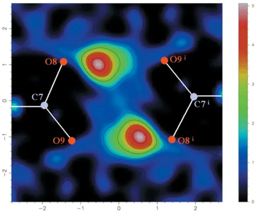
The molecules of (I) assemble to form a classical hydrogen-bonded dimer, in which the C7—O9 and C7—O8 bond lengths in the carboxylic acid group of 1.223 (2) and 1.3015 (18) A˚ , respectively, indicate a well ordered hydrogen bond. This is supported by the lack of H-atom disorder observed in the
Fourier difference map (calculated with the program
MAPVIEW, part of theWinGXsuite; Farrugia, 1999) through the dimer group (Fig. 2). The single crystallographically
unique hydrogen bond,viz. O8—H1 O9i [symmetry code:
(i) 3x, 1y,z], exhibits a typical O O separation for benzoic acid dimers of 2.651 (2) A˚ . The remainder of the contacts lie outside the sum of the van der Waals radii of the two atoms involved, but these very weak interactions can still be used to describe the remainder of the structure. The dimers
assemble into extended ribbons through C—H
inter-actions of 3.658 A˚ for C4—H4 C12ii[symmetry code: (ii)x,
y1,z] (Fig. 3a), and these ribbons form stacks defined by a
– contact of 3.446 A˚ between atoms C13 and C16iii
[symmetry code: (iii)x 1, y, z] (Fig. 3b). The stacks pack
together with C—H interactions of 3.697 A˚ for C12—
H12 C5iv[symmetry code: (iv)x1,y+ 1,z] (Fig. 4). The most striking difference between the molecular struc-ture presented here and that of MIGPAT (Field &
Venkata-organic papers
Acta Cryst.(2005). E61, o2280–o2282 Parkinet al. C
13H10O3
o2281
Figure 2 [image:2.610.45.297.68.274.2] [image:2.610.315.565.69.470.2]Fourier difference map section through the carboxylic acid dimer plane defined by atoms C7, O8, O9, C7i, O8iand O9i[symmetry code: (i) 3x, 1 y, z]. There is clearly only a single peak associated with each hydrogen bond, corresponding to an ordered H atom.
Figure 1
A drawing of the molecule of (I), showing the atomic numbering scheme. Ellipsoids for non-H atoms are shown at the 30% probability level. All H atoms take their number from the parent C atom, except for H1.
Figure 3
[image:2.610.43.298.333.547.2]raman, 2002) is the geometry of the carboxylic acid group. In the title compound, it is clear from the bond lengths that the
C O double bond is C7 O9, involving the O atom closest to
the ether group. By contrast, the shorter C—O bond in MIGPAT is that further from the ether O atom, although the difference between the two bond lengths is much less than we report here. As the two chemically different carboxylic acids in MIGPAT are crystallographically identical, it is possible that there is some correlated structural disorder between the C—O
and C O bonds; this might explain the very similar C—O
bond lengths in MIGPAT.
Experimental
The title compound was used as received from Aldrich. Crystals of diffraction quality were grown from an acetone solution.
Crystal data
C13H10O3 Mr= 214.22
Triclinic,P1
a= 5.2736 (5) A˚
b= 7.7366 (6) A˚
c= 13.6863 (10) A˚
= 89.184 (6)
= 83.433 (6)
= 74.640 (6) V= 534.84 (8) A˚3
Z= 2
Dx= 1.330 Mg m
3
MoKradiation Cell parameters from 8180
reflections
= 2–28
= 0.10 mm1
T= 293 K Block, colourless 0.300.150.10 mm
Data collection
Bruker APEX2 diffractometer
’and!scans
Absorption correction: multi-scan (SADABS; Sheldrick, 1996)
Tmin= 0.98,Tmax= 0.99
8180 measured reflections 2565 independent reflections
1516 reflections withI> 2(I)
Rint= 0.028 max= 28.5 h=6!7
k=10!10
l=18!18
Refinement
Refinement onF2 R[F2> 2(F2)] = 0.054
wR(F2) = 0.164 S= 0.93 2565 reflections 148 parameters
H atoms treated by a mixture of independent and constrained refinement
w= 1/[2(F2) + 0.08
+ 0.08P],
whereP= [max(Fo2,0) + 2Fc2]/3
(/)max< 0.001 max= 0.38 e A˚
3
[image:3.610.45.296.73.154.2] [image:3.610.313.566.88.274.2]min=0.29 e A˚ 3
Table 1
Selected geometric parameters (A˚ ,).
C1—C2 1.397 (2) C1—C6 1.390 (2) C1—C7 1.477 (2) C2—O10 1.371 (2) C2—C3 1.389 (3) O10—C11 1.387 (2) C11—C12 1.346 (3) C11—C16 1.372 (4) C12—C13 1.373 (3)
C13—C14 1.343 (4) C14—C15 1.328 (4) C15—C16 1.379 (3) C3—C4 1.367 (3) C4—C5 1.376 (3) C5—C6 1.378 (3) C7—O9 1.223 (2) C7—O8 1.3015 (18)
C2—C1—C6 117.52 (16) C2—C1—C7 122.57 (15) C6—C1—C7 119.91 (14) C1—C2—O10 117.58 (15) C1—C2—C3 120.32 (16) O10—C2—C3 122.08 (15) C2—O10—C11 119.76 (14) O10—C11—C12 119.5 (2) O10—C11—C16 120.0 (2) C12—C11—C16 120.14 (19) C11—C12—C13 119.2 (2)
C12—C13—C14 121.1 (2) C13—C14—C15 119.9 (2) C14—C15—C16 120.9 (3) C15—C16—C11 118.8 (2) C2—C3—C4 120.45 (17) C3—C4—C5 120.44 (19) C4—C5—C6 119.18 (18) C1—C6—C5 122.08 (17) C1—C7—O9 124.03 (14) C1—C7—O8 114.22 (15) O9—C7—O8 121.75 (16)
Table 2
Hydrogen-bond geometry (A˚ ,).
D—H A D—H H A D A D—H A
O8—H1 O9i
0.90 (2) 1.75 (2) 2.651 (2) 175 (2)
Symmetry code: (i) 3x;1y;z.
H atoms were positioned geometrically and refined as riding groups, with C—H = 1.0 A˚ andUiso(H) = 1.2Ueq(C), except for atom
H1, which was located in a Fourier difference map and refined with an O—H distance restraint of 0.90 (5) A˚ and a fixedUiso(H) = 0.05 A˚2.
Data collection:APEX2(Bruker, 2005); cell refinement:APEX2; data reduction:APEX2; program(s) used to solve structure:SIR92
(Altomareet al., 1994); program(s) used to refine structure: CRYS-TALS (Betteridge et al., 2003); molecular graphics: ORTEP3 for Windows (Farrugia, 1997) and MERCURY (Bruno et al., 2002); software used to prepare material for publication:CRYSTALS.
This paper is the result of an optional undergraduate class project entitled ‘Frontiers of Crystallography’, designed to show some of the sort of research that can be undertaken in crystallography. The data collection, structure solution, refinement and post-refinement analysis of the unknown title structure were all undertaken in parallel by the undergraduate students, who are all co-authors, and the collated information has resulted in this paper.
References
Allen, F. H. (2002).Acta Cryst.B58, 380–388.
Altomare, A., Cascarano, G., Giacovazzo, C., Guagliardi, A., Burla, M. C., Polidori, G. & Camalli, M. (1994).J. Appl. Cryst.27, 435.
Betteridge, P. W., Carruthers, J. R., Cooper, R. I., Prout, C. K. & Watkin, D. J. (2003).J. Appl. Cryst.36, 1487.
Bruker (2005).APEX2. Bruker AXS Inc., Madison, Wisconsin, USA. Bruno, I. J., Cole, J. C., Edgington, P. R., Kessler, M., Macrae, C. F., McCabe, P.,
Pearson, J. & Taylor, R. (2002).Acta Cryst.B58, 389–397. Farrugia, L. J. (1997).J. Appl. Cryst.30, 565.
Farrugia, L. J. (1999).J. Appl. Cryst.32, 837–838.
Field, J. E. & Venkataraman, D. (2002).Chem. Commun.pp. 306–307. Sheldrick, G. M. (1996).SADABS. University of Go¨ttingen, Germany.
organic papers
o2282
Parkinet al. C13H10O3 Acta Cryst.(2005). E61, o2280–o2282
Figure 4
supporting information
sup-1 Acta Cryst. (2005). E61, o2280–o2282
supporting information
Acta Cryst. (2005). E61, o2280–o2282 [https://doi.org/10.1107/S1600536805019495]
2-Phenoxybenzoic acid at room temperature
Andrew Parkin, Suzanne M. Harte, Donnie Carmichael, Scott Currie, Lorna Drummond, Adam
Haahr, Kevin Haggarty, Tracey Hunter, Andr
é
Lamarque, Loretta Lawton, Craig Martin, Jennifer
E. Mathieson, Jennifer S. Mathieson, Thomas McGlone, Julie McGregor, Liam McMillan, Louise
Robertson, Robert Thatcher, Steven Vance and Chick C. Wilson
2-Phenoxybenzoic acid
Crystal data
C13H10O3
Mr = 214.22
Triclinic, P1 Hall symbol: -P 1
a = 5.2736 (5) Å
b = 7.7366 (6) Å
c = 13.6863 (10) Å
α = 89.184 (6)°
β = 83.433 (6)°
γ = 74.640 (6)°
V = 534.84 (8) Å3
Z = 2
F(000) = 224
Dx = 1.330 Mg m−3
Mo Kα radiation, λ = 0.71073 Å Cell parameters from 8180 reflections
θ = 2–28°
µ = 0.10 mm−1
T = 293 K Block, colourless 0.30 × 0.15 × 0.10 mm
Data collection
Bruker APEX2 diffractometer
Graphite monochromator
φ and ω scans
Absorption correction: multi-scan (SADABS; Sheldrick, 1996)
Tmin = 0.98, Tmax = 0.99 8180 measured reflections
2565 independent reflections 1516 reflections with I > 2σ(I)
Rint = 0.028
θmax = 28.5°, θmin = 1.5°
h = −6→7
k = −10→10
l = −18→18
Refinement
Refinement on F2 Least-squares matrix: full
R[F2 > 2σ(F2)] = 0.054
wR(F2) = 0.164
S = 0.93 2565 reflections 148 parameters 1 restraint
Primary atom site location: structure-invariant direct methods
Hydrogen site location: inferred from neighbouring sites
H atoms treated by a mixture of independent and constrained refinement
w = 1/[σ2(F2) + 0.08 + 0.08P], where P = [max(Fo2,0) + 2Fc2]/3 (Δ/σ)max = 0.000159
supporting information
sup-2 Acta Cryst. (2005). E61, o2280–o2282
Fractional atomic coordinates and isotropic or equivalent isotropic displacement parameters (Å2)
x y z Uiso*/Ueq
C1 1.0774 (3) 0.2871 (2) 0.12595 (11) 0.0538 C2 0.8810 (4) 0.3698 (2) 0.20060 (13) 0.0663 O10 0.8556 (4) 0.54655 (19) 0.22196 (13) 0.1192 C11 0.6892 (5) 0.6279 (2) 0.30383 (16) 0.0787 C12 0.4462 (5) 0.7320 (3) 0.29248 (17) 0.0909 C13 0.2911 (5) 0.8241 (4) 0.3725 (2) 0.0970 C14 0.3793 (5) 0.8138 (3) 0.46118 (19) 0.0911 C15 0.6204 (6) 0.7129 (4) 0.47235 (19) 0.1149 C16 0.7826 (5) 0.6184 (4) 0.3938 (2) 0.1112 C3 0.7150 (4) 0.2753 (3) 0.24872 (15) 0.0760 C4 0.7430 (4) 0.1000 (3) 0.22415 (16) 0.0772 C5 0.9367 (4) 0.0147 (3) 0.15164 (16) 0.0781 C6 1.1009 (4) 0.1085 (2) 0.10356 (14) 0.0673 C7 1.2568 (3) 0.3820 (2) 0.07145 (11) 0.0558 O9 1.2443 (3) 0.54022 (17) 0.08452 (10) 0.0788 O8 1.4326 (3) 0.28285 (19) 0.00646 (10) 0.0819
H12 0.3789 0.7416 0.2267 0.1076*
H13 0.1095 0.9007 0.3647 0.1114*
H14 0.2637 0.8818 0.5187 0.1056*
H15 0.6847 0.7053 0.5386 0.1326*
H16 0.9651 0.5443 0.4023 0.1243*
H3 0.5743 0.3357 0.3018 0.0891*
H4 0.6217 0.0335 0.2590 0.0934*
H5 0.9583 −0.1138 0.1341 0.0941*
H6 1.2412 0.0460 0.0508 0.0791*
H1 1.535 (3) 0.348 (2) −0.0246 (12) 0.0500*
Atomic displacement parameters (Å2)
U11 U22 U33 U12 U13 U23
supporting information
sup-3 Acta Cryst. (2005). E61, o2280–o2282
O8 0.0886 (10) 0.0656 (8) 0.0801 (9) −0.0197 (7) 0.0387 (8) −0.0198 (7)
Geometric parameters (Å, º)
C1—C2 1.397 (2) C15—C16 1.379 (3)
C1—C6 1.390 (2) C15—H15 1.000
C1—C7 1.477 (2) C16—H16 1.000
C2—O10 1.371 (2) C3—C4 1.367 (3)
C2—C3 1.389 (3) C3—H3 1.000
O10—C11 1.387 (2) C4—C5 1.376 (3)
C11—C12 1.346 (3) C4—H4 1.000
C11—C16 1.372 (4) C5—C6 1.378 (3)
C12—C13 1.373 (3) C5—H5 1.000
C12—H12 1.000 C6—H6 1.000
C13—C14 1.343 (4) C7—O9 1.223 (2)
C13—H13 1.000 C7—O8 1.3015 (18)
C14—C15 1.328 (4) O8—H1 0.901 (17)
C14—H14 1.000
C2—C1—C6 117.52 (16) C16—C15—H15 119.7
C2—C1—C7 122.57 (15) C15—C16—C11 118.8 (2)
C6—C1—C7 119.91 (14) C15—C16—H16 120.6
C1—C2—O10 117.58 (15) C11—C16—H16 120.6
C1—C2—C3 120.32 (16) C2—C3—C4 120.45 (17)
O10—C2—C3 122.08 (15) C2—C3—H3 119.8
C2—O10—C11 119.76 (14) C4—C3—H3 119.8
O10—C11—C12 119.5 (2) C3—C4—C5 120.44 (19)
O10—C11—C16 120.0 (2) C3—C4—H4 119.8
C12—C11—C16 120.14 (19) C5—C4—H4 119.8
C11—C12—C13 119.2 (2) C4—C5—C6 119.18 (18)
C11—C12—H12 120.4 C4—C5—H5 120.4
C13—C12—H12 120.4 C6—C5—H5 120.4
C12—C13—C14 121.1 (2) C1—C6—C5 122.08 (17)
C12—C13—H13 119.6 C1—C6—H6 119.0
C14—C13—H13 119.3 C5—C6—H6 118.9
C13—C14—C15 119.9 (2) C1—C7—O9 124.03 (14)
C13—C14—H14 120.0 C1—C7—O8 114.22 (15)
C15—C14—H14 120.1 O9—C7—O8 121.75 (16)
C14—C15—C16 120.9 (3) C7—O8—H1 110.1 (10)
C14—C15—H15 119.5
Hydrogen-bond geometry (Å, º)
D—H···A D—H H···A D···A D—H···A
O8—H1···O9i 0.90 (2) 1.75 (2) 2.651 (2) 175 (2)