Original Article
Overexpression of FoxP1 is a novel biomarker of
malignant human pancreatic cancer
Dapeng Qiu, Feng Han, Hao Zhuang, Qingjun Li, Xianzhou Zhang
Department of Hepato-Biliary-Pancreatic Surgery, Henan Tumor Hospital (The Tumor Hospital Affiliated to Zheng
-zhou University), Zheng-zhou 450008, Henan, China
Received November 13, 2015; Accepted February 2, 2016; Epub June 15, 2016; Published June 30, 2016
Abstract: Recent studies have shown that FOXP3 plays a significant role in the pancreatic cancer cell metastasis, but whether FOXP1 plays a role in mediating tumor metastasis in pancreatic cancer has not been explored. Here, Co-immunoprecipitation assays were utilized to detect that FOXP1 was physically associated with the EMT inducer ZEB2. Most important, CAPAN-1 cells transfected with FOXP1 shRNA displayed a reversed epithelial morphology and reduced the expression of N-cadherin, while increase the expression of E-cadherin, and the PANC-1 cells transfected with FOXP1 displayed the contrary tendency. Transwell invasion assay, as well as colony formation together shown that FOXP1 promoted cell epithelial-mesenchymal transition and tumorigenesis in vitro and in vivo. Endothelial tube formation assays indicated FOXP1 might regulate the angiogenesis of pancreatic cancer through transcriptional activation of VEGF. qChIP, ChIP as well as Luciferase reporter assays suggested FOXP1 binding on the promoter of E-cadherin and VEGF. The changes in the level of protein and RNA implied that suppression of downstream E-E-cadherin or activation of VEGF was two distinct important mechanisms by which FOXP1 triggered EMT and angiogenesis. Fur-ther, patient samples collected shown that FOXP1 was significantly increased in pancreatic cancer samples, and its higher expression is correlated with poor prognosis, worse over all survivals. Together, our experiments revealed the mechanism for FOXP1 in facilitating tumorigenesis, angiogenesis and metastasis of pancreatic cancer cells, sug-gesting that FOXP1 might be a novel biomarker of malignant human pancreatic cancer and a potential therapeutic target for treating pancreatic cancer.
Keywords: FOXP1, ZEB2, EMT, E-cadherin, VEGF
Introduction
Pancreatic cancer is one of the most aggres-sive human malignancies. Metastasis is a major clinical event which is the main cause of disease-related death, and it often has a relation with angiogenesis, and is usually cor-related with the low 5-year survival rates [1, 2]. Epithelial-mesenchymal transition (EMT) are believed to be the initial step of pancreatic can-cermetastasis [3], during the process of EMT, epithelial cells with a cobblestone phenotype lost their differentiated characteristics, and acquire mesenchymal cell feature, such as spindle-shaped, motility and invasiveness [4, 5]. Epithelial-cadherin (E-cadherin; encoded by CDH1) is a member of the classical cadherins, it usually works as a tumor suppressor in the cancer progression [6, 7]. Generally downregu-lation of E-cadherin and upregudownregu-lation of
N-cadherin are considered as the mark of EMT [8-10]. Which play important roles in the pro-cess of embryonic development as well as in tumor metastasis?
Furthermore, angiogenesis, the process of new blood vessels formation, is another important step in tumor metastasis, which involves sev-eral signaling between growth factors and endothelial cell receptors [11]. Among them, vascular endothelial growth factor (VEGF), by inducing the formation of new blood vessels, was reported by several groups [12, 13], to get an important role in angiogenesis and tumor metastasis.
pan-creatic cancer progression was well discussed [14, 15], although the sequence of FOXP1 shares high similarity with FOXP3, but the role of FOXP1 was still poorly understood.
To our particular aspects of knowledge, ZEB2/ SIP1, which is a potent repressor of E-cadherin expression [16], is considered to be one of the key factors of EMT. Oppositely, vimentin, anoth-er mesenchymal markanoth-er, is reported to be up-regulated by ZEB2, which is associated with breast tumor cell EMT [17].
Materials and methods
Cells and cell culture
Cell lines PANC-1 and CAPAN-1 cells were acquired from the American Type Culture Collection (ATCC). They were cultured in DMEM or RPMI 1640 (Gibco) respectively, supple-mented with 10% fetal bovine serum (FBS)
(HyClone) in a humidified atmosphere with 5%
CO2 at 37°C. CAPAN-1 medium was in addition supplemented with 1 mM glutamine. Cell lines hTERT-HPNE were from Shanghai institutes for Biological Sciences (CAS), and were cultured in MEGM BulletKit.
Reagents
Rabbit human FOXP1 antibody, ZEB2
anti-body, E-cadherin antianti-body, VEGF antianti-body,
β-actin antibody and secondary antibody were all
purchased from Santa Cruz (CA, USA). α-catenin
antibody, N-cadherin antibody, Vimentin
anti-body, were from abcam (USA). Specific shRNA
targeting FOXP1, ZEB2 were from Sigma-Aldrich (CA, USA). A negative control shRNA (SCR) was also used as control (Sigma-Aldrich). Matrigel was purchased from BD Biosciences (CA. USA). Lipofectamine2000 (Invitrogen) was used for transfection.
Patients and specimens
30 pancreatic cancer samples of patients and the adjacent normal tissues were obtained in Henan Tumor Hospital from 2001 to 2008. Samples were chosen with completely clinico-pathologic information. Patients who were received radiation therapy or chemotherapy prior to the surgery were excluded. The survival times were calculated based on the operation day to death, via the evaluation of metastasis or recurrence. This study has been approved by the hospital ethical committee.
Quantitative real-time PCR (qRT-PCR)
Total RNA of cell lysates were extracted with Trizol solution (Invitrogen, Carlsbad, CA, USA), and 1 μg of total RNA were reverse
transcrib-ed to cDNA using with M-MLV Reverse Tran- scriptase, according to the manufacturer’s instructions (TransGen Beijing, China). Real-time RT-PCR primers were as follows: FOXP1, forward 5-CTACCACAAGATGAATGGGC-3, rever- se 5-CCTGTTAACATTGTGCAGCT-3; E-cadherin, forward 5-AAATCACATCCTACACTGCC-3, reverse 5-GCAACTGGAGAACCATTGTC-3; VEGF, forward 5-GACATCTTCCAGGAGTACCC-3, reverse 5-TCT- TTCTTTGGTCTGCATTCAC-3; ZEB2, forward 5-C- CAGTCCAGACCAGTATTCC-3, reverse 5-AGCAAT- TCTCCCTGAAATCC-3; GAPDH, forward 5-CATTT- CCTGGTATGACAACGA-3, reverse 5-GTCTACAT- GGCAACTGTGAG-3, Real-time PCR was per-formed on an ABI 7500 sequence detection system (Applied Biosystems), using SYBR (Roche). All experiments were performed in three times. GAPDH was used as a normaliza-tion control.
Co-immunoprecipitation
For immunoprecipitation assays, cells were washed with cold PBS and lysed with cold lysis buffer on a rotator at 4°C for 45 min. Whole cell lysates were incubated with appropriate prima-ry antibodies or normal rabbit/mouse immuno-globin G (IgG) as negative controls on a rotator overnight at 4°C, then added protein A/G Sepharose CL-4B beads for 2 h at 4°C. Beads were then washed 5 times with lysis buffer (50 mM Tris-Cl, pH 7.4, 0.5% NP-40, 150 mM NaCl, 1 mM EDTA, 0.5% sodium deoxycholate and protease inhibitor cocktail). The immune com-plexes were subjected to SDS-PAGE (Invitrogen), followed by immunoblotting with secondary antibodies.
Western blot
Cells were harvested 48 h to 72 h after trans-fection with the indicated shRNA or recombi-nant plasmid. Protein concentration was mea-sured using the bicinchoninic acid kit (BCA). Subsequently, 30 μg proteins were run by 10%
SDS-PAGE gel and then transferred on to NC membranes. 5% skim milk were used to block the NC membranes for 1 h, then incubated with primary antibodies, overnight at 4°C, including FOXP1 (1:1000), ZEB2 (1:1000), E-cadherin
(1:1000), Vimentin (1:500), VEGF (1:1000) and
β-Actin (1:500). Goat anti-Rabbit secondary
antibodies (1:5000), Goat anti-Mouse second-ary antibodies (1:3000) and Western blotting Luminal reagent (Santa Cruz Biotechnology) were used to visualize the protein bands. Colony formation assay
After infected the related lentivirus for 3 days, a total of 1000 PANC-1 cells were seeded into 6-well plates and the medium was changed every 3 days. After 10 days of culturing, the colonies formed were collected, washed with
PBS and fixed in 4% paraformaldehyde at 37°C
for 15 min, after then, the colonies were stained with Coomassie for 15 min, washed and then air-dried. The colonies were counted using the microscopy (Olympus, Tokyo, Japan). The exper-iment was performed in triplicate.
ChIP and qChIP
Chromatin immunoprecipitation (ChIP) assays were carried out according to the manufactur-er’s protocol (Upstate). The chromatins were
incubated with 4 μg of FOXP1 antibody, ZEB2
antibody or normal Rabbit IgG as negative con-trol (Santa Cruz Biotechnology), at 4°C on a rotator overnight. Immunoprecipitated DNA
was purified with the Qiagen PCR purification
kit. qChIP was analyzed by quantitative PCR
using specific primers as follows. For common ChIP assays, the final target DNA sequence was amplified and resolved on standard agarose
DNA gels.
Primers used for common ChIP: E-cadherin: 5-AGGGTCACCGCGTCTATG-3 (forward) and 5- CTTCCGCAAGCTCACAGG-3 (reverse), VEGF: 5- AAGGAGGAAAGTTAGTGGCTTCCCT-3 (forward) and 5-TGTTGCTGTGGTTTGGTGGA-3 (reverse); qChIP primers: E-cadherin: 5-GCAGGTCCCATAA- CCCACCTA-3 (forward) and 5-CATAGACGCGG- TGACCCTCTA-3 (reverse), VEGF: 5-AGCCTAGAG- CATGGAGCCCAGGTGA-3 (forward) and 5-CCAT- CGGTATGGTGTCCTAA-3 (reverse).
Luciferase reporter assay
For E-cadherin or VEGF reporter construction, the sequence of the relative promoter and par-tial exon was obtained by PCR. The pGL3 basic vector (Promega) was utilized to ligate these PCR products to generate pGL3-E-cadherin or pGL3-VEGF luciferase reporter construct. PANC-1 cells were transfected with promoter
luciferase reporter, Renilla luciferase plasmid, and the indicated expression constructs, using Lipofectamine LTX-Plus (Invitrogen) in 96-well plates. By addition of empty vector in each transfection, the amount of DNA was retained constant. Renilla plasmid was used as a
nor-malization control of transfection efficiency. 48 hours after transfection, the firefly and Renilla
luciferases were assayed according to the man-ufacturer’s protocol (Promega). Each experi-ment was conducted in triplicate.
Transwell invasion assay
The invasive ability of the CAPAN-1 cells was
investigated using Transwell assay (8-μm pore
size; for 24-well plate. Millipore), matrigel used
were from BD Biosciences (50 μg/ml). First,
100 μl matrigel was added onto the surface of
the chamber, incubated in room temperature
for 2 hours for solidification. A total of 100 μl of
CAPAN-1 cell suspension (5×10⁴ cells) was in addition to the upper chamber, with serum-free medium. The lower compartment was incubat-ed with 500 μl of RPMI 1640 containing 10%
fetal bovine serum. After incubation for 24 hours, the cells invaded into the lower surface
were fixed with 2% paraformaldehyde, stained
with crystal violet. Photos were taken using an inverted microscope (Olympus Corp. Japan) at 100× magnifications.
Tube formation assay
Angiogenesis in vitro was performed utilizing the endothelial tube formation assay kit (Cell Biolabs, CA), according to the instruction. The extracellular matrix gel was prepared from
Engelbreth-Holm-Swarm tumor cells (200 μl/
well). HUVECs (ten thousand cells/well) infect-ed with retroviruses or culturinfect-ed with CM from PANC-1 cells that were infected with the indi-cated retroviruses. The mixture was added
onto solidified extracellular matrix gel in 600 μl
medium. After incubation for 18 h, endothelial cell tube formation was assessed and tube number was counted.
Statistical analysis
All observations were confirmed by at least
three independent experiments. The data was presented as mean ± SD. One-way ANOVA was
used to analyze the statistical significance of
the mean values. Bivariate correlation was
was utilized to test the prognostic significance of factors. P < 0.05 was considered
signifi-cant. Results
FOXP1 is physically associated with ZEB2 in different pancreatic cancer cell lines
In order to investigate the role of FOXP1 during
the progress of pancreatic cancer, we first
As showed in Figure 2A, CAPAN-1 cells were infected with control shRNA (SCR), shFOXP1#1, or shFOXP1#2 groups. Both mRNA level and protein level in different groups were detected.
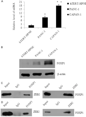
[image:4.612.92.382.79.470.2]We found that mRNA level of FOXP1 was signifi -cantly reduced when cells were infected with shFOXP1#1, or shFOXP1#2 (P < 0.05. Figure 2A left panel). Concomitantly, in western blot analysis, FOXP1 was also greatly lowered in shFOXP1-infected CAPAN-1 cells as compared Figure 1. FOXP1 is physically associated with ZEB2 in different pancreatic
can-cer cell lines. A. The expression level of FOXP1 was determined in hTERT-HPNE, PANC-1 and CAPAN-1. Quantitative real-time PCR were repeated three times. GAPDH was used as a normalization control. Error bars represent average ± SD, *P < 0.05, **P < 0.01. B. Western blotting was used to detect the expres-sion of FOXP1 in hTERT-HPNE, PANC-1 and CAPAN-1 cells; β-actin was used as a normalization control. C. Co-IP analysis of the association between FOXP1 and ZEB2. Whole cell lysates of PANC-1 were immunoprecipitated with antibod-ies against FOXP1, with normal IgG as the negative control, while the whole lysates as positive control. The immunocomplexes were then immunoblotted using ZEB2, then inversely. D. Whole cell lysates of CAPAN-1 were further used to detect the association between FOXP1 and ZEB2.
examine the expression of FOXP1 in two pancreatic cancer cell lines (PANC-1 and CAPAN-1) and the nor-mal pancreas cell lines hTERT-HPNE by using qRT-PCR (Figure 1A) and west-ernblot (Figure 1B). The expression of FOXP1 was
significantly increased in
PANC-1 and CAPAN-1, com-paring with hTERT-HPNE. Then we used co-immuno-precipitation experiments to reveal that FOXP1
copu-rified with ZEB2 (Figure 1C and 1D), a key factor of epithelial-mesenchymal tr- ansition. Total protein ex- tracts from PANC-1 cells
were prepared, first,
Imm-unoprecipitation (IP) with anti-FOXP1 followed by immunoblotting (IB) with the anti-ZEB2 indicated that FOXP1 was co-immu-noprecipitated with ZEB2 (Figure 1C left panel), again, IP with anti-ZEB2 fol-lowed by IB with anti-FOXP1 (Figure 1C right panel). To further support the in vivo interaction between FOXP1 and ZEB2, this interaction
is also confirmed with
endogenous proteins in CAPAN-1 cells (Figure 1D). We also detected another transcription factor, such as snail1 and twist1, both of them had no interaction with FOXP1, data were not shown.
FOXP1 promotes
with the control shRNA-infected cells (Figure 2A right panel). The results suggested both the two relative shRNA were suc-cessfully constructed in CAPAN-1 cells. Interestingly, when CAPAN-1 were trans-fected with the relative shRNA, As shown in Figure 2B, while control CAPAN-1 cells maintained a
spindle-like, fibroblastic morpholo -gy, knock down of FOXP1 became organized cell-cell adhesion and cobble stone-like epithelial appearance. Considering the expression of FOXP1 in PANC-1 was lower than that of CAPAN-1, we choose FOXP1 “loss of function” experiment in CAPAN-1 cells, the expres-sion of the epithelial mark-ers such as E-cadherin, a-catenin were up regulat-ed; while the mesenchymal markers N-cadherin, Vimen- tin were raised as mea-sured by real-time RT PCR or western blotting (Figure 2C). When FOXP1 was over-expressed in PANC-1 cells, the opposite result was shown. E-cadherin, a-cat- enin were decreased while N-cadherin, Vimentin were up regulated (Figure 2D).
FOXP1 enhances pancre-atic cancer cells invasion
and angiogenesis in vitro
[image:5.612.92.380.70.589.2]Considering EMT as the ini-tiation step of metastasis, we further focus our atten-tion on whether FOXP1 could also take part in the metastasis; transwell as- say was performed to assess the effect of FOXP1 on cell invasion. Migrated CAPAN-1 cells infected with shFOXP1 were counted for Figure 2. FOXP1 promotes pancreatic cancer cells EMT in vitro. A. Knockdown
one third than those infected with control, while FOXP1 over expression was related with 3.5 times increase of the invasion cells, which indi-cated FOXP1 might take a role in the metasta-sis of pancreatic cancer (Figure 3A).
[image:6.612.95.522.76.547.2]SCR, ShFOXP1, vector, or FOXP1, (Figure 3B), or
[image:7.612.93.522.76.593.2]shFOXP1 or cultured in the CM with knockdown
FOXP1, formed significantly fewer tubes than
those in the SCR or vector group, whereas HUVECs with FOXP1 ovexpression or that which were cultured in the CM with FOXP1 ovexpres-sion generated more tubes than the control. To further explore the tumorigenic ability of FOXP1, in vitro, colony formation was per-formed, the average number of colonies in the SCR group or vector group was similar to each other, whereas the number of colonies in FOXP1
was reasonable to suppose there was function-al consistency between ZEB2 and FOXP1. To further support the argument, luciferase report-er activity assays wreport-ere carried out in PANC-1 cells with E-cadherin or VEGF promoter-driven luciferase reporter under FOXP1 over expres-sion or depletion. These experiments indicated that FOXP1 over expression or knockdown resulted in repressed or enhanced E-cadherin reporter activity, contrary to E-cadherin, on the promoter of VEGF, FOXP1 over expression or
silencing led to a significant effect on the acti -Figure 5. FOXP1 is a potential cancer biomarker and enhances MRI Phenotyping
detection. A. The expression of FOXP1 was determined by real-time PCR in 30 pairs of pancreatic cancer samples and adjacent noncancerous tissue (NT). Er-ror bars represent standard erEr-ror of the mean, Student’s t-test, *P < 0.05; **P < 0.01. B. Correlation between FOXP1 and E-cadherin in the resected pancre-atic cancer samples from 30 patients. *P < 0.05. C. Correlation between FOXP1 and VEGF in the resected pancreatic cancer samples from 30 patients. *P < 0.05. D. Clinical data were plotted using Kaplan-Meier curves, and the 5-year survival rate was compared using the Cox log-rank test (**P < 0.001). The y-axis represents the survival probability, and the x-y-axis represents the survival in months.
knockdown was only 30%, while FOXP1 overexpres-sion was about 2.7 fold (P < 0.05). The data indicated there was an obviously increase in colony forma-tion because of the high expression of FOXP1 (Figu- re 3D).
The molecular mechanism of FOXP1 in promoting pancreatic cancer cell
invasion and angiogenesis
To further understand the molecular mechanism of FOXP1 in regulating tran-scription, quantitative ChIP (qChIP) assays were per-formed in PANC-1 cells, several key genes in differ-ent pathways which were involved in metastasis were chosen to detect, such as
E-cadheirn, α-catenin,
[image:8.612.94.382.65.431.2]vation or repression of the VEGF reporter activ-ity (Figure 4B). The up regulation or down regu-lation of ZEB2 got the similar results on the two promoters (Figure 4C).
Consistent with the promoter occupancy, in FOXP1, ZEB2 overexpressed PANC-1 cells, the mRNA (Figure 4D) expression of E-cadherin decreased, to the contrary. VEGF increased. Respectively, while FOXP1 or ZEB2 was knock-down in CAPAN-1 cells, in mRNA level, E- cadherin increased, VEGF decreased, which further supporting the notion that both FOXP1 and ZEB2 have dual transcriptional activities (Figure 4E).
FOXP1 is a potential cancer biomarker and enhances MRI Phenotyping detection
In order to understand the clinical significance
of FOXP1 during the progress of pancreatic can-cer, we collected 30 pancreatic cancer sam-ples, and paired with adjacent noncancerous samples. The expression levels of FOXP1 were examined, using qRT-PCR. The results showed that the expression level of FOXP1 in
pancreat-ic cancer samples was signifpancreat-icantly higher than
the noncancerous tissues (P < 0.01, Figure 5A). Further, we performed correlation tests in the resected patient samples. An inverse cor-relation was observed between FOXP1 and E-cadherin (R = -0.4859; P < 0.001; Figure 5B), as to FOXP1 and VEGF, there was a strong posi-tive correlation (R = 0.6232; P < 0.0001; Figure 5C). Further, depending on the median FOXP1 expression level, patients were divided in to two groups, the group of FOXP1 over expres-sion was found to have a poorer prognosis for an overall 5-year survival (P < 0.05) (Figure 5D). Discussion
In conclusion, we found that the expression of FOXP1 in pancreatic cancer was obviously high-er than that of the noncanchigh-erous samples. Furthermore, the expression of E-cadherin was negatively associated with FOXP1 in the tumor tissues, while the expression of VEGF was posi-tively correlated with FOXP1. The concept of ZEB2 as a powerful repressor of E-cadherin has been widely accepted; here we reported not only the physical interaction between FOXP1 and ZEB2, but also the functional consistency between the two factors. We declared that FOXP1 exerts its dual transcriptional regulatory
function via activate the expression of genes implicated in angiogenesis, including VEGF and suppress the expression of E-cadherin. Further, the over expression of FOXP1 meaning for a worse overall survival than that of patients with FOXP1 low expression, it indicates FOXP1 may act as an independent prognostic factor. Especially, Tube formation assay reveal
overex-pression of FOXP1 may influence the depth of
tumor invasion and angiogenesis which are associated with the worst prognosis.
We demonstrate that FOXP1 promotes pancre-atic cancer cell angiogenesis and invasion in vitro and in vivo, implies that FOXP1 promotes EMT and enhances the invasive capacity of pancreatic cancer. Together, our experiments reveal the mechanism for FOXP1 in facilitating EMT and tumorigenesis of pancreatic cancer, uncover the distinct role of FOXP1 resulting in altered expression of the downstream target genes, suggesting that FOXP1 might be a potential therapeutic target of pancreatic can-cer for treating the angiogenesis and metasta-sis. It is well known that the changes in the development of pancreatic cancer should not be regarded as the alternations of a small a small part of genes. Thus, we predict other tar-gets of FOXP1 that are related to pancreatic cancer angiogenesis and invasion will be dis-covered in the future. Although FOXP1 and FOXP3 share high similarity sequences, the function of FOXP3 needs further discussion. So it is necessary for further exploration of the potential role of other fork head family num-bers which might contribute to the pancreatic cancer.
Disclosure of conflict of interest
None.
Address correspondence to: Feng Han, Department of Hepato-Biliary-Pancreatic Surgery, Henan Tumor Hospital (The Tumor Hospital Affiliated to Zhengzhou University), No. 127 dongming Road, Zhengzhou 450008, Henan, China. E-mail: xianzhouzhang007@ sina.com
References
[2] Feig C, Gopinathan A, Neesse A, Chan DS, Cook N and Tuveson DA. The pancreas cancer microenvironment. Clin Cancer Res 2012; 18: 4266-4276.
[3] Cano CE, Motoo Y and Iovanna JL. Epithelial-to-mesenchymal transition in pancreatic adeno-carcinoma. Sci World J 2010; 10: 1947-1957. [4] Yang J and Weinberg RA.
Epithelial-mesenchy-mal transition: At the crossroads of develop-ment and tumor metastasis. Dev Cell 2008; 14: 818-829.
[5] Maier HJ, Schmidt-Strassburger U, Huber MA, Wiedemann EM, Beug H and Wirth T. NF-kap-paB promotes epithelial-mesenchymal transi-tion, migration and invasion of pancreatic car-cinoma cells. Cancer Lett 2010; 295: 214-228. [6] Kalluri R and Weinberg RA. The basics of epi-thelial-mesenchymal transition. J Clin Invest 2009; 119: 1420-1428.
[7] Adhikary A, Chakraborty S, Mazumdar M, Ghosh S, Mukherjee S, Manna A, Mohanty S, Nakka KK, Joshi S, De A, Chattopadhyay S, Sa G, Das T. Inhibition of epithelial to mesenchy-mal transition by E-cadherin up-regulation via repression of slug transcription and inhibition of E-cadherin degradation: dual role of scaf-fold/matrix attachment region-binding protein 1 (SMAR1) in breast cancer cells. J Biol Chem 2014; 289: 25431-25444.
[8] Mallini P, Lennard T, Kirby J and Meeson A. Epithelial-to-mesenchymal transition: What is the impact on breast cancer stem cells and drug resistance. Cancer Treat Rev 2014; 40: 341-348.
[9] Park KS, Dubon MJ and Gumbiner BM. N-cad-herin mediates the migration of MCF-10A cells undergoing bone morphogenetic protein 4-me-diated epithelial mesenchymal transition. Tu-mour Biol 2015; 36: 3549-56.
[10] Kang Y, Ling J, Suzuki R, Roife D, Chopin-Laly X, Truty MJ, Chatterjee D, Wang H, Thomas RM, Katz MH, Chiao PJ, Fleming JB. SMAD4 regu-lates cell motility through transcription of N-cadherin in human pancreatic ductal epitheli-um. PLoS One 2014; 9: e107948.
[11] Hogendorf P, Durczynski A, Kumor A and Strzel-czyk J. Pancreatic head carcinoma and vascu-lar endothelial growth factor (VEGF-A) concen-tration in portal blood: its association with cancer grade, tumor size and probably poor prognosis. Arch Med Sci 2014; 10: 288-293. [12] Jamison S, Lin Y and Lin W. Pancreatic
endo-plasmic reticulum kinase activation promotes medulloblastoma cell migration and invasion through induction of vascular endothelial growth factor A. PLoS One 2015; 10: e0120- 252.
[13] Ferrara N. Binding to the extracellular matrix and proteolytic processing: two key mecha-nisms regulating vascular endothelial growth factor action. Mol Biol Cell 2010; 21: 687-690. [14] Hiraoka N, Onozato K, Kosuge T and Hirohashi
S. Prevalence of FOXP3+ regulatory T cells in-creases during the progression of pancreatic ductal adenocarcinoma and its premalignant lesions. Clin Cancer Res 2006; 12: 5423-5434.
[15] Jiang Y, Du Z, Yang F, Di Y, Li J, Zhou Z, Pillari-setty VG, Fu D. FOXP3+ lymphocyte density in pancreatic cancer correlates with lymph node metastasis. PLoS One 2014; 9: e106741. [16] Comijn J, Berx G, Vermassen P, Verschueren K,
van Grunsven L, Bruyneel E, Mareel M, Huylebroeck D, van Roy F. The two-handed E box binding zinc finger protein SIP1 downregu -lates E-cadherin and induces invasion. Mol Cell 2001; 7: 1267-1278.