Original Article
The value of empiric anti-tuberculosis therapy in the
differential diagnosis between intestinal tuberculosis
and Crohn’s disease in China
Yao He1*, Yujun Chen1,2*, Baili Chen1, Yun Wu1, Fang Chen1, Ren Mao1, Shenghong Zhang1, Qiao Yu1, Zhirong
Zeng1, Pinjing Hu1, Minhu Chen1
1Department of Gastroenterology, The First Affiliated Hospital of Sun Yat-sen University, Guangzhou, PR China; 2Department of Medical Ultrasonics, The First Affiliated Hospital of Sun Yat-sen University, Guangzhou, PR China. *Equal contributors.
Received October 18, 2015; Accepted March 24, 2016; Epub June 15, 2016; Published June 30, 2016
Abstract: Objective: To find out the criteria, the suitable duration and the efficacy of using empiric anti-tuberculosis to differentiate intestinal tuberculosis (ITB) from Crohn’s disease (CD). Methods: Forty-eight patients receiving em-piric anti-tuberculosis therapy were retrospectively analyzed and underwent 4-10 years follow-up. Among them, 28 were confirmed as ITB and 20 as CD at final evaluation. Clinical manifestations, endoscopic changes and the response to anti-tuberculosis therapy were compared between ITB and CD groups. Results: 1) The baseline clinical characteristics anal fistula/perianal abscess and aphthous ulcers were significantly different between 2 groups. Strong positive skin test (PPD test) result was also not comparable between 2 groups; 2) Clinical symptoms im-proved in both groups after 3 mo empiric anti-tuberculosis therapy. The non-improvement rate in ITB group was 0%, whereas that of CD group was 15.0% (P=0.007); 3) Endoscopic appearance improved significantly in ITB group but not CD group after 3 mo or 6 mo empiric anti-tuberculosis therapy, Ulcer disappearance rate in ITB group was 90.9% (20/22) in 3 mo and 100% (28/28) in 6 mo. Nodular transformation disappearance rate of in ITB group was 58.8% (10/17) in 3 mo and 76.5% (13/17) in 6 mo. There was no obvious improvement of active ulcer or nodular transformation in CD group at any time point (P<0.001 compared with ITB). Conclusions: Three months of empiric anti-tuberculosis therapy was a proper duration for the purpose of differentiating ITB from CD. Disappearance of active ulcer and nodular transformation endoscopically, together with clinical cure or obviously improvement are engraved as effective.
Keywords: Intestinal tuberculosis, Crohn’s disease, empiric anti-tuberculosis therapy, differential diagnosis
Introduction
Intestinal tuberculosis (ITB) and Crohn’s dis-ease (CD) are not rare in China, but the differ-ential diagnosis of the two diseases is still very challenging [1]. In Chinese consensus on diag-nosis and treatment of inflammatory bowel dis-ease, key points for differentiating these two diseases were raised. The consensus also em- phasized that empiric anti-tuberculosis should be employed when there was difficulty in the differential diagnosis [2]. However there was still no consensus on how to evaluate the effi-cacy of empiric anti-tuberculosis therapy. As we know, patients’ clinical symptoms improved in both ITB and CD patients after short-term
anti-tuberculo-sis treatment in patients who was suspected as ITB originally, and confirmed as ITB or CD at the end of study, to find out the criteria, the suitable duration and the efficacy of using empiric anti-tuberculosis to differentiate ITB from CD.
Materials and methods
Patients
A retrospective analysis of cases from May 2005 to March 2011 was conducted on 48 consecutive patients who received empiric anti-tuberculosis therapy and underwent 4-10 years follow-up at the Inflammatory Bowel Disease Center of Sun Yat-Sen University, China. All the following criteria must be fulfilled in patients enrolled in our study: ① Clinical fea-tures including abdominal pain, diarrhea, fever, weight loss, or abdominal mass; ② Ulcers
found in segmental or regional distribution in ileo-colon area under colonoscopy; ③ Patho- logically confirmed ITB (caseating granuloma) or clinically suspected ITB, or hard to differenti-ate ITB from CD clinically; ④ Patients who needed anti-tuberculosis therapy or empiric therapeutic trial; ⑤ All patients received anti-tuberculosis treatment for at least 3 months. Colonoscopy was enrolled in 3 mo, 6 mo, and 12 mo; ⑥ Confirmed diagnosis of ITB or CD was based on pathology or cases follow-up (cri-teria appear below). The recommended regi-mens for anti-tuberculosis in our study was 2 HRZE/7~10 HR, or adjusted according to pul-monary tuberculosis chemotherapy regimens (WHO, 1988).
Methods
[image:2.612.93.522.85.454.2]Patients evaluation and follow-up: Patients’ ge- neral date, epidemiological data, symptoms
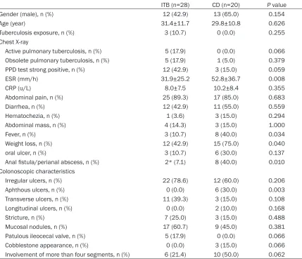
Table 1. Baseline clinical and colonoscopic characteristics in CD and ITB patients
ITB (n=28) CD (n=20) P value
Gender (male), n (%) 12 (42.9) 13 (65.0) 0.154
Age (year) 31.4±11.7 29.8±10.8 0.626
Tuberculosis exposure, n (%) 3 (10.7) 0 (0.0) 0.255
Chest X-ray
Active pulmonary tuberculosis, n (%) 5 (17.9) 0 (0.0) 0.066 Obsolete pulmonary tuberculosis, n (%) 5 (17.9) 1 (5.0) 0.379 PPD test strong positive, n (%) 12 (42.9) 3 (15.0) 0.059
ESR (mm/h) 31.9±25.2 52.8±36.7 0.008
CRP (u/L) 8.0±7.5 10.2±8.4 0.355
Abdominal pain, n (%) 25 (89.3) 17 (85.0) 0.683
Diarrhea, n (%) 12 (42.9) 11 (55.0) 0.559
Hematochezia, n (%) 1 (3.6) 3 (15.0) 0.294
Abdominal mass, n (%) 4 (14.3) 3 (15.0) 1.000
Fever, n (%) 3 (10.7) 8 (40.0) 0.034
Weight loss, n (%) 12 (42.9) 15 (75.0) 0.040
oral ulcer, n (%) 3 (10.7) 6 (30.0) 0.137
Anal fistula/perianal abscess, n (%) 2* (7.1) 8 (40.0) 0.010 Colonoscopic characteristics
Irregular ulcers, n (%) 22 (78.6) 12 (60.0) 0.206
Aphthous ulcers, n (%) 0 (0.0) 6 (30.0) 0.003
Transverse ulcers, n (%) 11 (39.3) 3 (15.0) 0.108
Longitudinal ulcers, n (%) 0 (0.0) 2 (10.0) 0.168
Stricture, n (%) 7 (25.0) 3 (15.0) 0.488
Mucosal nodules, n (%) 17 (60.7) 9 (45.0) 0.381
Patulous ileocecal valve, n (%) 5 (17.9) 0 (0.0) 0.066
Cobblestone appearance, n (%) 0 (0.0) 3 (15.0) 0.066
Involvement of more than four segments, n (%) 6 (21.4) 10 (50.0) 0.062
CD, extra-intestinal manifestations and compli-cations were retrospectively analyzed. Results of colonoscopy, chest X-ray, PPD-test, PPD-lgG, ESR, and CRP were collected as baseline data. Clinical evaluation was carried out in 1 wk, 2 wk, 1 mo, 3 mo, 6 mo and 12 mo after the start of anti-tuberculosis treatment.
Clinical assessments of the efficacy of anti-tuberculosis therapy. Cured: Disappearance of symptoms; obviously improved: severity of symptoms reduced up to half or above; non-improved: Severity of symptoms reduced less than half, symptoms aggravated or recurred. Endoscopic assessments of the efficacy of anti-tuberculosis therapy. Colonoscopy was performed at baseline, and 3 mo, 6 mo, 12 mo after the start of anti-tuberculosis therapy. Endoscopic features including ulcers, mucosal nodules, pseudopolyps, and luminal strictures, as well as their distribution and range were evaluated. Healed: Disappearance of all ulcers, with/without polypoid changes; Improved: Mu- cosa ulcer area reduced equaled or more than half of the previous; Non-improved: Mucosa ulcer area reduced less than half of the previ-ous, unchanged or aggravated.
Diagnostic criteria for ITB and CD: A final con-firmed diagnosis of ITB or CD was based on pathology of surgical specimens, or endoscopic results and disease course after a 12 mo follow-up.
Criteria for confirmed ITB: ① Formal anti-tuber-culosis therapy for 6 mo, clinically cured, endo-scopically healed, and no recurred in 12 mo follow-up. ② Caseating granuloma on mucosal biopsy or/and an acid fast stain positive before
intestinal perforation; ④ Surgery on any cause, pathology of excised intestine or lymph node showed no evidence of caseating granuloma and evidence that supporting CD.
Clinical confirmation: two or more out of criteria ①, ②, ③ were met; Pathological confirmation: ④ was met.
Statistical analysis
Statistical analysis was performed with SPSS- 18.0 software. Results of quantitative variables were reported as median ± SD, whereas quali-tative and ranked variables were reported as frequency or percentages. Fisher exact test was used to compare the categorical variables between ITB and CD group, Kruskal-Wallis test was used for intergroup comparison of non-parametric variance, Wilcoxon test was used for comparisons of endoscopic assessments during follow-up. A significant result is indicated by a P<0.05.
Results
Forty-eight cases were enrolled, among which 28 were ITB (3 cases were confirmed by pathol-ogy before empiric anti-tuberculosis therapy), 20 were CD.
Clinical findings
[image:3.612.93.371.99.233.2]Patients’ general date, laboratory findings and clinical features were summarized in Table 1. The percentage of active pulmonary tuberculo-sis in ITB and CD group was 17.9% and 0% respectively, and strong positive skin test was 42.9% and 9.0% respectively, but no significant difference was found between 2 groups for these two relatively specific indexes.
Table 2. Clinical assessments after anti-tuberculosis therapy, n (%)
Follow-up duration Clinical assessment ITB (n=28) CD (n=20) P value
1~2 week Cured 2 (7.2) 3 (15.0) 0.885
Obviously improved 25 (89.2) 15 (75.0) Non-improved 1 (3.6) 2 (10.0)
1 mo Cured 6 (21.4) 4 (20.0) 0.471
Obviously improved 22 (78.6) 14 (70.0) Non-improved 0 (0.0) 2 (10.0)
3 mo Cured 20 (71.4) 7 (35.0) 0.007
Obviously improved 8 (28.6) 10 (50.0) Non-improved 0 (0.0) 3 (15.0)
Patients with a tentative diagnosis of intestinal tuberculosis demonstrated at least one of the following criteria: ① evidence of caseating gra- nulomas; ② positive acid-fast staining; ③ ac- tive extra-intestinal tuberculosis; ④ strong pos-itive skin test. Among all 28 ITB patients, 13 cases fulfilled one criteria (46.4%), 4 cases ful-filled two, 1 case fulful-filled three at their initial work-up. Only three cases in CD group had strong positive skin test.
Colonoscopy findings
As shown in Table 1, patulous ileocecal valve and transverse ulcers were more common in ITB, but no statistical significance was found when compared with those in CD. Longitudinal ulcers, cobblestone appearance, involvement
of more than four segments and aphthous ulcers were more frequent in CD, but no statis-tical significance was found when compared with those characteristics in ITB except aph-thous ulcers.
Clinical outcomes after empiric anti-tuberculo-sis therapy
[image:4.612.92.523.84.257.2]As shown in Table 2, clinical symptoms of most patients in both groups improved after 1 to 2 week on anti-tuberculosis treatment, the Cured-Obviously improved rate was 96.4% and 90.0% in ITB and CD group respectively, no sta-tistical significance was found. In 1 mo, the Cured-Obviously improved rate reached 100% in ITB group, but still 90.0% in CD group, no sta-tistical significance was found between two
Table 3. Endoscopic assessments after anti-tuberculosis therapy
ITB CD
3 mo
n=22 n=286 mo 12 mon=24 3 mon=16 6 mon=8 12 mon=5
Active ulcers 22 28 24 16 8 5
Disappeared, n (%) 20 (90.9) 28 (100.0) 24 (100.0) 0 (0.0) 0 (0.0) 0 (0.0) Lesions area reduced ≥1/2, n (%) 2 (9.1) 0 (100.0) 0 (0.0) 0 (0.0) 0 (0.0) 0 (0.0) Lesions area reduced ≤1/4, unchanged or aggravated, n (%) 0 (0.0) 0 (0.0) 0 (0.0) 16 (100.0) 8 (100.0) 5 (100.0)
Mucosal nodules 17 17 17 13 8 5
Disappeared, n (%) 10 (58.8) 13 (76.5) 15 (88.2) 0 (0.0) 0 (0.0) 0 (0.0) Lesions range reduced ≥1/2, n (%) 7 (41.2) 4 (23.5) 2 (11.8) 0 (0.0) 0 (0.0) 0 (0.0) Lesions range reduced ≤1/4, unchanged or aggravated, n (%) 0 (0.0) 0 (0.0) 0 (0.0) 13 (100) 8 (100.0) 5 (100.0)
Pseudopolyps 12 12 12 7 5 2
Disappeared, n (%) 0 (0.0) 5 (41.7) 8 (66.7) 0 (0.0) 0 (0.0) 0 (0.0) Lesions range reduced ≥1/2, n (%) 4 (33.3) 4 (33.3) 3 (25.0) 0 (0.0) 0 (0.0) 0 (0.0) Lesions range reduced ≤1/4, unchanged or aggravated, n (%) 8 (66.7) 3 (33.3) 1 (8.3) 7 (100) 5 (100.0) 2 (100.0)
Comparison between CD and ITB: Active ulcers: 3 mo P<0.001 (χ2=34.716); 6 mo P<0.001 (χ2=35.000); 12 mo P<0.001 (χ2=28.000). Mucosal nodules: 3 mo
P<0.001 (χ2=24.587); 6 mo P<0.001 (χ2=19.048); 12 mo P<0.001 (χ2=16.500). Pseudopolyps: 3 mo P=0.094 (χ2=2.800); 6 mo P=0.011 (χ2=6.537); 12 mo P=0.025
(χ2=5.056).
[image:4.612.91.524.305.462.2]groups. In 3 mo, the Cured rate was 71.4% in ITB group and 35.0% in CD group, the Non-improved rate was 0 in ITB group and 15.0% in CD group (P=0.007).
Endoscopic assessments in the course of anti-tuberculosis therapy
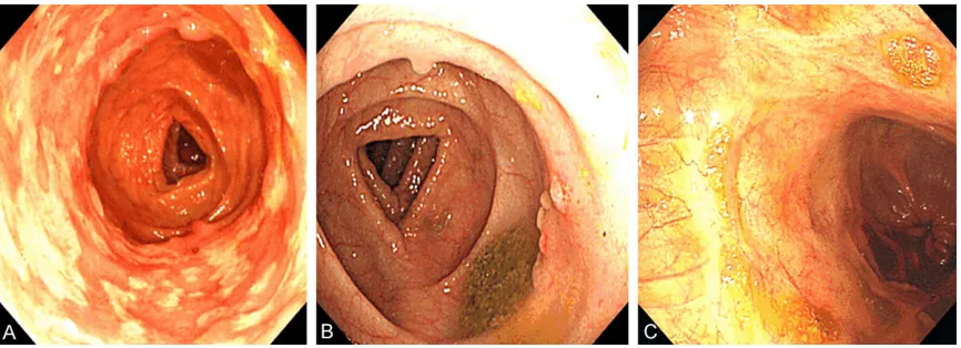
(Table 3; Figures 1-3). As shown in Table 3, for the assessments of active ulcers, the Healed rate and Improve rated in ITB group was 90.9% (20/22) and 9.1% (2/22) respectively in 3 mo after empiric therapy, and 100% (28/28) and 0 respectively in 6 mo. Statistical significance was found in both 3 mo and 6 mo when com-pared with the changes in CD group in which all cases remained Non-improved during the empiric therapy (P<0.001). For the assess-ments of mucosal nodules, the disappeared rate in ITB group was 58.8% in 3 mo and 76.5%
[image:5.612.87.525.71.224.2]in 6 mo after empiric therapy. As evaluated the rest 23.5% cases with still existed lesions in 6 mo, the amounts of mucosal nodules reduced obviously when compared with the baseline changes. For the assessments of pseudopol-yps, in 3 mo after empiric therapy, some cases in ITB group had fewer lesions, some had more, and some remained unchanged when com-pared with the baseline lesions. But in 6 mo and 12 mo, the amount of pseudopolyps re- duced obviously in 75.0% and 91.7% cases respectively in ITB group. Three cases in ITB group had luminal stricture before therapy, which leading to the failure of completing colo-noscopy examination. All strictures improved 3 mo after anti-tuberculosis therapy with suc-cessful pass of colonoscope. But one new situ-ation emerged which was narrowing of ileoce-cal valve with the failure of pass of colonscope. All the above lesions in CD group, including
Figure 2. A female intestinal tuberculosis patient aged 26. A. Before tuberculosis therapy; B. 3 mo after anti-tuberculosis therapy; C. 12 mo after anti-anti-tuberculosis therapy.
[image:5.612.93.523.283.438.2]mucosal nodules, pseudopolyps and strictures, did not improve in both 3 mo and 6 mo after anti-tuberculosis therapy.
The follow-up
Duration of following-up was 4 to 10 years since the end of empiric anti-tuberculosis in ITB group. No patients diagnosed as ITB recurred so far. As for the outcome of the 20 patients diagnosed and treated as CD, 1 received sur-gery because of intestinal obstruction, 1 devel-oped abdominal abscess in 4 mo but refused surgery, the other 18 cases improved obviously on medicines for treating CD, 10 of them received colonoscopy with mucosal healing or improvement.
Discussion
A number of reports have documented the dif-ferential diagnosis between ITB and CD [1-5]. Some characteristics are specific for ITB, such as caseating granulomas or acid-fast bacilli identified in the histological specimens; Co- existing active extra-intestinal tuberculosis or strong positive skin test suggests the necessity of empiric anti-tuberculosis. Other characteris-tics are likely to be found in CD, such as anal fistula/perianal abscess, extra-intestinal mani-festations, intestinal fistula and abdominal abscess [2]. However above characters are absent in most patients and not golden stan-dards for diagnosing CD or ITB [5, 6]. In our study, only 3 out of 28 ITB patients (10.7%) were confirmed at the initial work-up by histo-logical evidence. Less than half ITB patients (13/28) met any one of the following criteria: caseating granulomas, positive acid-fast stain-ing, active extra-intestinal tuberculosis, strong positive PPD-test. Perianal abscess which was inclined to CD diagnosis existed in two ITB patients. Eight CD patients (40.0%) had peri-anal abscess, but still received empiric anti-tuberculosis therapy because of the consider-ation of the possibility of ITB based on other manifestations in this patient. From above we can see, due to the lack of specific characteris-tics for differentiating ITB from CD, adequate course of empiric anti-tuberculosis therapy should be considered in patients with difficulty in differential diagnosis between ITB and CD. The endoscopic findings for distinction between ITB and CD have been well described recent years [5-12]. Segmental longitudinal ulcers and cobble stone appearance are regarded as
typi-cal colonoscopy features described in patients with tuberculous colitis. By contrast, transverse or linear ulcers are considered as typical chang-es in CD. We reviewed the endoscopic findings in our study, longitudinal ulcers and/or a cob-blestone appearance existed in about 1/3 cases in CD group, transverse or linear ulcers was found in about 1/3 cases in ITB group. Hence judge based on combining endoscopic and clinical features would make empiric anti-tuberculosis treatment more targeted. For bet-ter differentiating CD from ITB, the direction of our further study should be focus on standard-izing and quantstandard-izing the criteria of disease fea-tures [1].
between CD and ITB. Disappearance or obvi-ous improvement of active ulcer accompanied by the improvement of nodular transformation is engraved as responding to anti-tuberculosis therapy, and the therapy should be continued. For those with obvious improvement of ulcers instead of disappearance in 3 mo, the lesions are supposed to disappear in 6 mo after thera-py or the diagnosis should be reconsidered. For the management of pseudopolyps, the dura-tion of therapy should be long enough till pseu-dopolyps disappear. On the contrary, those with non-improvement of ulcers in 3 mo after anti-tuberculosis should be considered as inef-fective empiric therapy and the strategy should be shift to CD therapy.
Our study showed that the clinical symptoms started to improve within 2 weeks and improved obviously in 1 mo after anti-tuberculosis thera-py in ITB group. The obviously improved rate reached 100% in 1 mo. Cured was achieved in 3 mo in most patients. Noteworthily, clinical symptoms also improved obviously in a propor-tion of CD patients in the early stage of anti-tuberculosis and this could last 3 mo or longer after the stop of empiric therapy. But eventually recurrence took place thereafter. These CD patients may be misdiagnosed as ITB if judged only by symptoms improvement. Therefore, to evaluate the effectiveness of the empiric thera-py, we should combine colonoscopy with toms evaluation instead of evaluating symp-toms only. Endoscopic healed together with the evidence of clinical cured or nearly cured is engraved as effective. From our study we can also see that an understanding of how the intestine responds to the treatment of TB or CD is fundamental to decide whether empiric ther-apy should be chosen or not.
Although we have well-organized follow-up sys-tem and file management for CD and ITB patients, and our results were similar to those from Korea [4], the limitations of this study due to retrospective reason were needed to be solved. Further large sample prospective inves-tigations should be carried out to achieve more reliable conclusions.
Disclosure of conflict of interest
None.
Address correspondence to: Pinjing Hu and Minhu Chen, Department of Gastroenterology, The First
Affiliated Hospital of Sun Yat-Sen University, 58 Zhongshan II Road, Guangzhou 510080, P. R. China. Tel: +86-20-8775-5766-8172; Fax: +86-20-8733-2916; E-mail: [email protected] (PJH); [email protected] (MHC)
References
[1] Zhou Z and Luo H. Differential diagnosis be-tween Crohn’s disease and intestinal tubercu-losis in China. Int J Clin Pract 2006; 60: 212-214.
[2] Ouyang Q, Hu P, Qian J, Zheng J and Hu R. Chi-nese concensus on diagnosis and treatment standard of inflammatory bowel disease. Wei Chang Bing Xue 2007; 12: 488-495.
[3] Misra SP, Misra V, Dwivedi M and Gupta SC. Colonic tuberculosis: clinical features, endo-scopic appearance and management. J Gas-troenterol Hepatol 1999; 14: 723-729. [4] Park YS, Jun DW, Kim SH, Lee HH, Jo YJ, Song
MH, Kim NI and Lee JS. Colonoscopy evalua-tion after short-term anti-tuberculosis treat-ment in nonspecific ulcers on the ileocecal area. World J Gastroenterol 2008; 14: 5051-5058.
[5] Gu Q, Ouyang Q, Zhang W and Li G. [A com-parison of clinical and pathologic characteris-tics between Crohn’s disease and intestinal tuberculosis]. Zhonghua Nei Ke Za Zhi 2009; 48: 291-294.
[6] Li X, Liu X, Zou Y, Ouyang C, Wu X, Zhou M, Chen L, Ye L and Lu F. Predictors of clinical and endoscopic findings in differentiating Crohn’s disease from intestinal tuberculosis. Dig Dis Sci 2011; 56: 188-196.
[7] Lee Y, Yang S, Byeon J, Myung S, Chang H, Hong S, Kim K, Lee G, Jung H and Hong W. Analysis of colonoscopic findings in the differ-ential diagnosis between intestinal tuberculo-sis and Crohn’s disease. Endoscopy 2006; 38: 592-597.
[8] Pulimood A, Ramakrishna B, Kurian G, Peter S, Patra S, Mathan V and Mathan M. Endoscopic mucosal biopsies are useful in distinguishing granulomatous colitis due to Crohn’s disease from tuberculosis. Gut 1999; 45: 537-541. [9] Kirsch R, Pentecost M, Hall Pde M, Epstein DP,
Watermeyer G and Friederich PW. Role of colo-noscopic biopsy in distinguishing between Crohn’s disease and intestinal tuberculosis. J Clin Pathol 2006; 59: 840-844.
[10] Pulimood AB, Peter S, Ramakrishna B, Chacko A, Jeyamani R, Jeyaseelan L and Kurian G. Seg-mental colonoscopic biopsies in the differenti-ation of ileocolic tuberculosis from Crohn’s disease. J Gastroenterol Hepatol 2005; 20: 688-696.
RP, Sreenivas V and Gupta SD. Clinical, endo-scopic, and histological differentiations be-tween Crohn’s disease and intestinal tubercu-losis. Am J Gastroenterol 2010; 105: 642-651. [12] Yu H, Liu Y, Wang Y, Peng L, Li A and Zhang Y.
Clinical, endoscopic and histological differen-tiations between Crohn’s disease and intesti-nal tuberculosis. Digestion 2012; 85: 202-209.