Original Article
Post-extraction socket changes in diabetic patients - a
retrospective study
Sijia Zhang1, Shuyan Wang2, Hongbo Wei1, Dehua Li1, Yingliang Song1
1State Key Laboratory of Military Stomatology, Department of Implant Dentistry, School of Stomatology, The Fourth
Military Medical University, Xi’an 710032, Shaan Xi, P. R. China; 2State Key Laboratory of Military Stomatology,
Department of Preventive Dentistry, School of Stomatology, The Fourth Military Medical University, Xi’an 710032, Shaan Xi, P. R. China
Received March 14, 2016; Accepted June 8, 2016; Epub July 15, 2016; Published July 30, 2016
Abstract: It is very difficult to determine the time point of inserting implants for diabetic patients. And less we know about how diabetic sockets change with time. Based on the previous studies on non-diabetic socket healing, we analysed pre- and post-extraction cone beam computed tomography images of type 2 diabetes patients (n=93) and, age and sex matched non-diabetic controls (n=93) to investigate post-extraction socket dimensional changes. Post-extraction follow-up time ranged from three to 24 months. Socket dimensions (socket width and height, alveolar ridge width and height) were compared between the groups using computed tomography images. No significant differences between the groups were observed in pre-extraction socket dimensions. Post-extraction socket width and depth were significantly higher in diabetic group compared to non-diabetic group in all follow-up time points (3, 4-6, 7-12 and 13-24 months). Differences in alveolar ridge width and height between the groups at all the post-extraction time points were not statistically significant. An unfavourable, delayed early post-post-extraction socket healing was observed in diabetic patients. Therefore, implant insertion could be delayed four to six months post-extraction in diabetic patients for improved prognosis of prosthetic management.
Keywords: Socket dimensions, post-extraction socket, dental implants, diabetes
Introduction
Loss of dentition results in morphological changes in both hard and soft tissues in the alveolar region. Extraction of teeth is followed by horizontal and vertical changes in the dimen-sion of the alveolar socket/bone [1, 2]. These changes occur more rapidly in the first 3-6 months of the post-extraction period [3, 4]. Maintenance and preservation of alveolar bone after extraction is necessary to ensure both functional stability and aesthetics of the pros-thesis/restoration. The time and magnitude of these post-extraction socket changes are criti -cal for planning implant-based treatment strat-egies to manage edentulous space [5]. The long-term success of an implant is dependent on effective osseointegration, which is influ -enced by the amount of healthy bone at the implant placement site. Hence, the ridge dimensions of the edentulous space are critical for implant placement [6]. If the socket has
severe bone defects, it cannot provide ade-quate bone to ensure primary stability and osseointegration of the implant-based prosthe-sis [7]. Local factors that influence residual ridge resorption include location, trauma/surgi-cal technique during extraction, the number of teeth extracted, bone density, infection, and habits such as smoking. Bone healing and resorption can also be affected by systemic fac-tors, such as age, gender, metabolic disorders, and hormonal dysregulation, including that associated with diabetes mellitus.
lowering bone mineral density and increasing the risk for bone fracture [13]. It also increases the risk for osteopenia, osteoporosis, poor osseous healing, and impaired bone regenera-tion [14]. Reports on diabetic animal models have suggested delayed healing of post-extrac-tion sockets with unfavourable socket dimen -sions and healing response post-implantation [15-18]. However, the reports based on obser -vation in humans (i.e., diabetic patients and/or diabetic patients on glycaemic management) indicate no deleterious effects of diabetes on post-extraction socket healing [19-21]. Further-more, there is lack of adequate studies with detailed analysis providing an objective assess-ment regarding post-extraction socket dimen -sions over time, which would be critical for implant-based prosthetic strategies.
Few studies have reported changes in post-extraction socket dimensions in diabetic pa-tients. However, more studies are required for a clinical community to derive effective treat-ment planning for implant-based prosthesis in management of single-tooth edentulous space. The objective of this study is to evaluate the change in horizontal and vertical post-extrac-tion socket dimensions over time (healing) and the post-extraction socket morphology in dia -betic patients. The study also intends to deter-mine a favourable implant insertion time, based on the post-extraction socket dimen -sions in diabetic patients.
Materials and methods
Subjects and study design
The current retrospective cohort study was approved by the Ethics Committee of the Fourth Military Medical University (Ethics Approval Number: 2015 #kq-001) and was performed in accordance with the institutional ethical guide-lines. Information from the records of patients who sought implant-based prosthesis following tooth extractions at the Department of Implant Dentistry, School of Stomatology, The Fourth Military Medical University in China from 2013-2015 were included in the study. Data relevant to the study were obtained only from the records of those subjects who had already pro-vided informed consent for the use of data for research studies. The patient cohort included those with and without type 2 diabetes melli-tus. Non-diabetic patients were categorized as the control group (NG; n=93) and those with
type 2 diabetes formed the diabetes group (DG; n=93).
Patients’ records included information such as age, gender, permanent residence (urban ver-sus rural), smoking, other habits, occupation, systemic disease, and other relevant medical history. In addition, cone beam computed tomography (CBCT) images of both the tooth to be extracted (prior to extraction) and the post-extraction socket were essential for the current study.
Inclusion criteria: (1) For DG - confirmation of type 2 diabetes for more than three years, with a fasting blood glucose value ≥7.0 mmol/L or 2 hours plasma glucose ≥11.1 mmol/L [22] and with maximum HbA1c levels under 8.0% both before and after extraction [23]; (2) For NG - fasting blood glucose less than 7.0 mmol/L or 2 hrs plasma glucose less than 11.1 mmol/L, and maximum HbA1c levels under 6.0% before and after extraction [24]; (3) no missing teeth or bone defects on either side of the extraction site and absence of third molar if the tooth to be extracted was a second molar; (4) minimally invasive extraction or extraction with minimal trauma; (5) no record of post-operative infec-tion; (6) pre- and post-extraction CBCT images, along with relevant medical history; (7) written informed consent.
Exclusion criteria: (1) progressive periodontitis; (2) tooth extraction due to malignancy, peri-odontal conditions or complex facial trauma; (3) osteoporosis and/or other metabolic or nutritional deficiencies; (4) hematopoietic, digestive or autoimmune diseases; (5) steroidal therapy; (6) smoking. Control subjects (NG) included in the study were matched to mem-bers of the DG for age, gender, tooth position, post-extraction follow-up visits, location of per-manent residence, and socket dimensions. Socket dimensions included alveolar ridge width (ARW), alveolar ridge height (ARH), socket width (SW) and socket depth (SD). Similar resi -dential location and socket dimensions, in addition to other matched variables, were cho-sen to reduce variation in analysis caused by these factors.
Clinical parameters
and SD) and socket morphology as described eleswhwere [25]. ARW, ARH, SD, SW, and sock -et morphology were independently measured and analyzed with GALAXIS (Galileos, Sirona, Shanghai, China)software by two dentists who were blinded to the patients’ information pre- and post-extraction. The mean values of the measurements were recorded [26]. In the event of any disagreement on the measurements, the data were then evaluated by two additional dentists. All four dentists then took additional measurements as needed, until consensus was reached.
Alveolar ridge width and height measurements
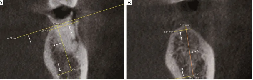
Pre-extraction: Buccolingual orientation of the CBCT images were used to measure alveolar ridge width (ARW) and alveolar ridge height (ARH). First, a reference line connecting the buccal and lingual alveolar bone peaks was drawn and termed the “ideal surface” (Figure 1A, arrow 1). The distance between the points connecting the outer surface of the buccal and lingual cortical plates parallel to and 1 mm below the ideal surface was considered the ARW(Figure 1A, arrow 2). ARH was measured perpendicular to the ideal surface (Figure 1A, arrows 3 and 4). In the anterior portion of the maxilla, ARH was measured as the distance from the nasal floor to the ideal surface. In the posterior portion of the maxilla, ARH was mea-sured as the distance from the floor of the max -illary sinus to the ideal surface. In the anterior portion of the mandible, ARH was measured as the distance from the inferior margin of the
mandible to the ideal surface. Whereas, in the posterior portion of the mandible, ARH was measured as the distance from the superior margin of the mandibular nerve canal to the ideal surface (Figure 1A, arrows 3 and 4).
[image:3.612.93.523.72.208.2]Post-extraction: The socket morphology and the planned implant direction influence the measurement of post-extraction ARW and ARH. In the presence of well-defined post-extraction socket morphology, intersecting reference lines are drawn based on tentative implant direction and the ideal surface; in the absence of well-defined post-extraction socket morphology, intersecting reference lines are drawn based on the tentative implant direction (Figure 1B, arrow 1) and the alveolar ridge surface. The dis-tance between the points connecting the outer surface of the buccal and lingual cortical plates parallel to and 1 mm below the ideal surface and perpendicular to the tentative implant direction/axis was considered the post-extrac-tion ARW(Figure 1B, arrow 2). ARH was mea-sured along the tentative implant direction/axis (Figure 1B, arrow 3). In the anterior portion of the maxilla, ARH was measured as the distance from the nasal floor to the intersection, and, in the posterior portion, it was measured as the distance from the floor of the maxillary sinus to the intersection. In the anterior portion of the mandible, ARH was measured as the distance from the inferior margin of the mandible to the intersection; in the posterior portion of the mandible, ARH was measured as the distance from the superior margin of the mandibular nerve canal to the intersection [27].
Socket width and depth measurements
Pre-extraction: Socket width (SW) and socket depth (SD) were measured in buccal-lingual and medial-distal directions. The “ideal sur-face” reference line was drawn as described earlier (Figure 2A, arrow 1), in this case be- tween the bucco-lingual and mesio-distal bone peaks. The distance between the points con -necting the inner surface of the buccal and lin-gual cortical plates parallel to and 1 mm below the ideal surface was considered the SWbl. Similar measurements from the edges of the socket made in the mesio-distal direction were considered the SWmd (Figure 2A, arrow 2). The higher of these two measurements (SWbl and SWmd) was considered as the SW. The measure-ment perpendicular to the ideal surface from the nadir point of the socket to the ideal sur -face was considered the SD (Figure 2A, arrow 3).
Post-extraction: In the presence of well-defined post-extraction socket morphology, intersect -ing reference lines were drawn based on tenta-tive implant direction (Figure 2B, arrow 1) and the ideal surface, drawn between the buccal-lingual bone peaks (Figure 2B, arrow 2). In the absence of well-defined post-extraction socket morphology, intersecting reference lines were drawn based on the tentative implant direction and the ideal surface as drawn between the mesio-distal bone peaks (Figure 1B and arrow 3). The distance between the points connecting the buccal-lingual/mesio-distal bone peaks 1 mm below the ideal surface and perpendicular to the tentative implant direction/axis was con-sidered the post-extraction SW (Figure 2B, arrow 3). SD was measured along the implant direction from the socket floor to the intersec -tion between tentative implant direc-tion axis and the ideal surface (Figure 3, arrow 1). For second molar post-extraction sockets, the ideal surface was drawn by referencing the alveolar bone of the retro-molar pad distally and the bony peak of the first molar mesially [27]. When the SD was less than 1 mm, the SW and SD were recorded as 0. Since the SD was less than 3 mm in many cases, SW was only measured at distance of 1 mm, instead of 1, 3 and 5 mm apical to the crest [2, 28].
Post-extraction follow-ups
The post-extraction socket dimension assess -ments were based on the follow-up schedule determined by the patients. Subsequent visits by patients were between three to 24 months. Therefore, the post-extraction socket dimen -Figure 2. Measurement of the socket width and depth. A. Representative image of pre-extraction socket (tooth posi-tion 36; DG), with arrows indicating: (1) the ideal surface; (2) socket width; and (3) socket depth. B. Representative image of post-extraction socket-nine months post-extraction (tooth position 36; DG) with well-defined post-extrac-tion socket morphology. Arrows indicate: (1) tentative implant direcpost-extrac-tion; (2) ideal surface; (3) socket width; and (4) reference line. Image captured using GALILEOS Implant, Sirona, Shanghai, China.
sion evaluation was categorized into four groups: three months, -four to six months, -seven to 12 months and 13 to 24 months. The difference in post-extraction evaluation be- tween the groups did not exceed 2 weeks.
Statistical analyses
The data were compiled using Microsoft Excel 2011, and SPSS 17.0 (SPSS Inc., Chicago, Illinois, USA) was used for statistical analyses. All the data were normally distributed. Chi-square tests and t-tests were used to deter-mine the statistical difference between the test groups. P < 0.05 was considered to be statisti-cally significant.
Results
Clinical information and CBCT images of pre- and post-extraction socket dimensions from a total of 93 subjects for each group (NG and DG) were analysed in the current retrospective study. The age in years for NG and DG were not significantly different (Table 1). Groups were matched for sex and residential location (Table 1), as well as for type/location and number of extracted teeth and post-extraction evaluation time (Table 2). Pre-extraction socket dimen -sions (SW, SD, ARW and ARH) did not differ sig-nificantly between the NG and DG (Table 3). However, post-extraction socket dimensions (SW and SD) were significantly different
be-tween the NG and DG (Table 3). Compared to the NG, the DG had higher SW and SD values in the post-extraction evaluations (at all follow-up time points; Table 3). In contrast, ARH values in the DG were lower in the post-extraction evalu-ations (at all follow-up time points) compared to the NG (Table 3), but the difference was not statistically significant. Similarly, no significant difference was observed in ARW in all the post-extraction evaluation time points (Table 3). A decrease in socket closure, as evidenced by a significantly lower difference between pre- and post-extraction SW and SD, was observed in the DG compared to the NG (Table 4). A signifi -cantly higher difference in post-extraction ARH was also observed in the DG compared to the NG (Table 4). However, the difference between pre- and post-extraction ARW was not signifi -cant between the DG and the NG (Table 4).
Discussion
The traditional recommendation for endosse-ous dental implant surgery requires that the thickness of alveolar bone be at least 5.5 mm in the buccal-lingual direction and the thick -ness of the buccal/labial and lingual bone be at least 1 mm to ensure both function and aes-thetics [29]. Studies have reported that bone and soft tissue characteristics and success rates following immediate implant placement or delayed implant (on healed ridges) place-ment are similar [30-32]. However, bone resorp
-Table 1. Study cohort details of diabetic and non-diabetic groups
Groups (n)
Gender Age (year) Post-extraction follow-up (months)
male female (χ ± SD) 3 4-6 7-12 13-24
DG (93) 72 21 55.9±7.6 35 6 2 50
NG (93) 72 21 56.0±6.9 35 6 2 50
t 0.081
p 0.372>0.05
NG-Non-diabetic group; DG-Diabetic group; SD-standard deviation.
Table 2. Extraction and post-extraction follow-up details in diabetic and non-diabetic groups
Groups (n)
Tooth position (FDI notation) Residence
Anterior teeth (18) Posterior teeth (75)
Urban Rural
11 12 21 31 41 42 16 17 26 27 36 37 46 47
DG 4 1 10 1 1 1 14 5 10 2 14 8 13 9 66 27
NG 4 1 10 1 1 1 14 5 10 2 14 8 13 9 67 26
χ2 0.026
P 0.871>0.05
tion was observed both at control and implant
sites, especially on the buccal socket [33]. Hence, buccal bone thickness and horizontal socket dimensions should be considered dur
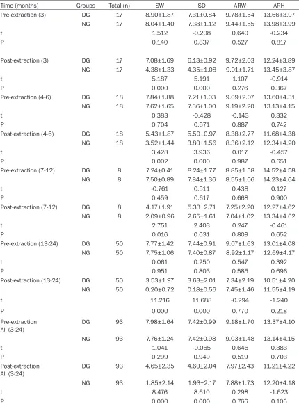
-Table 3. Socket dimensions-Pre-extraction and 3-24 months post-extraction (χ ± SD mm)
Time (months) Groups Total (n) SW SD ARW ARH
Pre-extraction (3) DG 17 8.90±1.87 7.31±0.84 9.78±1.54 13.66±3.97
NG 17 8.04±1.40 7.38±1.12 9.44±1.55 13.98±3.99
t 1.512 -0.208 0.640 -0.234
P 0.140 0.837 0.527 0.817
Post-extraction (3) DG 17 7.08±1.69 6.13±0.92 9.72±2.03 12.24±3.89
NG 17 4.38±1.33 4.35±1.08 9.01±1.71 13.45±3.87
t 5.187 5.191 1.107 -0.914
P 0.000 0.000 0.276 0.367
Pre-extraction (4-6) DG 18 7.84±1.88 7.21±1.03 9.09±2.07 13.60±4.31
NG 18 7.62±1.65 7.36±1.00 9.19±2.20 13.13±4.15
t 0.383 -0.428 -0.143 0.332
P 0.704 0.671 0.887 0.742
Post-extraction (4-6) DG 18 5.43±1.87 5.50±0.97 8.38±2.77 11.68±4.38
NG 18 3.52±1.44 3.80±1.56 8.36±2.12 12.34±4.20
t 3.428 3.936 0.017 -0.457
P 0.002 0.000 0.987 0.651
Pre-extraction (7-12) DG 8 7.24±0.41 8.24±1.77 8.85±1.58 14.52±4.58
NG 8 7.50±0.89 7.84±1.36 8.55±1.06 14.23±4.64
t -0.761 0.511 0.438 0.127
P 0.459 0.617 0.668 0.900
Post-extraction (7-12) DG 8 4.17±1.91 5.33±2.71 7.25±2.20 12.27±4.62
NG 8 2.09±0.96 2.65±1.61 7.04±1.02 13.34±4.62
t 2.751 2.403 0.247 -0.461
P 0.016 0.031 0.809 0.652
Pre-extraction (13-24) DG 50 7.77±1.42 7.44±0.91 9.07±1.63 13.01±4.08
NG 50 7.75±1.06 7.40±0.87 8.92±1.17 12.69±4.17
t 0.061 0.250 0.547 0.392
P 0.951 0.803 0.585 0.696
Post-extraction (13-24) DG 50 3.53±1.97 3.63±2.01 7.34±2.19 10.51±4.20
NG 50 0.20±0.72 0.18±0.56 7.45±1.46 11.55±4.19
t 11.216 11.688 -0.294 -1.240
P 0.000 0.000 0.770 0.218
Pre-extraction
All (3-24) DG 93 7.98±1.64 7.42±0.99 9.18±1.70 13.37±4.10
NG 93 7.76±1.24 7.42±0.98 9.03±1.48 13.14±4.15
t 1.041 -0.065 0.646 0.383
P 0.299 0.949 0.519 0.703
Post-extraction
All (3-24) DG 93 4.65±2.35 4.60±2.04 7.97±2.43 11.21±4.22
NG 93 1.85±2.14 1.93±2.17 7.88±1.73 12.20±4.18
t 8.476 8.610 0.298 -1.623
P 0.000 0.000 0.766 0.106
NG - Non-diabetic group; DG - Diabetic group; Socket width (SW); Socket depth (SD); Alveolar ridge width (ARW); Alveolar ridge
[image:6.612.92.522.84.665.2]ing implant placement [34]. The alveolar ridge undergoes horizontal and vertical reduction by 3.8 mm and 1.24 mm respectively, six months post-extraction [4]. In humans, horizontal bone dimension was reduced by 29-63% and vertical by 11-22%, six months post-extraction, and the observed decrease was rapid during the first three to six months post-extraction [3]. The ver -tical and horizontal bone dimensions at the implant site decreased by 0.5-1.0 mm during 4-12 months follow-up after immediate implant insertion [35]. In this study, the mean SW at three months post-extraction in the DG was 7.08±1.69 mm and 4.38±1.33 in the NG. The results suggest that the SW poses no risk to implant surgery in patients with healthy blood glucose levels, because there are implants with sufficiently large diameters from which to choose. However, for diabetic patients, it is dif-ficult to find an appropriate implant. If an implant of 4.5 mm diameter is normally chosen for such patients, there would be a mean of 2.5 mm of empty space around the implant neck that would need to be filled or left empty. Although the empty space may not affect pri-mary implant stability, it also may affect
osseo-integration around the implant neck, which can cause bone resorption and aesthetic risk [36]. Thus, healing time must be extended to achieve ideal socket healing for diabetic patients. Guided bone regeneration technique could be used to fill the empty space around the implant neck, which could cause marginal bone loss. Meanwhile, if implantation is not delayed to allow for slower healing in diabetic patients, dif-ficulties in finding appropriate implants and positioning can also lead to aesthetic compro-mises [6, 37].
Compared to healthy people, diabetic patients have a higher probability of periodontitis and of alveolar bone loss secondary to tooth loss [38].
[image:7.612.92.524.85.373.2]In vivo studies have reported unfavourable socket healing and alveolar bone destruction in diabetic animals [15-18]. Expression of TGF-beta isoforms and TGF-TGF-beta receptor genes, essential for wound healing, were significantly downregulated in the diabetic animals following tooth extraction [39, 40]. Decreased differenti -ation of osteoblasts and mineral apposition rates were observed in diabetic animals, con-tributing to delayed healing [18, 41]. Insulin-like
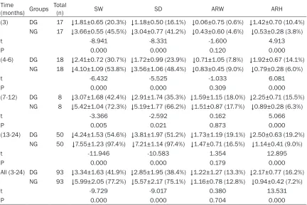
Table 4. Mean difference in post-extraction socket dimensions (χ ± SD mm) (rate of change)
Time
(months) Groups Total (n) SW SD ARW ARH
(3) DG 17 ↓1.81±0.65 (20.3%) ↓1.18±0.50 (16.1%) ↓0.06±0.75 (0.6%) ↓1.42±0.70 (10.4%) NG 17 ↓3.66±0.55 (45.5%) ↓3.04±0.77 (41.2%) ↓0.43±0.60 (4.6%) ↓0.53±0.28 (3.8%)
t -8.941 -8.331 -1.600 4.913
P 0.000 0.000 0.120 0.000
(4-6) DG 18 ↓2.41±0.72 (30.7%) ↓1.72±0.99 (23.9%) ↓0.71±1.05 (7.8%) ↓1.92±0.67 (14.1%) NG 18 ↓4.10±1.09 (53.8%) ↓3.56±1.06 (48.4%) ↓0.83±0.45 (9.0%) ↓0.79±0.28 (6.0%)
t -6.432 -5.525 -1.033 6.081
P 0.000 0.000 0.309 0.000
(7-12) DG 8 ↓3.07±1.68 (42.4%) ↓2.91±1.74 (35.3%) ↓1.59±1.15 (18.0%) ↓2.25±0.71 (15.5%) NG 8 ↓5.42±1.04 (72.3%) ↓5.19±1.77 (66.2%) ↓1.51±0.87 (17.7%) ↓0.89±0.28 (6.3%)
t -3.366 -2.592 0.162 5.066
P 0.005 0.021 0.873 0.000
(13-24) DG 50 ↓4.24±1.53 (54.6%) ↓3.81±1.97 (51.2%) ↓1.73±1.19 (19.1%) ↓2.50±0.63 (19.2%) NG 50 ↓7.55±1.23 (97.4%) ↓7.21±1.14 (97.4%) ↓1.47±0.71 (16.5%) ↓1.14±0.41 (9.0%)
t -11.946 -10.583 1.354 12.895
P 0.000 0.000 0.179 0.000
All (3-24) DG 93 ↓3.34±1.63 (41.9%) ↓2.85±1.95 (38.4%) ↓1.22±1.27 (13.3%) ↓2.17±0.77 (16.2%) NG 93 ↓5.99±2.05 (77.2%) ↓5.57±2.17 (75.1%) ↓1.16±0.78 (12.8%) ↓0.94±0.42 (7.2%)
t -9.729 -9.017 0.380 13.531
P 0.000 0.000 0.704 0.000
NG - Non-diabetic group; DG - Diabetic group; Socket width (SW); Socket depth (SD); Alveolar ridge width (ARW); Alveolar ridge
growth factor I (IGF-I) increases the differentia -tion of osteoblasts and mineraliza-tion of bone. Thus, treatment with IGF-1 improved alveolar bone morphology in diabetic rats exhibiting de- creased alveolar bone formation [15]. Similarly, metformin administration or Ellagic acid com-bined with statins improved post-extraction socket healing in rats [17, 42]. Mineral apposi -tion rate following immediate implant inser-tion was significantly decreased in the diabetic rats compared to the normal rats [41]. However, in humans, there have been favourable reports on post-extraction socket healing and implant outcomes in diabetic patients, especially in those with controlled hyperglycaemia [19-21, 43, 44]. Furthermore, improved post-extraction healing characteristics were observed with the use of plasma-rich growth factor (PRGF) in sockets of diabetic patients [45]. Bone quality has significant positive correlations with HbA1c level [46]. The results in our study were obtained from diabetic patients with good blood glucose control. However, in patients without good gly-cemic control, dental implant therapy must accommodate for delays to ensure adequate osseointegration [47].
However, the SD and SW were statistically dif-ferent between the DG and the NG as early as 3 months post extraction (P < 0.05). The mean socket width 4-6 months post extraction of dia -betic group was 5.43±1.87 mm and 3.52±1.44 mm of non-diabetic group. Four to six months post-extraction is thus the best time to insert implants for diabetics. The ARH 4-6 months post-extraction reduced 1.92 mm, indicating that a longer healing period may increase aes-thetic risk. Therefore, we suggest intervention, such as alveolar ridge preservation techniques (e.g., guided bone regeneration), early on. Depending on pre-extraction bone loss, bone augmentation can be carried out [48, 49]. Many clinical trials confirm that alveolar ridge preservation techniques can reduce the height and width of reduction at extraction sites [27, 50, 51]. These techniques can reduce aesthet -ic risk and achieve required bone density for implant insertion [52]. Since all existing studies have focused on non-diabetic patients, the implications for diabetic patients are rather unclear.
Based on this study and others, we have the following recommendations for diabetic pa-tients: early intervention, minimally invasive
extraction, good blood glucose control, smok -ing cessation, and a 4-6 month delay in implant insertion [53]. Whether alveolar ridge preserva -tion techniques could be used in diabetics remains unknown, and delay of prosthesis will increase the risk of unfavourable aesthetics and a prolonged treatment period [54]. To opti -mize treatment strategies, reduce treatment time, and improve treatment outcome, the rel-evance of routine alveolar ridge preservation techniques in diabetics needs further study.
Acknowledgements
The authors acknowledge and thank the Na-tional Natural Science Foundation of China for funding this study (No. 81170984; 81470775). The authors also thank Dr. Wei Ma, Dr. Chao Xie and Dr. Yan Liu, for their assistance with statis-tics and language.
Disclosure of conflict of interest
None.
Address correspondence to: Yingliang Song, State Key Laboratory of Military Stomatology, Department of Implant Dentistry, School of Stomatology, The Fourth Military Medical University, Xi’an 710032, Shan Xi, P. R. China. E-mail: [email protected]
References
[1] Schropp L, Kostopoulos L and Wenzel A. Bone healing following immediate versus delayed placement of titanium implants into extraction sockets: a prospective clinical study. Int J Oral Maxillofac Implants 2003; 18: 189-199. [2] Lekovic V, Kenney EB, Weinlaender M, Han T,
Klokkevold P, Nedic M and Orsini M. A bone regenerative approach to alveolar ridge main-tenance following tooth extraction. Report of 10 cases. J Periodontol 1997; 68: 563-570. [3] Tan WL, Wong TL, Wong MC and Lang NP. A
systematic review of post-extractional alveolar hard and soft tissue dimensional changes in humans. Clin Oral Implants Res 2012; 23 Suppl 5: 1-21.
[4] Hammerle CH, Araujo MG and Simion M. Evidence-based knowledge on the biology and treatment of extraction sockets. Clin Oral Implants Res 2012; 23 Suppl 5: 80-82. [5] Hammerle CH, Chen ST and Wilson TG Jr.
[6] Mecall RA and Rosenfeld AL. Influence of re-sidual ridge resorption patterns on implant fix-ture placement and tooth position. 1. Int J Periodontics Restorative Dent 1991; 11: 8-23. [7] Heinemann F, Hasan I, Bourauel C, Biffar R
and Mundt T. Bone stability around dental im-plants: Treatment related factors. Ann Anat 2015; 199: 3-8.
[8] Guariguata L, Whiting DR, Hambleton I, Beagley J, Linnenkamp U and Shaw JE. Global estimates of diabetes prevalence for 2013 and projections for 2035. Diabetes Res Clin Pract 2014; 103: 137-149.
[9] Kaur G, Holtfreter B, Rathmann WG, Schwahn C, Wallaschofski H, Schipf S, Nauck M and Kocher T. Association between type 1 and type 2 diabetes with periodontal disease and tooth loss. J Clin Periodontol 2009; 36: 765-774. [10] Patel MH, Kumar JV and Moss ME. Diabetes
and tooth loss An analysis of data from the National Health and Nutrition Examination Survey, 2003-2004. J Am Dent Assoc 2013; 144: 478-485.
[11] Kim EK, Lee SG, Choi YH, Won KC, Moon JS, Merchant AT and Lee HK. Association between diabetes-related factors and clinical periodon-tal parameters in type-2 diabetes mellitus. Bmc Oral Health 2013; 13: 64.
[12] Adeghate E, Schattner P and Dunn E. An up-date on the etiology and epidemiology of dia-betes mellitus. Ann N Y Acad Sci 2006; 1084: 1-29.
[13] Vestergaard P. Discrepancies in bone mineral density and fracture risk in patients with type 1 and type 2 diabetes--a meta-analysis. Osteo-poros Int 2007; 18: 427-444.
[14] Retzepi M, Lewis MP and Donos N. Effect of diabetes and metabolic control on de novo bone formation following guided bone regen-eration. Clin Oral Implants Res 2010; 21: 71-79.
[15] Fang Y, Wang LP, Du FL, Liu WJ and Ren GL. Effects of insulin-like growth factor I on alveo-lar bone remodeling in diabetic rats. J Periodontal Res 2013; 48: 144-150.
[16] Devlin H, Garland H and Sloan P. Healing of tooth extraction sockets in experimental diabe-tes mellitus. J Oral Maxillofac Surg 1996; 54: 1087-1091.
[17] Inouye KA, Bisch FC, Elsalanty ME, Zakhary I, Khashaba RM and Borke JL. Effect of metfor-min on periimplant wound healing in a rat model of type 2 diabetes. Implant Dent 2014; 23: 319-327.
[18] Colombo JS, Balani D, Sloan AJ, Crean SJ, Okazaki J and Waddington RJ. Delayed osteo-blast differentiation and altered inflammatory response around implants placed in incisor sockets of type 2 diabetic rats. Clin Oral Implants Res 2011; 22: 578-586.
[19] Huang S, Dang H, Huynh W, Sambrook PJ and Goss AN. The healing of dental extraction sock-ets in patients with Type 2 diabetes on oral hy-poglycaemics: a prospective cohort. Aust Dent J 2013; 58: 89-93.
[20] Joshipura K. Glycemic control is not related to postextraction healing in patients with diabe-tes. J Evid Based Dent Pract 2011; 11: 187-188.
[21] Fernandes KS, Glick M, de Souza MS, Kokron CM and Gallottini M. Association between im-munologic parameters, glycemic control, and postextraction complications in patients with type 2 diabetes. J Am Dent Assoc 2015; 146: 592-599.
[22] Kerner W and Bruckel J. Definition, classifica-tion and diagnosis of diabetes mellitus. Exp Clin Endocrinol Diabetes 2014; 122: 384-386. [23] Association AD. Standards of medical care in
diabetes--2015: summary of revisions. Dia- betes Care 2015; 38 Suppl: S4.
[24] Wei X, E M and Yu S. A meta-analysis of pas-sive smoking and risk of developing Type 2 Diabetes Mellitus. Diabetes Res Clin Pract 2015; 107: 9-14.
[25] Hoang TN and Mealey BL. Histologic compari-son of healing after ridge preservation using human demineralized bone matrix putty with one versus two different-sized bone particles. J Periodomtol 2012; 83: 174-181.
[26] Kim TS, Obst C, Zehaczek S and Geenen C. Detection of bone loss with different X-ray techniques in periodontal patients. J Perio- domtol 2008; 79: 1141-1149.
[27] Zhan YL, Hu WJ, Zhen M, Xu T and Lu RF. Radiographic evaluation of ridge preservation after molar tooth extraction: a controlled clini-cal trial. Beijing Da Xue Xue Bao 2015; 47: 19-26.
[28] Araujo MG, Silva CO, Misawa M and Sukekava F. Alveolar socket healing: what can we learn? Periodontol 2000 2015; 68: 122-134. [29] Adell R, Lekholm U, Rockler B and Branemark
PI. A 15-year study of osseointegrated implants in the treatment of the edentulous jaw. Int J Oral Surg 1981; 10: 387-416.
[30] Cooper LF, Reside GJ, Raes F, Garriga JS, Tarrida LG, Wiltfang J, Kern M and De Bruyn H. Immediate provisionalization of dental im-plants placed in healed alveolar ridges and extraction sockets: a 5-year prospective evalu-ation. Int J Oral Maxillofac Implants 2014; 29: 709-717.
[31] Pal US, Dhiman NK, Singh G, Singh RK, Mohammad S and Malkunje LR. Evaluation of implants placed immediately or delayed into extraction sites. Natl J Maxillofac Surg 2011; 2: 54-62.
sites. Int J Oral Maxillofac Implants 2009; 24 Suppl: 186-217.
[33] Vignoletti F, Discepoli N, Muller A, de Sanctis M, Munoz F and Sanz M. Bone modelling at fresh extraction sockets: immediate implant placement versus spontaneous healing: an ex-perimental study in the beagle dog. J Clin Periodontol 2012; 39: 91-97.
[34] Ferrus J, Cecchinato D, Pjetursson EB, Lang NP, Sanz M and Lindhe J. Factors influencing ridge alterations following immediate implant placement into extraction sockets. Clin Oral Implants Res 2010; 21: 22-29.
[35] Lee CT, Chiu TS, Chuang SK, Tarnow D and Stoupel J. Alterations of the bone dimension following immediate implant placement into extraction socket: systematic review and meta-analysis. J Clin Periodontol 2014; 41: 914-926. [36] Chrcanovic BR, Albrektsson T and Wennerberg
A. Reasons for failures of oral implants. J Oral Rehabil 2014; 41: 443-476.
[37] Mecall RA and Rosenfeld AL. The influence of residual ridge resorption patterns on implant fixture placement and tooth position. 2. Presurgical determination of prosthesis type and design. Int J Periodontics Restorative Dent 1992; 12: 32-51.
[38] Khader YS, Dauod AS, El-Qaderi SS, Alkafajei A and Batayha WQ. Periodontal status of diabet-ics compared with nondiabetdiabet-ics: a meta-analy-sis. J Diabetes Complications 2006; 20: 59-68.
[39] Yamano S, Kuo WP and Sukotjo C. Down- regulated gene expression of TGF-betas in dia-betic oral wound healing. J Craniomaxillofac Surg 2013; 41: e42-48.
[40] Younis WH, Al-Rawi NH, Mohamed MA and Yaseen NY. Molecular events on tooth socket healing in diabetic rabbits. Br J Oral Maxillofac Surg 2013; 51: 932-936.
[41] Shyng YC, Devlin H and Ou KL. Bone formation around immediately placed oral implants in diabetic rats. Int J Prosthodont 2006; 19: 513-514.
[42] Al-Obaidi MM, Al-Bayaty FH, Al Batran R, Hussaini J and Khor GH. Impact of ellagic acid in bone formation after tooth extraction: an ex-perimental study on diabetic rats. Sci World J 2014; 2014: 908098.
[43] Aronovich S, Skope LW, Kelly JP and Kyriakides TC. The relationship of glycemic control to the outcomes of dental extractions. J Oral Maxillofac Surg 2010; 68: 2955-2961.
[44] Bell C and Bell RE. Immediate restoration of NobelActive implants placed into fresh extrac-tion sites in the anterior maxilla. J Oral Implantol 2014; 40: 455-458.
[45] Mozzati M, Gallesio G, di Romana S, Bergamasco L and Pol R. Efficacy of plasma-rich growth factor in the healing of postextrac-tion sockets in patients affected by insulin-de-pendent diabetes mellitus. J Oral Maxillofac Surg 2014; 72: 456-462.
[46] Nemtoi A, Ladunca O, Dragan E, Budacu C, Mihai C and Haba D. Quantitative and qualita-tive bone assessment of the posterior mandi-ble in patients with diabetes mellitus: a cone beam computed tomography study. Rev Med Chir Soc Med Nat Iasi 2013; 117: 1002-1008. [47] Oates TW, Huynh-Ba G, Vargas A, Alexander P
and Feine J. A critical review of diabetes, glyce-mic control, and dental implant therapy. Clin Oral Implants Res 2013; 24: 117-127.
[48] Chrcanovic BR, Albrektsson T and Wennerberg A. Diabetes and oral implant failure: a system-atic review. J Dent Res 2014; 93: 859-867. [49] Retzepi M and Donos N. Guided Bone
Regeneration: biological principle and thera-peutic applications. Clin Oral Implants Res 2010; 21: 567-576.
[50] Heinemann F, Hasan I, Schwahn C, Bourauel C and Mundt T. Bone level change of extraction sockets with Bio-Oss collagen and implant placement: a clinical study. Ann Anat 2012; 194: 508-512.
[51] Bartee BK. Extraction site reconstruction for alveolar ridge preservation. Part 2: membrane-assisted surgical technique. J Oral Implantol 2001; 27: 194-197.
[52] Kesmas S, Swasdison S, Yodsanga S, Sessirisombat S and Jansisyanont P. Esthetic alveolar ridge preservation with calcium phos-phate and collagen membrane: preliminary report. Oral Surg Oral Med Oral Pathol Oral Radiol Endod 2010; 110: e24-36.
[53] Holtzclaw D. Extraction site preservation using new graft material that combines mineralized and demineralized allograft bone: a case se-ries report with histology. Compend Contin Educ Dent 2014; 35: 107-112.