Original Article
Protective effects of tamoxifen on traumatic brain injury
in rats
Xiaodong Qing1, Peilong Jiang1, Li Tang2
1Department of Neurosurgery, Ningbo Yinzhou Second Hospital, Ningbo, P. R. China; 2Department of Renal
Medicine, Ningbo Yinzhou Second Hospital, Ningbo, P. R. China
Received September 18, 2015; Accepted March 29, 2016; Epub April 15, 2016; Published April 30, 2016
Abstract: Objective: To investigate the neuroprotective effects of tamoxifen on traumatic brain injury in rats and
po-tential mechanisms. Methods: The modified Feeney freefall method was used to establish a rat model of traumatic
brain injury. Male Sprague-Dawley (SD) rats were randomly dividedinto four groups: sham surgery group, traumatic brain injury group, traumatic brain injury + solvent treatment group, and traumatic brain injury + tamoxifen
treat-ment group. The experitreat-ment included two parts. During the first part, drugs were provided via intraperitoneal injec -tion at 1 and 8 hours after traumatic brain injury, a histopathological examina-tion of the brain tissue was performed at 24 hours after traumatic brain injury in rats, and the relevant mechanisms were explored. During the second part of the experiment, tamoxifen was provided at 1, 8, 24 and 72 hours after injury, and neurological function
was assessed after the rats survived for seven days. Results: Tamoxifen treatment significantly inhibited nuclear transfer of nuclear factor kappa-light-chain-enhancer of activated B cells (NF-κB) p65 in the brain tissue surround
-ing the traumatic brain injury site (P < 0.05) and significantly reduced the expression levels of tumor necrosis factor-α (TNF-α), interleukin 1 (IL-1) β and cleaved-caspas-3 (P < 0.05). In addition, tamoxifen significantly reduced
neuronal apoptosis after traumatic brain injury, the extent of neuronal damage, and neurological defects (P < 0.05). Conclusion: Tamoxifen exerts a neuroprotective effect on traumatic brain injury, which may be related to inhibiting
the NF-kB-mediated inflammatory response.
Keywords: Brain injury, tamoxifen, inflammatory response
Introduction
Traumatic brain injury has high prevalence, morbidity and mortality worldwide [1]. With the rapid development of the economy and society in China, the prevalence of traumatic brain inju-ry has risen over the years, which has heavily burdened society and patients’ families. At present, no drugs effectively treat traumatic brain injury [2]. Recent studies showed that tamoxifen features good neuroprotective effects [3-6], but few studies have investigated its protective effects in traumatic brain injury and potential mechanisms. In this study, we investigated the neuroprotective effects of tamoxifen in traumatic brain injury and the potential mechanisms to provide new ideas and methods for clinically treating patients with traumatic brain injury.
Materials and methods
Experimental animals and groups
For experiment 1, 45 adult healthy male
Sprague-Dawley (SD) rats were randomly assigned to one of four groups: the sham sur-gery group, traumatic brain injury group, trau-matic brain injury + solvent treatment group, and traumatic brain injury + tamoxifen
treat-ment group. Five rats died following traumatic
brain injury, and additional rats were randomly assigned to replenish the number of rats. None of the rats in the sham surgery group died. The
Feeney method was modified to establish a rat
ethanol disinfection, a 2 cm-long incision was made along the scalp midline. Next, an ortho-pedic drill was used to make a bone opening 5 mm in diameter at 3 mm right of the coronal suture midline and 3 mm posterior to the sagit-tal suture. The dura remained intact. A 40 g weight was dropped from 15 cm above and allowed to freefall vertically to strike the right dura pad, which caused an injury 3 mm deep and 4 mm in diameter. The rats in the sham surgery group were only subject to bone open-ing. A tamoxifen treatment regimen and dose were provided in reference to previous litera-ture [7]. Intraperitoneal injection of tamoxifen was provided 1 and 8 hours after successful establishment of the rat model of traumatic brain injury. Rats in the solvent group only received an intraperitoneal injection of solvent.
All rats were sacrificed under anesthesia 24
hours after traumatic injury, and their necks
were broken to obtain brain tissue. For experi -ment 2, 87 rats were randomly assigned to the sham surgery group, the traumatic brain injury group, the traumatic brain injury + solvent treat-ment group, and the traumatic brain injury + tamoxifen treatment group. All the rats received an intraperitoneal injection of tamoxifen 1, 8, 24 and 72 hours after traumatic brain injury, and their neurological function was assessed 1, 3, 5 and 7 days after traumatic brain injury. Seven rats died following traumatic brain injury, and additional rats were randomly assigned to replenish the number of rats. No rats died in the sham surgery group.
Neurological function assessment in rats after traumatic brain injury
After traumatic brain injury, the neurological
neurological severity scores (NSS) [8], which mainly included a motor function test, a senso-ry function test, a balance test, physiological
reflex defects, and abnormal movements. The
highest score was 18 points. The rats were given 1 point if they could not perform the task or lacked the appropriate response. Moreover, 13-18 points indicated a severe injury, 7-12 points indicated a moderate injury, and 1-6 points indicated a mild injury.
Western blot analysis
Twenty-four hours after traumatic brain injury, the rats were anesthetized with an intraperito-neal injection of chloralhydrate; the chest was then opened, and 100 ml of saline was injected into the left apex. When the liver turned white, the neck was quickly broken, and the brain was removed; the brain tissue surrounding the inju-ry site was then maintained in a freezer at -80°C for later use. The nuclear and cytoplas-mic proteins were extracted in strict accor-dance with the extract kit instructions (Biyuntian Biotech Co., Ltd.), and the Bradford assay was used to measure the protein extract concentra-tion. Next, the protein was mixed with 5 × load-ing buffer at a 1:4 ratio and boiled in water for 10 minutes; 35 micrograms of the denatured protein was then added to each well for electro-phoresis and transferred to a membrane. Next, the membrane was blocked for 1 hour; diluted primary anti-nuclear factor
kappa-light-chain-enhancer of activated B cells (NF-κB) p65, anti-H3, anti-cleaved-caspase-3, and anti-β-actin
antibodies (1:1,000, purchased from Cell Signaling Technology, Inc., USA) were then added, and the membrane was placed on a shaker at 4°C overnight. Next, the secondary antibody was added, and the sample was incubated followed by washing the membrane, adding enhanced chemiluminescence (ECL) agents, and gray-scale analysis with the Image J software.
Immunohistochemistry and terminal deoxy-nucleotidyl transferase dUTP nick end labeling (TUNEL) assay
Twenty-four hours after the injury, 10 rats in each group were perfused with paraformalde-hyde; their necks were broken, and their brains
were removed, embedded in paraffin and pre
-pared as 4 μm sections. For immunohisto -chemistry, the brain tissue sections were depa-Figure 1. Detection of the p65 and
[image:2.612.90.288.71.158.2]Table 1. Analysis of the assay results for each group in this study
Group N P65 caspase-3Cleavage (pg/mg)TNF-α (pg/mg)IL-1β of TUNEL-pos-The number
itive cells
Nissl stain-ing (cells)
The number of
cleaved-caspase-3-positive cells
Sham surgery group 10 0.12 ± 0.01 0.09 ± 0.01 42.1 ± 6.3 17.6 ± 3.6 2.3 ± 0.1 87.3 ± 9.6 9.2 ± 0.9
repaired in citrate buffer. Next, the slides were blocked with normal goat serum, and the anti-cleaved-caspase-3 antibody (1:50) was added. The slides were placed into a wet pot and incu-bated for 24 h at 4°C overnight followed by a PBS wash 3 times 5 minutes each. Next, horse-radish peroxidase-labeled goat anti-rabbit IgG and horseradish peroxidase-labeled streptavi-din-biotin were added; 3,3’-diaminobenzidine was used as the luminescence agent. The slides were mounted and observed under a light microscope; images were collected. Ten
fields were counted on each slide, and the mean value was used as the final result for the slide. Four slides were prepared for each rat. A
TUNEL staining kit was purchased from Roche (USA), and the assay was performed in strict accordance with the instructions.
Enzyme-linked immunosorbent assay of in-flammatory cytokine expression
Tumor necrosis factor-α (TNF-α) and interleukin 1 (IL-1) β enzyme-linked immunosorbent assay
kits were purchased from UNOCI Biotechnology
Co., Ltd. For each rat, 50 mg of brain tissue sur -rounding the injury site was collected, added into 500 ml of PBS, placed into a tissue homog-enizer for thorough homogenization, and then stored in a refrigerator at 4°C for later use. During the experiment, the sample was centri-fuged at 14,000 rpm for 10 minutes, the super-natant was collected, and the protein concen-tration was measured using the Bradford assay in strict accordance with the instructions.
Statistical analysis
SPSS 15.0 software was used to process the data, which were expressed as the mean ± standard deviation (x ± S). The neurological
using a Kruskal-Wallis test, and one-way analy-sis of variance was performed for the remaining multi-group comparisons.
Results
Effects of tamoxifen on the NF-κB signaling pathway
Compared with the sham surgery group (Figure 1; Table 1), the NF-κB p65 protein expression level was significantly higher in the brain tissue
nuclei surrounding the injury site in the trau-matic brain injury group (P < 0.05); however,
tamoxifen significantly inhibited p65 protein
expression in the nucleus (P < 0.05).
Effects of tamoxifen on nerve cell apoptosis after traumatic brain injury
The Western blot analyses (Figure 1) showed that the expression level of cleaved-caspase-3, which is an apoptosis execution protein, was
significantly higher in brain tissue surrounding
the injury site after traumatic brain injury;
however, tamoxifen significantly reduced the
cleaved-caspas-3 protein expression level (P < 0.05). Immunohistochemical results show that compared with the sham surgery group,
trau-matic brain injury in rats yielded significantly
more cleaved-caspase-3 positive cells in brain tissue surrounding the injury site. However,
tamoxifen significantly reduced the number
of cleaved-caspase-3 positive cells (Figure 2; Table 1, P < 0.05), which is consistent with the Western blot analysis. TUNEL-positive cells showed positive results in the nucleus. This study (Figure 3; Table 1) showed significantly
more TUNEL-positive cells in brain tissue sur-rounding the injury site after traumatic brain
[image:4.612.94.520.77.186.2]Effects of tamoxifen on inflammatory cytokine expression after traumatic brain injury
Compared with the control group (Table 1), the
expression levels of inflammatory cytokines TNF-α and IL-1β significantly increased in brain
tissue surrounding the injury site after traumat-ic brain injury (P < 0.05); furthermore, com-pared with the solvent treatment group,
tamoxi-fen significantly reduced the TNF-α and IL-1β
expression levels (P < 0.05).
Nissl staining results
Nissl staining showed that normal neurons were round with no staining, but the damaged
neurons exhibited an irregular shape or shrank with staining. Compared with the sham surgery group (Figure 4; Table 1), the traumatic brain injury and solvent treatment groups (P < 0.05) exhibited fewer undamaged neurons; however,
tamoxifen significantly increased the number of
undamaged neurons (P < 0.05).
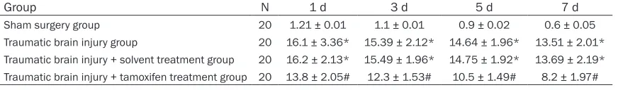
Effects of tamoxifen treatment on the neuro-logical function assessment score after trau-matic brain injury in rats
Compared with the sham surgery group, the
[image:5.612.93.522.79.198.2]neurological defect score was significantly high -er 1, 3, 5 and 7 days aft-er traumatic brain injury in the traumatic brain injury and solvent treat-Figure 3. The effects of tamoxifen treatment on apoptosis after injury.
Figure 4. Nissl staining results.
Table 2. The effects of tamoxifen on the neurological function assessment score after traumatic brain injury in rats
Group N 1 d 3 d 5 d 7 d
Sham surgery group 20 1.21 ± 0.01 1.1 ± 0.01 0.9 ± 0.02 0.6 ± 0.05
[image:5.612.95.522.245.354.2] [image:5.612.90.526.425.489.2]ment groups (Table 2; P < 0.05); however,
tamoxifen treatment significantly reduced the
neurological defect score (Table 2; P < 0.05). Discussion
Traumatic brain injury can be categorized as primary or secondary; primary brain injury refers to brain damage directly caused by an external force, which is beyond the scope of clinical interventions, and secondary brain inju-ry occurs a few hours to a few days after injuinju-ry, which is the focus of traumatic brain injury clini-cal treatments [9]. Previous studies have shown that a variety of pathological factors, such as
oxidative stress, the inflammatory response
and apoptosis, are involved in secondary brain
injury after traumatic brain injury. Furthermore,
early interventions to reduce the level of
oxida-tive stress and the extent of the inflammatory response can significantly reduce the extent of
traumatic brain injury [10]. Tamoxifen is a syn-thetic partial agonist of the estrogen receptor with certain estrogen-like effects and is now widely used to clinically treat breast cancer. Tamoxifen is convenient to administer and is quickly absorbed into the body after oral admin-istration. Its serum concentration peaks as soon as 4-7 hours after oral administration and its elimination half-life is approximately 7 days [11]. Recent studies have shown that tamoxifen exerts good neuroprotective effects, which are
related to its anti-inflammatory and antioxidant
role [5, 12]. This study showed that tamoxifen
significantly reduced neuronal apoptosis and
the number of damaged neurons after
traumat-ic brain injury. In addition, tamoxifen signiftraumat-icant -ly improved neurological defects after traumat-ic brain injury, whtraumat-ich suggests that tamoxifen exerts good neuroprotective effects after trau-matic brain injury.
Tamoxifen not only reduces lipopolysaccharide-induced activation of microglias, thus exerting
an anti-inflammatory effect on in vitro experi -ments, but also reduces the extent of the
inflammatory response and pathological dam
-age to brain tissue. Thus, it exerts significant
neuroprotective effects in animal models of spinal cord injury and subarachnoid
hemor-rhage [5, 6, 13]. After the data confirmed that
tamoxifen exerts good neuroprotective effects after traumatic brain injury, we further explored the potential mechanisms with a focus on the
effects of tamoxifen on NF-κB-mediated inflam
-matory responses. Previous studies have
shown that NF-κB plays an important role in
secondary brain injury after traumatic brain
injury; NF-κB activity was significantly increased
in brain tissue surrounding the injury site after
traumatic brain injury, and inhibiting NF-κB activity significantly reduced the extent of brain
injury [14]. Once transferred into the nucleus,
NF-κB p65 binds a specific target gene sequence to regulate downstream inflammato
-ry cytokine expression (TNF-α and IL-1β). The TNF-α and IL-1β expression levels significantly increased in brain tissue, cerebrospinal fluid
and blood after traumatic brain injury, which are closely related to a patient’s prognosis.
Inhibiting TNF-α and IL-1β expression signifi -cantly reduced the extent of pathological dam-age after traumatic brain injury [15]. Caspase-3 is a critical apoptosis execution protein after traumatic brain injury and is expressed in ani-mal models of traumatic brain injury and hu- man brain tissue. Inhibiting cleaved-caspase-3
(mature caspase-3 protein) expression signifi -cantly reduced nerve injury [16]. This study
showed that the NF-κB p65, cleaved-caspas-3, TNF-α and IL-1β expression levels significantly
increased in brain tissue surrounding the injury
site. However, tamoxifen significantly reduced the nuclear transfer of NF-κB p65 and the expression levels of TNF-α and IL-1β in brain tis -sue surrounding the injury site, which is consis-tent with previous studies and suggests that
inhibition of NF-κB-mediated inflammatory sig -naling pathways may be one mechanism under-lying the neuroprotective effects of tamoxifen.
In summary, tamoxifen plays a significant neu -roprotective role in the animal model of trau-matic brain injury, which may be related to
inhi-bition of NF-κB-mediated inflammatory path
-ways. Based on its safety profile in human and
the results of this study, tamoxifen may be used to treat patients with traumatic brain injury in
clinical practice. Future studies should include
a more detailed and comprehensive investiga-tion of the mechanisms underlying tamoxifen’s neuroprotective effects to lay a solid theoreti-cal foundation for its clinitheoreti-cal application.
Disclosure of conflict of interest
None.
1 Qianhe Road, Yinzhou District, Ningbo 315100, Zhejiang, P. R. China. Tel: +86 574-55662011; 137-
32119148; Fax: +86 574-55662011;
1373211-9148; E-mail: tangli508@126.com
References
[1] Chantsoulis M, Mirski A, Rasmus A, Kropotov JD, Pachalska M. Neuropsychological rehabili-tation for traumatic brain injury patients. Ann Agric Environ Med 2015; 22: 368-79.
[2] Bergold PJ. Treatment of traumatic brain injury
with anti-inflammatory drugs. Exp Neurol
2016; 275 Pt 3: 367-80.
[3] Wei HY, Ma X. Tamoxifen reduces infiltration of inflammatory cells, apoptosis and inhibits IKK/ NF-kB pathway after spinal cord injury in rats.
Neurol Sci 2014; 35: 1763-8.
[4] Franco Rodríguez NE, Dueñas Jiménez JM, De
la Torre Valdovinos B, López Ruiz JR, Hernández
Hernández L, Dueñas Jiménez SH. Tamoxifen
favoured the rat sensorial cortex regeneration after a penetrating brain injury. Brain Res Bull 2013; 98: 64-75.
[5] Salgado IK, Torrado AI, Santiago JM, Miranda JD. Tamoxifen and Src kinase inhibitors as neuroprotective/neuroregenerative drugs af-ter spinal cord injury. Neural Regen Res 2015; 10: 385-90.
[6] Sun X, Ji C, Hu T, Wang Z, Chen G. Tamoxifen as an effective neuroprotectant against early
brain injury and learning deficits induced by
subarachnoid hemorrhage: possible
involve-ment of inflammatory signaling. J Neuro-inflammation 2013; 10: 157.
[7] Wang GH, Zhang XG, Jiang ZL, Li X, Peng LL, Li YC, Wang Y. Neuroprotective effects of hyper-baric oxygen treatment on traumatic brain in-jury in the rat. J Neurotrauma 2010; 27: 1733-43.
[8] Si D, Li J, Liu J, Wang X, Wei Z, Tian Q, Wang H, Liu G. Progesterone protects blood-brain barri-er function and improves neurological out-come following traumatic brain injury in rats. Exp Ther Med 2014; 8: 1010-1014.
[9] Paterniti I, Cordaro M, Navarra M, Esposito E, Cuzzocrea S. Emerging pharmacotherapy for treatment of traumatic brain injury: targeting
hypopituitarism and inflammation. Expert Opin
Emerg Drugs 2015; 20: 583-96.
[10] Gyoneva S, Ransohoff RM. Inflammatory reac -tion after traumatic brain injury: therapeutic potential of targeting cell-cell communication by chemokines. Trends Pharmacol Sci 2015; 36: 471-80.
[11] Morad SA, Cabot MC. Tamoxifen regulation of sphingolipid metabolism-Therapeutic impli-cations. Biochim Biophys Acta 2015; 1851: 1134-1145.
[12] Boulos AS, Deshaies EM, Dalfino JC, Feustel
PJ, Popp AJ, Drazin D. Tamoxifen as an effec-tive neuroprotectant in an endovascular ca-nine model of stroke. J Neurosurg 2011; 114: 1117-26.
[13] Sun XB and Ma C. Neuroprotective effect of tamoxifen on early brain injury in rat model with subarachnoid hemorrhage. Jiangsu Medi- cal Journal 2013; 39: 1888-1890.
[14] Yang L, Tao LY, Chen XP. Roles of NF-kappaB in
central nervous system damage and repair. Neurosci Bull 2007; 23: 307-13.
[15] Corps KN, Roth TL, McGavern DB. Inflammation
and neuroprotection in traumatic brain injury. JAMA Neurol 2015; 72: 355-62.
[16] Keane RW, Kraydieh S, Lotocki G, Alonso OF,
Aldana P, Dietrich WD. Apoptotic and antiapop-totic mechanisms after traumatic brain injury.