Original Article
Resveratrol inhibited the formation
of NLRP3 inflammatory body by activating
autophagy signal pathway in atherosclerosis
Yue Wang*, Li Fan*, Junfeng Zhang, Yuqi Fan, Qizhi Chen, Kan Chen, Lin Gao, Huasu Zeng, Changqian Wang#, Zhihua Han#
Department of Cardiology, No. 9 People’s Hospital, School of Medicine, Shanghai Jiao Tong University, 639 Zhizaoju Road, Shanghai 200011, P.R. China. *Equal contributors and co-first authors. #Equal contributors. Received February 9, 2017; Accepted November 14, 2017;Epub December 15, 2017; Published December 30, 2017
Abstract: Atherosclerosis (AS) is a common disease and it seriously endangers human health. Studies have found that inflammation theory is a possible pathogenesis of AS. Howevere, the specific mechanism between AS and in-flammation remains to be elaborated. Therefore the objective of this study was to explore the association between NLRP3 inflammatory body and autophagy signal pathway in AS and evaluate the therapeutic effect of resveratrol (RSV) on AS and its underlying mechanism. In the study, we detected the function of RSV on the expression of inflammation related genes and activated autophagy related genes in vitro. And we tested RSV’s effect on the oc-currence and development of AS in vivo. From the results, we found that RSV could inhibit the mRNA expression of SIRT1, NF-kB and NLRP3 and activate the mRNA expression of mTOR, p70S6Kaxl and LC3-I/II with a concentration dependent. Consistently, western bloting showed that RSV could decrease the protein expression of pNF-kB and NLRP3 and increased the protein expression of LC3-II/I, Beclin1 and mTOR with a concentration dependent. In ad-dition, RSV decreased the IL-1β level in serum of ApoE-/- mice. Moreover, the fluorescence intensity of Dil-OX-LDL was significantly enhanced in RSV+ group in Raw cells at per concentration of Dil-OX-LDL, including 6.25, 12.5, 50, 100 μg/ml. And animal experiment also indicated that RSV attenuated AS of ApoE-/- mice. In conclusion, RSV may inhibit the formation of NLRP3 inflammatory body and attenuate inflammation by activating autophagy signal pathway in AS.
Keywords: Resveratrol, NLRP3, inflammation, autophagy, mTOR
Introduction
Atherosclerosis (AS) is a common disease, which seriously endangers human health, and is the main pathological basis of ischemic car-diovascular and cerebrovascular diseases such as coronary heart disease, cerebrovascular dis-ease and thromboembolic disdis-ease [1, 2]. So far, the pathogenesis of AS is not yet fully un-
derstood. There are a variety of theories, which involve a variety of risk factors, but still lack
effective clinical medicine for treatment of AS. A large number of basic and clinical studies and
investigations have indicated variety of risk fac -tors for AS, including hyperlipidemia [3], hyper-tension [4], hyperglycemia (diabetes) [5], hyper-
fibrinogenemia [6], hyperhomocysteinemia [7],
hyperuricemia [8], obesity [8], renin-angioten-
smoking, coagulation hyperthyroidism (tissue
factor, thrombin) [10], metabolic disorders of trace elements (iron, copper, zinc, selenium, chromium, manganese, germanium, etc.) [11], autologous bioactive substances (such as se-
rotonin, NO, endothelin-1) [12]. Moreover, the
theory of the pathogenesis of AS includes lipid
infiltration theory [13], retention theory [14],
vascular smooth muscle cell cloning theory [15], oxidative stress theory [16], platelet hy- perfunction theory, thrombosis theory [17], Ca2+ super Load theory [18], immune dysfunc-tion theory, the theory of shear stress, injury
NLRP3, fully named nucleotide binding and
oligomerization domain-like receptor family pyrin domain-containing 3, is a famous inflam
-mation-related gene, which encodeds the key protein of NLRP3 inflammatory body and plays a crucial role in the process of inflammation. And the NLRP3 inflammatory body consists of NLRP3, apoptosis-associated speck-like pro -tein containing a caspase recruitment domain
(ASC) and caspase-1 or caspase-5 [19]. Over
the two decades, emerging evidences have de-
monstrated that the NLRP3 inflammatory body
was associated with AS [20, 21]. Autophagy is
a kind of self-stabilizing mechanism of eukary -otic organisms, which could degrade intracel-lular dysfunctional organelles, misclassify pro-teins and other harmful macromolecules to maintain normal function of cells. Autophagy process is regulated by the
autophagy-associ-ated gene (ATG), including microtubule associ
-ated protein 1 light chain 3 (LC3A and LC3B) and target of rapamycin (TOR), in which, autoph -agy cell could apply its double - layer membrane structure to wrap the obsolete, damaged pro-teins or organelles and degrade them after fus-ing with lysosomes into autophagy lysosomes [22]. Macrophages, endothelial cells (ECs) and smooth muscle cells (SMCs), which are
consid-ered as the three types of key cells in formation
and stability of AS, were reported to promote the development of AS by regulating the
forma-tion of complex regulatory network through the
expression of adhesion molecules and
secre-tion of cytokine interacsecre-tions [23].
In this study, we aimed to explore the
associa-tion between NLRP3 inflammatory body and
autophagy signal pathway in AS and evaluate the therapeutic effect of resveratrol (RSV) on AS and investigate its underlying mechanism.
Materials and methods Cell culture
RAW264.7 cells and a mouse monocyte/mac-rophage cell line were obtained from American
Type Culture Collection (ATCC) (Manassas, VA,
USA) and maintained in an atmosphere with
5% CO2 in Dulbecco’s modified Eagle’s medium (DMEM; Gibco-BRL, Grand Island, NY, USA) sup -plemented with 10% heat-inactivated fetal bo-
vine serum (FBS; HyClone, Logan, UT, USA) and
1% antibiotic-antimycotic (Invitrogen, Grand
Island, NY, USA). The RAW264.7 cells were maintained by weekly passage, and the cells
were utilized for experimentation at 60-80%
confluence. In addition, LPS-stimulated
RAW-264.7 cells were established by the activation of lipopolysaccharide (LPS).
RSV concentration administration
According to the concentration of RSV, all cells
were divided into four groups, including 0 μM, 1 μM, 10 μM, 100 μM group, of which, 0 μM
group was wild RAW264.7 cells and the other groups were LPS-stimulated RAW264.7 cells.
Animal study
All the protocol of animal experiment was approved by the Laboratory Animal Admin-
istration Committee of Shanghai Jiao Tong
University and was carried out in accordance with the Guidelines for Animal Experimentation.
A total of 20 ApoE-/- mice (10-week-old males, C57BL/6J background) were randomly divided
into two groups (n = 10 per group), including
high-fat diet group and high-fat diet +RSV (100)
group, were fed a chow diet of 1.25%
choles-terol for 20 weeks. 10 wild-type mice (C57BL/ 6J, 10 week-old males) were served as a con
-trol group. After 20 weeks feed, all mice were sacrificed by decapitated and the aortas tis -sues of mice were isolated and collected for the further investigations. And aortas tissue of per mice was divided into two portions, including the upper (aortic root) portion for histologic analysis and the abdominal/thoracic aorta for
mRNA and protein expression analyses. Blood
was immediately obtained for analyses.
Histology
The heart and whole aorta were immediately extracted when mice were sacrificed by decapi
-tated. The aorta was embedded in optimal cut
-ting temperature (OCT) embedding medium (Tissue-Tek, Sakura Finetek USA, Torrance, CA). Then hematoxylin-eosin (HE) staining was used
to determine the morphology of atherosclerotic
plaque. The aorta (except for the aortic root)
from each mouse from all the groups were removed and stored in -80°C.
RNA isolation and real-time PCR
In accordance with the manufacturer’s instruc -tions, total RNA was isolated from the cells
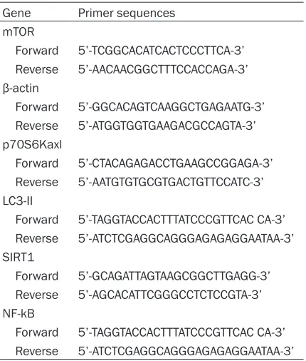
Table 1. All primer sequences for RT-PCR
Gene Primer sequences mTOR
Forward 5’-TCGGCACATCACTCCCTTCA-3’ Reverse 5’-AACAACGGCTTTCCACCAGA-3’ β-actin
Forward 5’-GGCACAGTCAAGGCTGAGAATG-3’ Reverse 5’-ATGGTGGTGAAGACGCCAGTA-3’ p70S6Kaxl
Forward 5’-CTACAGAGACCTGAAGCCGGAGA-3’ Reverse 5’-AATGTGTGCGTGACTGTTCCATC-3’ LC3-II
Forward 5’-TAGGTACCACTTTATCCCGTTCAC CA-3’ Reverse 5’-ATCTCGAGGCAGGGAGAGAGGAATAA-3’ SIRT1
Forward 5’-GCAGATTAGTAAGCGGCTTGAGG-3’ Reverse 5’-AGCACATTCGGGCCTCTCCGTA-3’ NF-kB
Forward 5’-TAGGTACCACTTTATCCCGTTCAC CA-3’ Reverse 5’-ATCTCGAGGCAGGGAGAGAGGAATAA-3’
Reverse Primer (0.6 μl), cDNA (2 μl), ROX Re-ference Dye II (0.4 μl), SYBR Premix Ex Taq (10 μl), and ddH2O (6.6 μl), and tested by the DA7600 Real-time Nucleic Acid Amplification Fluorescence Detection System (Bio-Rad).
GA-PDH was used as internal control in present
study. The primers used for mTOR, p70S6Kaxl, LC3-II, IL-1β, SIRT1, NF-kB and NLRP3 in this
study were recorded in Table 1. The levels of relative expression were quantified using the
2-ΔΔCT threshold cycle method.
Quantification of released IL-1β, IFN-γand
TNF-α
Concentration of interleukin (IL)-1β, interferon-γ (IFN-γ), tumor necrosis factor-α (TNF-α) in mice serum were determined using mouse IL-1β, IFN-γ and TNF-α enzyme-linked immuno sorbent as-say (Elisa) kit (4A Biotech, China) according to the manufacturer’s protocol.
Western blotting
Cells were collected and lysed by RIPA buffer
and BCA assay was used to detect the protein
concentration, and equal amount of protein were electrophoresed on 10% sodium dodecyl
sulfate-polyacrylamide gel (Bio-Rad, USA) and transferred onto poly vinylidene difluoride mem-branes (PVDF, Millipore). After blocking for non
-specific binding by 5% skim milk, the mem
-brane was incubated with specific primary anti
-bodies against mTOR (1:1000; Abcam),
beclin-1 (beclin-1:beclin-1000; Abcam), LC3-I/II (beclin-1:beclin-1000; Abcam),
pNF-kB (1:1000; Abcam) and NLRP3 (1:1000;
Abcam) overnight at 4°C. After washing with tris
buffered saline Tween (TBST) 3 times, mem -branes were incubated with secondary antibod-ies (Abcam) for 1 h at room temperature. Immunofluorescence assay
Cells were grown on glass slices and fixed in 4%
formaldehyde for 10 min, permeabilized th-
rough 0.3% Triton X-100. Then the slices were blocked in goat serum for 15 min, 37°C and incubated overnight at 4°C with anti-LC3B (1:80, Bioworld, MN, USA), anti-NLRP3 (1:80, Bioworld, MN, USA). Samples were washed three times before incubated with goat TRITC labeled secondary antibody (1:70, Bioworld,
MN, USA) at 37°C for 1 h. DAPI (GenviewInc, Shanghai, China) was used for counterstaining.
Then the cells were examined under a laser scanning microscope (TCSSP2-AOBS-MP, Leica
Microsystems CMS). Dil-ox-LDL uptake
Cells were incubated with 1 µg/ml Dil-ox-LDL for 2 hours. Upon completion of incubation,
cells were gently washed with 1× PBS three
times to remove free Dil-ox-LDL and analyzed
using fluorescent microscope
(TCSSP2-AOBS-MP, Leica Microsystems CMS).
Statistical analysis
Continuous variables are presented as mean and standard deviation (SD). Data analysis was conducted by SPSS 19.0 software. Statistical analysis was carried out by GraphPad Prism5.0 (San Diego, CA, USA). Comparisons between
two groups were made using the Student’s
t-test. P value <0.05 was considered
statisti-cally significant. All data were obtained from at
least three independent experiments.
Results
RSV inhibited the mRNA expression of inflam
-mation related genes
As shown in Figure 1, compared with that in 0
Figure 2. Effect of RSV on IL-1β, IFN-γ, TNF-α serum levels of mice by Elisa. Compared with the 0 mM group, *P<0.05, **P<0.01, ***P<0.001, differenc-es were statistically significant.
Figure 1. The mRNA expression of inflammation related genes were determined by QPCR after add-ing RSV. Compared with the 0 mM group, *P<0.05, ***P<0.001, differences were statistically signifi-cant.
0.11±0.02 vs. 1.0±0.10, P<0.05, P<0.001, P<
0.001, respectively). Moreover, the mRNA ex-
pression of NF-KB were remarkably decreased in 0.1 μM, 1 μM and 100 μM group compared with 0 μM group (0.90±0.13, 0.48±0.13, 0.17± 0.03 vs. 1.0±0.09, P<0.05, P<0.001, P<0.001,
respectively), and the mRNA expression of NL-
RP3 was remarkably decreased in 0.1 μM, 1 μM and 100 μM group compared with 0 μM group (0.85±0.14, 0.40±0.11, 0.11±0.02 vs. 1.0±0.10, P<0.05, P<0.001, P<0.001, respec
-tively). These results indicated that RSV inhibit
-ed the mRNA expression of inflammation relat -ed genes with a concentration dependent.
RSV decreased the IL-1β, IFN-γ and TNF-α
lev-els in serum of mice
To further investigate the effect of RSV on inflammation, Elisa assay was used to detect the expression levels of IL-1β, IFN-γ and TNF-α
in serum of mice. As shown in Figure 2,
com-pared with that in 0 μM group, the IL-1β, IFN-γ
and TNF-α levels were significantly inhibited in 0.1 μM, 1 μM and 100 μM group, respectively. In addition, the IL-1β, IFN-γ and TNF-α levels among 0.1 μM, 1 μM and 100 μM group had an obvious statistically significant (P<0.05).
The-se results also demonstrated that RSV could
decrease inflammation with a concentration
dependent.
RSV inhibited the protein expression of inflam
-mation related genes
To validate the effect of RSV on protein expres
-sion of inflammation related gene, western blot
was applied for detecting the protein
expres-sion of inflammation related gene, including NF-kB and NLRP3. As shown in Figure 3, com
-pared with that in 0 μM group, the protein expression of NLRP3 were significantly inhibit
[image:4.612.90.290.75.182.2]-ed in 0.1 μM, 1 μM and 100 μM group, respec -tively (P<0.05). In addition, the expression dif-ference of protein expression of NLRP3 among Figure 3. Western Blotting was used to evaluate the function of RSV inhibiting theprotein expression of inflammation related genes and activated autophagy related genes. GAPDH was used as a loading control.
[image:4.612.89.290.265.372.2] [image:4.612.325.524.277.388.2]ly significant (P<0.05). A
consistent conclusion sh- owed that the levels of
IL-1β, TNF-α and IFN-γ
de-creased with the increase in RSV concentration.
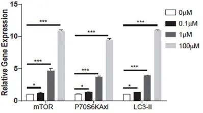
RSV activated
autoph-agy in LPS-stimulated
RAW264.7 cells
As shown in Figure 4, compared with that in
0 μM group, the mRNA expression of mTOR we-re significantly incwe-reas- increas-ed in 0.1 μM, 1 μM and 100 μM group, respec
-tively (1.21±0.12, 4.67± 0.34, 10.89±0.19 vs. 1.0±0.02, P<0.05).
More-over, the mRNA expres-sion of p70S6Kaxl was
remarkably overexpress-ed in 0.1 μM, 1 μM and 100 μM group compared with 0 μM group (1.30± 0.13, 3.67±0.15, 9.48± 0.23 vs. 1.0±0.01, P<
0.05), and the express- ion difference among 0.1
μM, 1 μM and 100 μM
group had an obvious
sta-tistically significant (P<
0.05, P<0.001, P<0.001). In addition, the mRNA ex- pression of LC3-II was
remarkably elevated in 0.1 μM, 1 μM and 100 μM group compared with 0 μM group (1.31±0.05, 3.89±0.11, 11.02±0.25 vs. 1.0±0.02, P<0.05, P<
[image:5.612.92.397.69.221.2]0.001, P<0.001). More- over, we further investi-gated the effect of RSV on the protein expression Figure 6. RSV promoted Raw cells to swallow Dil-OX-LDL. Compared with the RSV-
group, ***P<0.001, differences were statistically significant. (Original magnifica-tion: ×200).
0.1 μM, 1 μM and 100 μM group had an obvi
-ous statistically significant (P<0.05). Compared with that in 0 μM group, the protein expression of pNF-kB were significantly inhibited in 0.1 μM, 1 μM and 100 μM group, respectively (P<0.05).
In addition, the expression difference of
pro-tein expression of pNF-kB among 0.1 μM, 1 μM and 100 μM group had an obvious
statistical-of autophagy related genes. As showed in
Fig-ure 3, compared with that in 0 μM group, the protein expression of mTOR were significantly increased in 0.1 μM, 1 μM and 100 μM group,
respectively (P<0.05, P<0.001, P<0.001). Simi- lar results showed in the protein expression of
[image:5.612.91.393.275.566.2]LC3-II/I and Beclin1. In addition, immunofluo -rescence assay showed that RSV could inhibit Figure 5. Effects of RSV on autophagy and inflammation related genes were
Over the past several
de-cades, pathological exami-nation revealed that AS had the basic
characteris-tics of inflammatory
res-ponse, such as degenera-tion, exudation and hyper-plasia [27, 28]. According to the different of nature of
inflammation and inflam
-matory substances, inflam -matory response related to AS could be divided
into biological inflamma
[image:6.612.89.387.72.212.2]-tion, immune inflammation
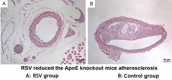
Figure 7. RSV attenuated AS in ApoE-/- mice. (Original magnification: ×200).
the formation of inflammatory body NLRP3 and activate the autophagy protein LC3B, and this
effect also had a concentration dependent ( Fig-ure 5). These results indicated that RSV acti -vated autophagy in LPS-stimulated RAW264.7 cells.
RSV promoted Raw cells to swallow Dil-OX-LDL
As shown in Figure 6, compared with that in
RSV- group, the fluorescence signal of Dil-OX-LDL was significantly enhanced in RSV+ group
in Raw cells at per concentration, including
6.25, 12.5, 25, 50, 100 μg/ml. These results
indicated that RSV promoted Raw cells to
swal-low Dil-OX-LDL.
RSV attenuates AS of ApoE-/- mice
As shown in Figure 7, compared with control group, AS was obviously attenuated in RSV group in ApoE-/- mice.
Discussion
As we all known, AS is one type of common dis -ease and seriously threats human health [24]. As the mayor pathological basis of coronary heart disease, cerebrovascular disease and thromboembolic disease, the pathogenesis of AS is not yet fully understood, which leads to
the lack of effective clinical targeted medicines [25, 26]. Therefore, it is crucial for us to explore
the underlying mechanism of pathogenesis of
AS and find more effective medicines for the
patients with AS. In this study, we found that
there was a close association between inflam -mation, autophagy and AS, and RSV might in-
hibit the formation of NLRP3 inflammatory body
by activating autophagy signal pathway in AS.
and chemical inflammation, of which, immuno
-logical inflammation has a crucial role in the formation of AS [29-31]. Of note, inflammatory
response associated with AS is regulated by a
series of inflammation-related genes and pa-thways, including NLRP3 and NF-kB [32-34].
Paramel et al. [35] showed that mRNA
expres-sion of CARD8 was significantly
overexpress-ed in AS plaques comparoverexpress-ed with normal
ves-sels, which could promote inflammation. In
our study, we found that RSV could inhibit the
mRNA and protein expression of SIRT1, NF-kB
and NLRP3 with a concentration dependent.
And immunofluorescence assay also showed that RSV could inhibit the formation of inflam -matory bodies NLRP3. Moreover, Elisa assay
indicated that RSV decreased the IL-1β, IFN-γ and TNF-α levels in serum of ApoE-/- mice. As we all known, IL-1β, IFN-γ and TNF-α were close -ly associated with incidence and development of AS [36-39]. Similarly, Alarcón et al. [40] found that the strawberry played a protective effect on thromboembolic-related disorders and
anti-inflammation through decreasing IL-1β level. In
addition, we also found that RSV could
obvi-ously attenuate AS of ApoE-/- mice. These
re-sults indicated that RSV could attenuate in-
flammation and plaque formation of AS and play a beneficial role in AS.
Over the past decades, more and more evi -dences showed that cell autophagy played a crucial role in the progression of AS [41, 42].
Through degradation of intracellular damage structure to adapt to oxidation, inflammation,
and other cell damage [43-45]. After knockout of Beclin1 and Atg5 gene, inflammatory mark
-ers in the plaque increased significantly, which confirmed that there was a close link between inflammation and autologous deletion [46].
Liao et al. [47] found that inhibition of
autopha-gy by silencing ATG5 enhanced apoptosis and
NADPH oxidase-mediated oxidative stress, whi- ch increased apoptosis and oxidative stress in advanced lesioned macrophages, promoted plaque necrosis, and worsened lesioned effero-cytosis. Moreover, autophagy has been shown to play an important role in the development of AS, which could degrade lipid droplets in the cell and regulate lipid metabolism [48, 49].
Wang et al. [50] found that the deficiency of Pdcd4 gene significantly improved oxidized low-density lipoproteins-impaired autophagy efflux,
which could promote autophagy-mediated lipid degradation and prevent macrophage conver-sion into foam cells. In our study, the results indicated that RSV could increase the mRNA
expression of mTOR, p70S6Kaxl and LC3-I/II and protein expression of LC3-II/I, Beclin1 and mTOR with a concentration dependent. Similar-ly, immunofluorescence assay showed that RSV could activate the autophagy protein LC3B.
Mo-reover, RSV promoted Raw cells to swallow
Dil-OX-LDL. These results demonstrated that RSV
might attenuate AS by activating autophagy signal pathway in AS.
In conclusion, our study found that there was a
close association between inflammation and
autophagy in AS, and RSV may inhibit the
for-mation of NLRP3 inflammatory body by activat -ing autophagy signal pathway in AS.
Acknowledgements
The work was supported by Shanghai health
development planning commission (Grant
num-ber: ZY3-CCCX-3-3006), the national natural
science foundation of China (Grant number: 81500392), and the national natural science foundation of China (Grant number: 813000- 87).
Disclosure of conflict of interest
None.
Address correspondence to: Zhihua Han and Changqian Wang, Department of Cardiology, No. 9 People’s Hospital, School of Medicine, Shanghai
Jiao Tong University, 639 Zhizaoju Road, Shanghai 200011, P.R. China. E-mail: changqianwang123@ 163.com
References
[1] Arbab-Zadeh A and Fuster V. The risk con- tinuum of atherosclerosis and its implications for defining CHD by coronary angiography. J Am Coll Cardiol 2016; 68: 2467-2478.
[2] Chan NC, Bhagirath V and Eikelboom JW. Profile of betrixaban and its potential in the prevention and treatment of venous thrombo-embolism. Vasc Health Risk Manag 2015; 11: 343-351.
[3] Montserrat-de la Paz S, Bermudez B, Cardelo MP, Lopez S, Abia R and Muriana FJ. Olive oil and postprandial hyperlipidemia: implications for atherosclerosis and metabolic syndrome. Food Funct 2016; 7: 4734-4744.
[4] Hurtubise J, McLellan K, Durr K, Onasanya O, Nwabuko D and Ndisang JF. The different fac-ets of dyslipidemia and hypertension in athero-sclerosis. Curr Atheroscler Rep 2016; 18: 82. [5] Sugimoto T, Sato M, Dehle FC, Brnabic AJ,
Weston A and Burge R. Lifestyle-related meta- bolic disorders, osteoporosis, and fracture risk in asia: a systematic review. Value Health Reg Issues 2016; 9: 49-56.
[6] Badeinikova KK, Mazaev AP, Toguzova ZA, Mamedov MN and Didigova RT. [Detection of early markers of atherosclerosis in men with various levels of risk of cardiovascular compli-cations]. Kardiologiia 2014; 54: 35-39. [7] Li H, He C, Wang J, Li X, Yang Z, Sun X, Fang L
and Liu N. Berberine activates peroxisome proliferator-activated receptor gamma to in-crease atherosclerotic plaque stability in Apoe-/- mice with hyperhomocysteinemia. J Diabetes Investig 2016; 7: 824-832.
[8] Liu Y, Liu C, Shi X, Lin M, Yan B, Zeng X, Chen N, Lu S, Liu S, Yang S, Li X and Li Z. Correlations of non-alcoholic fatty liver disease and serum uric acid with subclinical atherosclerosis in obese Chinese adults. J Diabetes 2017; 9: 586-595.
[9] Nehme A, Cerutti C and Zibara K. Transcriptomic analysis reveals novel transcription factors as-sociated with renin-angiotensin-aldosterone system in human atheroma. Hypertension 2016; 68: 1375-1384.
[10] Erem C, Suleyman AK, Civan N, Mentese A, Nuhoglu I, Uzun A, Ersoz HO and Deger O. Ischemia-modified albumin and malondialde-hyde levels in patients with overt and subclini-cal hyperthyroidism: effects of treatment on oxidative stress. Endocr J 2015; 62: 493-501. [11] El Dib R, Gameiro OL, Ogata MS, Modolo NS,
Beletate V. Zinc supplementation for the pre-vention of type 2 diabetes mellitus in adults with insulin resistance. Cochrane Database Syst Rev 2015; Cd005525.
[12] Yoon JJ, Lee YJ, Han BH, Choi ES, Kho MC, Park JH, Ahn YM, Kim HY, Kang DG and Lee HS. Protective effect of betulinic acid on early ath-erosclerosis in diabetic apolipoprotein-E gene knockout mice. Eur J Pharmacol 2017; 796: 224-232.
[13] Caligiuri G. [Role of the immune response in atherosclerosis and acute coronary syndro- mes]. Med Sci (Paris) 2004; 20: 175-181. [14] Camelon KM, Hadell K, Jamsen PT, Ketonen
KJ, Kohtamaki HM, Makimatilla S, Tormala ML and Valve RH. The plate model: a visual meth-od of teaching meal planning. DAIS project group. Diabetes atherosclerosis intervention study. J Am Diet Assoc 1998; 98: 1155-1158. [15] Groves J, Wang Z and Newman WH. Two dis-tinct phenotypes of rat vascular smooth mus-cle cells: growth rate and production of tumor necrosis factor-alpha. Am Surg 2005; 71: 546-550; discussion 550-541.
[16] Lankin VZ and Tikhaze AK. Role of oxidative stress in the genesis of atherosclerosis and diabetes mellitus: a personal look back on 50 years of research. Curr Aging Sci 2017; 10: 18-25.
[17] Sosa I, Strenja Linic I, Bajek S, Cuculic D, Crncevic-Orlic Z, Grubesic A, Cvijanovic O and Bosnar A. Corticosteroids provoke acute endo-thelial injury -- an ideal ground for thrombosis in multiple sclerosis. J Biol Regul Homeost Agents 2012; 26: 131-134.
[18] Lebeau P, Al-Hashimi A, Sood S, Lhotak S, Yu P, Gyulay G, Pare G, Chen SR, Trigatti B, Prat A, Seidah NG and Austin RC. Endoplasmic re- ticulum stress and Ca2+ depletion differen- tially modulate the sterol regulatory protein PCSK9 to control lipid metabolism. J Biol Chem 2017; 292: 1510-1523.
[19] Qiu YY and Tang LQ. Roles of the NLRP3 in-flammasome in the pathogenesis of diabetic nephropathy. Pharmacol Res 2016; 114: 251-264.
[20] Ozaki E, Campbell M and Doyle SL. Targeting the NLRP3 inflammasome in chronic inflam-matory diseases: current perspectives. J In- flamm Res 2015; 8: 15-27.
[21] Chen Z, Martin M, Li Z and Shyy JY. Endothelial dysfunction: the role of sterol regulatory ele-ment-binding protein-induced NOD-like recep-tor family pyrin domain-containing protein 3 inflammasome in atherosclerosis. Curr Opin Lipidol 2014; 25: 339-349.
[22] Martinet W and De Meyer GR. Autophagy in atherosclerosis: a cell survival and death phe-nomenon with therapeutic potential. Circ Res 2009; 104: 304-317.
[23] Mach F, Schonbeck U, Sukhova GK, Bourcier T, Bonnefoy JY, Pober JS and Libby P. Functional CD40 ligand is expressed on human vascular endothelial cells, smooth muscle cells, and macrophages: implications for CD40-CD40 li-gand signaling in atherosclerosis. Proc Natl Acad Sci U S A 1997; 94: 1931-1936.
[24] Sage AP and Mallat Z. Readapting the adaptive immune response-therapeutic strategies for atherosclerosis. Br J Pharmacol 2017; 174: 3926-3939.
[25] Eleid MF, Lester SJ, Wiedenbeck TL, Patel SD, Appleton CP, Nelson MR, Humphries J and Hurst RT. Carotid ultrasound identifies high risk subclinical atherosclerosis in adults with low framingham risk scores. J Am Soc Echocardiogr 2010; 23: 802-808.
[26] Bates TR, Connaughton VM and Watts GF. Non-adherence to statin therapy: a major challenge for preventive cardiology. Expert Opin Ph- armacother 2009; 10: 2973-2985.
[27] Albini PT, Segura AM, Liu G, Minard CG, Coselli JS, Milewicz DM, Shen YH and LeMaire SA. Advanced atherosclerosis is associated with increased medial degeneration in sporadic as-cending aortic aneurysms. Atherosclerosis 2014; 232: 361-368.
[28] Kim EJ, Kim BH, Seo HS, Lee YJ, Kim HH, Son HH and Choi MH. Cholesterol-induced non-al-coholic fatty liver disease and atherosclerosis aggravated by systemic inflammation. PLoS One 2014; 9: e97841.
[29] He X, Liang Y, LaValley MP, Lai J and Ingalls RR. Comparative analysis of the growth and bio-logical activity of a respiratory and atheroma isolate of chlamydia pneumoniae reveals strain-dependent differences in inflammatory activity and innate immune evasion. BMC Microbiol 2015; 15: 228.
[30] Yang X, Gao F and Liu Y. Association of homo-cysteine with immunological-inflammatory and metabolic laboratory markers and factors in relation to hyperhomocysteinaemia in rheuma-toid arthritis. Clin Exp Rheumatol 2015; 33: 900-903.
[31] Matsuura E, Lopez LR, Shoenfeld Y and Ames PR. beta2-glycoprotein I and oxidative inflam-mation in early atherogenesis: a progression from innate to adaptive immunity? Autoimmun Rev 2012; 12: 241-249.
[32] Cheng L, Pan GF, Zhang XD, Wang JL, Wang WD, Zhang JY, Wang H, Liang RX and Sun XB. Yindanxinnaotong, a Chinese compound medi-cine, synergistically attenuates atherosclerosis progress. Sci Rep 2015; 5: 12333.
[34] Zheng F, Xing S, Gong Z, Mu W and Xing Q. Silence of NLRP3 suppresses atherosclerosis and stabilizes plaques in apolipoprotein E- deficient mice. Mediators Inflamm 2014; 2014: 507208.
[35] Paramel GV, Folkersen L, Strawbridge RJ, Elmabsout AA, Sarndahl E, Lundman P, Jansson JH, Hansson GK, Sirsjo A and Fransen K. CARD8 gene encoding a protein of innate immunity is expressed in human atherosclero-sis and associated with markers of inflamma-tion. Clin Sci (Lond) 2013; 125: 401-407. [36] Eun SY, Ko YS, Park SW, Chang KC and Kim HJ.
IL-1beta enhances vascular smooth muscle cell proliferation and migration via P2Y2 recep-tor-mediated RAGE expression and HMGB1 re-lease. Vascul Pharmacol 2015; 72: 108-117. [37] Mukohda M, Stump M, Ketsawatsomkron P,
Hu C, Quelle FW and Sigmund CD. Endothelial PPAR-gamma provides vascular protection from IL-1beta-induced oxidative stress. Am J Physiol Heart Circ Physiol 2016; 310: H39-48. [38] Jia G, Cheng G, Gangahar DM and Agrawal DK.
Insulin-like growth factor-1 and TNF-alpha reg-ulate autophagy through c-jun N-terminal ki-nase and akt pathways in human atheroscle-rotic vascular smooth cells. Immunol Cell Biol 2006; 84: 448-454.
[39] Li N, McLaren JE, Michael DR, Clement M, Fielding CA and Ramji DP. ERK is integral to the IFN-gamma-mediated activation of STAT1, the expression of key genes implicated in athero-sclerosis, and the uptake of modified lipopro-teins by human macrophages. J Immunol 2010; 185: 3041-3048.
[40] Alarcon M, Fuentes E, Olate N, Navarrete S, Carrasco G and Palomo I. Strawberry extract presents antiplatelet activity by inhibition of inflammatory mediator of atherosclerosis (sP-selectin, sCD40L, RANTES, and IL-1beta) and thrombus formation. Platelets 2015; 26: 224-229.
[41] Xue Z, Yuan W, Li J, Zhou H, Xu L, Weng J, Li X, Zhang X, Wang Z and Yan J. Cyclophilin A medi-ates the ox-LDL-induced activation and apopto-sis of macrophages via autophagy. Int J Cardiol 2017; 230: 142-148.
[42] Peng J, Yang Q, Li AF, Li RQ, Wang Z, Liu LS, Ren Z, Zheng XL, Tang XQ, Li GH, Tang ZH, Jiang ZS and Wei DH. Tet methylcytosine dioxy-genase 2 inhibits atherosclerosis via upregula-tion of autophagy in ApoE-/- mice. Oncotarget 2016; 7: 76423-76436.
[43] Li X, Xu M, Pitzer AL, Xia M, Boini KM, Li PL and Zhang Y. Control of autophagy maturation by acid sphingomyelinase in mouse coronary ar-terial smooth muscle cells: protective role in atherosclerosis. J Mol Med (Berl) 2014; 92: 473-485.
[44] Zhang T, Tian F, Wang J, Jing J, Zhou SS and Chen YD. Endothelial cell autophagy in athero-sclerosis is regulated by miR-30-mediated translational control of ATG6. Cell Physiol Biochem 2015; 37: 1369-1378.
[45] Mollace V, Gliozzi M, Musolino V, Carresi C, Muscoli S, Mollace R, Tavernese A, Gratteri S, Palma E, Morabito C, Vitale C, Muscoli C, Fini M and Romeo F. Oxidized LDL attenuates protec-tive autophagy and induces apoptotic cell death of endothelial cells: role of oxidative stress and LOX-1 receptor expression. Int J Cardiol 2015; 184: 152-158.
[46] Razani B, Feng C, Coleman T, Emanuel R, Wen H, Hwang S, Ting JP, Virgin HW, Kastan MB and Semenkovich CF. Autophagy links inflamma-somes to atherosclerotic progression. Cell Metab 2012; 15: 534-544.
[47] Liao X, Sluimer JC, Wang Y, Subramanian M, Brown K, Pattison JS, Robbins J, Martinez J and Tabas I. Macrophage autophagy plays a protective role in advanced atherosclerosis. Cell Metab 2012; 15: 545-553.
[48] Robinet P, Ritchey B and Smith JD. Physiological difference in autophagic flux in macrophages from 2 mouse strains regulates cholesterol es-ter metabolism. Ares-terioscler Thromb Vasc Biol 2013; 33: 903-910.
[49] Giardina B, Brix O, Colosimo A, Petruzzelli R, Cerroni L and Condo SG. Interaction of hemo-globin with chloride and 2,3-bisphosphoglycer-ate. A comparative approach. Eur J Biochem 1990; 194: 61-65.