Original Article
Theaflavins ameliorate palmitic acid-induced human
umbilical vein endothelial cells injury and
mitochondrial dysfunction
Guang-Xue Fu1, Ning Pan2, Xiao-Feng Qin2, Qing-Hui Li1, Yu-Dong Chen1
1Department of Cardiovascular Medicine, Central Hospital of Shengli Oil Field, Dongying 257000, China; 2Department of Emergency Medicine, Central Hospital of Shengli Oil Field, Dongying 257000, China
Received October 23, 2015; Accepted January 21, 2016; Epub February 15, 2016; Published February 29, 2016
Abstract: The study was performed to investigate the influence of theaflavins on cell apoptosis, mitochondrial dys -function and DNA damage in PA-induced atherosclerotic HUVECs injury model. The results indicated that treat-ment of HUVEC with PA induced cell death in a time-dependent manner, and the number of HUVECs was markedly
decreased in PA treatment group. However, co-incubated with PA and theaflavins in HUVECs, the cell viability was significantly increased, and apoptotic cell proportion was decreased. In addition, the levels of NO and p-eNOS were significantly suppressed when the cells were exposed to PA, in contrast to that co-incubation with theaflavins reversed the decreased level of NO and p-eNOS in HUVECs. Furthermore, PA combination with theaflavins could sig
-nificantly improve mitochondrial membrane potential and inhibit DNA damage in HUVECs. These results suggested that theaflavins could alleviate the mitochondrial dysfunction and DNA damage that were induced by palmitic acid. In conclusion, theaflavins had a potential protective effect against PA-induced mitochondrial dysfunction and DNA
damage in HUVECs, and the underlying mechanism was mediated, at least partially, through the activation of eNOS/ NO signaling pathway.
Keywords: Mitochondrial dysfunction, DNA damage, human umbilical vein endothelial cells, theaflavins, athero -sclerosis
Introduction
Palmitic acid (PA) is the most common saturat-ed free fatty acid and is known to induce endo-thelial dysfunction that leads to atherosclerosis [1, 2]. Endothelial cells are exposed to PA can
induce apoptosis, oxidative stress and inflam -mation, all factors that aggravate the vascular damage [3, 4]. In human umbilical vein endo-thelial cells (HUVECs), PA-induced increase in the generation of reactive oxygen species, the activation of NADPH oxidase, the up-regulation of inducible nitric oxide synthase (iNOS) and down-regulation of endothelial nitric oxide syn-thase (eNOS) [5]. Moreover, PA can inhibit the attachment, migration, and tube formation of endothelial progenitor cells (EPCs) through sup-pression of the Akt/endothelial nitric oxide (NO) synthase (eNOS) signaling pathway [6]. In vivo, PA induces apoptosis in mouse aortic endothe-lial cells and endotheendothe-lial dysfunction, and en- hances oxidative and ER stress [7]. Thus,
endo-thelial cell exposure to PA is a suitable cell model for the exploration of the molecular mechanisms by which free fatty acids induce vascular damage.
Theaflavins have been identified as the major
active ingredient in black tea and are mixture of
theaflavin 3-gallate (TF3G), theaflavin 3’-gallate (TF3’G), and theaflavin 3, 3’-digallate (TFDG) [8]. Theaflavins are characterized by the benz -otropolone ring structure and a bright red or orange color, and they contribute to the unique taste of black tea [9]. Pharmacological and
epi-demiological studies demonstrate that theafla
-vins possess many health beneficial properties including antioxidant [10, 11], anti-inflammato -ry [12], anti-cancer [13, 14] and cardioprotec-tive effects [15, 16]. It has been reported that
obser-percent change in activity compared to untreat-ed control.
Cell apoptosis was measured using an Anne- xin-V and Propidium Iodide (PI) Apoptosis De-
tection Kit (Beyotime, China) by a flow cytome -ter (Becton Dickinson, USA) according to the guidelines.
Nitric oxide quantification
HUVECs were plated and treated in 96-well plates and were stimulated with palmitic acid
and theaflavins. Forty-eight hours later centrif -ugate to obtain the supernatant, and the level of nitric oxide was measured by nitrite produc-tion using the Griess reagent (Invitrogen, USA) at 540 nm using an ELISA reader (BioTek, USA)
according to the manufacturer’s instructions.
Detection of Ca2+ concentrations
HUVECs were plated and treated in 12-well pla- tes and were incubated with palmitic acid and
theaflavins to detect changes in Ca2+ levels.
Cells were harvested and washed twice, and
re-suspension in Indo 1/AM (3 μg/ml) at 37°C for 30 min and analyzed by flow cytometry.
Determination of the mitochondrial membrane potential
The mitochondrial membrane potential was as-
sessed using a fluorometric probe, DiOC6 (Mo-lecular Probes). Briefly, cells were plated in 6-well culture dishes. After reaching conflu -ence, cells were treated with palmitic acid and
theaflavins. After incubation, cells were stained with DiOC6 (40 nM) for 15 min at 37°C. Cells
were collected, washed twice in PBS, and
ana-lyzed by FACScan flow cytometry.
The comet assay
Briefly, fully frosted slides were precoated on
each end with 100 mL of 0.8% agarose in phos-phate-buffered saline (pH = 7.4) and covered with a 22 mm × 22 mm glass coverslip and left at room temperature for 20 minutes. Then, 30 mL of the cell culture was mixed with 70 mL of 1% low-melting point agarose in
phosphate-buffered saline and maintained at 42°C on a
dry-bath incubator. The mixture was immedi-ately spread onto each end of a precoated slide and covered with a fresh glass coverslip, and the comets were captured with an Olympus
vations document that theaflavins possess the
protective effect on injuring-induced cell injury in vitro. However, the protective effect of the-
aflavins on PA-induced injury in HUVEC and
mitochondrial has not been clearly delineated. Mitochondria are important organelles with di- verse functions, not only in ATP production and calcium homeostasis, but also in reactive oxy-gen species (ROS) oxy-generation [18]. Mitochon- drial damage is closely related to the develop-ment of cardiovascular diseases [19]. In endo-thelial cells, mitochondria are essential for maintaining cell homeostasis and functions, and mitochondrial damage can induce cell apoptosis [20]. The present study was under-taken to determine the protective potential of
theaflavin, a major polyphenol derived from
black tea, against mitochondrial injury in HU- VECs.
Materials and methods
Cell culture
The human umbilical vein endothelial cells (HU- VECs) was obtained from the Cell Resource Center, Shanghai Institutes for Biological Sci- ences (SIBS, China), and maintained in RPMI-1640 (Invitrogen, USA) supplemented with 10%
FBS (Invitrogen, USA) at 37°C in a humidified
incubator (Thermo, USA), 5% CO2, 95% air atmo- sphere.
Cell viability detection by CCK8
HUVECs (5.0 × 10³/well) were plated and
treat-ed in 96-well plates (three wells per group) with
palmitic acid (50 μM) for 1 day, 2 days or 3 days
respectively, and different concentrations (0, 5,
10, or 20 mg/L) of theaflavins were added. After the combination treatment, 10 μL of Cell
Counting Kit-8 (CCK-8) was added to the cells, and the OD value of the cells was measured at 450 nm using an ELISA reader (BioTek, USA)
according to the manufacturer’s instructions.
Caspase-3 activity and cell apoptosis assay
HUVECs lysates were prepared and incubated with anti-caspase 3. Immunocomplexes were incubated with peptide substrate in assay
buf-fer for 2 h at 37°C. Release of p-nitroaniline
was measured at 405 nm using an ELISA read-er (MD SpectraMax M5, USA) according to the
microscope equipped with a CCD camera
con-nected to the fluorescent microscope.
Western blotting
HUVECs were homogenized and extracted in
NP-40 buffer, followed by 5-10 min boiling and centrifugation to obtain the supernatant. Sam-
ples containing 30 μg of protein were separat -ed on 10% SDS-PAGE gel, transferr-ed to nitro-cellulose membranes (Bio-Rad Laboratories, Hercules, CA, USA). After saturation with 5% (w/v) non-fat dry milk in TBS and 0.1% (w/v) Tween 20 (TBST), the membranes were incu-bated with the following antibodies: Bax, Bcl-2,
caspase-3, caspase-9 and p-eNOS (Santa Cruz
Biotechnoogy, CA, USA). After three washes with TBST, The membranes were next incubat-ed with the appropriate HRP (horseradish per-
oxidase)-conjugated antibody visualized with
chemiluminescence (Thermo, USA).
Statistical analysis
The data from these experiments were report-ed as mean ± standard errors of mean (SEM) for each group. All statistical analyses were performed by using PRISM version 4.0 (Graph-
Pad). Inter-group differences were analyzed by one-way ANOVA, and followed by Tukey’s multi -ple comparison test as a post test to compare the group means if overall P < 0.05. Differences with P value of < 0.05 were considered
statisti-cally significant.
Results
Effect of theaflavins on PA-induced apoptosis
in HUVECs
To investigate the potential cell death of PA in
HUVECs, we first examined the effect of PA on
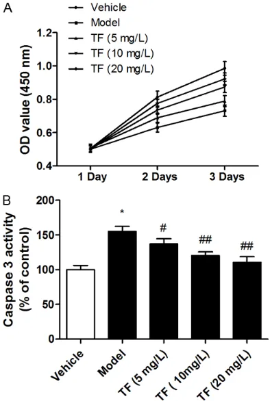
cell survival by CCK-8 assay. The results indi-cated that treatment of HUVEC with PA induced cell death in a time-dependent manner, and the number of HUVECs was markedly decreased in PA treatment group. The inhibition rate was about 30% with PA treatment at day 3 (Figure 1A). However, co-incubated with PA and theafla -vins (10 mg/L and 20 mg/L) in HUVECs, the cell
viability was significantly increased in day 2 and
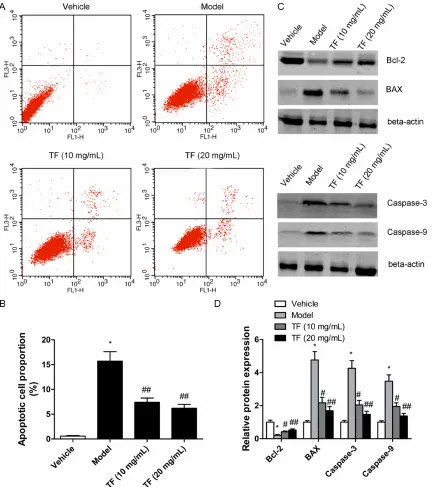
day 3 (Figure 1A). Moreover, we examined whe- ther PA induced apoptosis in HUVECs through an apoptotic mechanism. Caspase-3 activity assay and Annexin V-PI double-labeling were measured after HUVECs exposure to PA for 48 h. The results indicated that HUVECs with PA
treatment showed significant cell apoptosis as
compared to that of the vehicle-treated group, and the caspase-3 activity in co-incubated with
PA and theaflavins (10 mg/L and 20 mg/L) was significantly lower than that of the PA-treated
group (Figures 1B, 2A and 2B). These results
demonstrated that theaflavins could increase
the cell viability and reverse PA-induced apop-tosis in HUVECs. Furthermore, the apoptotic response was further investigated by measur-ing apoptosis-related proteins with western
blot. Treatment with 50 μM PA led to increase
the pro-apoptotic BAX level and decrease the anti-apoptotic Bcl-2 level in HUVECs. Simultane- ously, the protein expression of caspase-3 and
caspase-9 was significantly up-regulated in PA
group as compared to vehicle-treated group (Figure 2C and 2D). However, treatment with
theaflavins dose-dependent reversed the
PA-induced down-regulation of anti-apoptotic pro-Figure 1. HUVECs were incubated with palmitic acid
and theaflavins, and the cell viability was examined
by CCK8 assay (A). HUVECs were incubated with
palmitic acid and theaflavins for 48 h, and caspase
3 activity was examined by caspase 3 ELISA assay (B). Values were expressed as mean ± SEM, n = 3 in each group. *P < 0.05 versus control group; #P <
[image:3.612.90.287.72.365.2]tein and up-regulation of pro-apoptotic proteins expression in HUVECs (Figure 2C and 2D).
Theaflavins reverse PA-induced HUVECs injury
by regulating NO and iNOS
The ability to generate NO has served as a marker for healthy endothelia, and nanomolar
concentrations of NO have anti-inflammatory
and protective effects on endothelial cell [1, 21]. To assess PA-induced secretory dysfunc-tion of HUVECs and the moduladysfunc-tion effect of
theaflavins, we measured the levels of NO and
p-eNOS in HUVECs. As shown in Figure 3, the
[image:4.612.93.525.71.558.2]levels of NO and p-eNOS were significantly sup -pressed when the cells were exposed to PA at Figure 2. HUVECs were incubated with palmitic acid and theaflavins for 48 h, the percentage of apoptotic cells was also analyzed by flow cytometric analysis of annexin V/PI double staining (A) and bar graphs represent the percent -age of apoptotic cells (B). The protein expression of BAX, Bcl-2, caspase-3 and caspase-9 was measured by western
blotting (C) and densitometric quantification normalized to β-actin protein bands (D). Values were expressed as
the concentration of 50 μM (Figure 3A and 3B).
However, co-incubation with theaflavins (10
mg/L and 20 mg/L) for 48 hours reversed the decreased level of NO and p-eNOS in HUVECs (Figure 3A and 3B). These results suggested
that theaflavins could modulate the PA-induced
secretory dysfunction of endothelial cells.
Theaflavins inhibit PA-induced mitochondrial
dysfunction and DNA damage
Mitochondrial dysfunction and DNA damage are involved in injuring-induced cell prolifera-tion inhibiprolifera-tion and apoptosis in HUVECs [22, 23]. To further explore whether PA-induced cell
apoptosis was mediated through mitochondrial dysfunction and DNA damage and the potential
protective effects of theaflavins was
investiga-ted.
We determined the mitochondrial membrane potential with the mitochondria-sensitive dye,
DiOC6, using flow cytometry. As shown in Figure 4, treatment of HUVECs with PA induced the loss of the mitochondrial membrane potential as compared to vehicle-treated group (Figure 4A). PA combination with theaflavins could sig
-nificantly improve mitochondrial membrane
potential in HUVECs when the concentration of
theaflavins was more than 10 mg/L (Figure 4A). Next, we assessed the effect of PA on the
mobilization of Ca2+. When HUVECs were
treat-ed with PA, Ca2+ levels were significantly
[image:5.612.91.285.69.457.2]in-creased as compared with the vehicle-treated group (Figure 4B). The results demonstrated that PA promoted the secretory dysfunction of Ca2+ in HUVECs. Intriguingly, treatment of HU-
Figure 3. HUVECs were incubated with palmitic acid
and theaflavins for 48 h, and the NO concentration
was detected by ELISA assay (A). The protein expres-sion of p-eNOS was measured by western blotting (B). Values were expressed as mean ± SEM, n = 3 in each group. *P < 0.05 versus control group; #P <
0.05, ##P < 0.01 versus model group.
Figure 4. HUVECs were incubated with palmitic acid
and theaflavins for 48 h, the mitochondrial mem -brane potential (A) and the release of Ca2+ (B) were
examined by flow cytometry. Values were expressed
as mean ± SEM, n = 3 in each group. *P < 0.05
ver-sus control group; #P < 0.05, ##P < 0.01, ###P < 0.001
[image:5.612.326.520.76.373.2]VECs with theaflavins inhibited PA-induced cal -cium releasing. DNA damage has been found in HUVECs with injuring-induced. In the present study, the tail length in the PA-treated group was markedly higher than in the control group.
However, tail length was significantly sup
-pressed by theaflavins co-incubated HUVECs
(Figure 5). These results suggested that the-
aflavins could alleviate the mitochondrial dys -function and DNA damage that were induced by palmitic acid.
Discussion
Endothelial dysfunction is a driving force in the initiation and development of atherosclerosis [22]. Although atherosclerosis development appears to be the result of multiple factors, a particularly important risk factor in the patho-genesis of atherosclerosis is saturated free fatty acid, which contributes to endothelial dys-function [3, 24]. As a major component of dietary saturated fat and 20% of the total serum free fatty acids, PA is often used to in-
apoptosis plays an important role in the devel-opment of atherosclerosis, and palmitate induces apoptosis in mouse aortic endothelial cells through the promotion of oxidative and ER stress [7]. These observations document that
theaflavins could inhibit PA-induced endothelial
cells apoptosis via regulating apoptosis-related protein expression.
In vitro and in vivo studies show that exposure to free fatty acids for prolonged periods causes endothelial dysfunction, including a reduction of endothelial cell NO levels, which has served as a marker for healthy endothelia and protects against injuring-induced endothelial dysfunc-tion [5, 7]. Endothelial-derived NO is produced by eNOS and regulates vascular tone, and NO
has anti-inflammatory and protective effects through the inhibition of the activation of NF-κB
[26, 27]. Our results indicated that PA inhibited NO production and eNOS phosphorylation.
However, co-incubation with theaflavins (10
[image:6.612.92.391.75.385.2]mg/L and 20 mg/L) for 48 hours reversed the decreased level of NO and p-eNOS in HUVECs. Figure 5. HUVECs were incubated with palmitic acid and theaflavins for 48 h,
and the cell DNA damage was measured by the comet assay.
duce endothelial
dysfunc-tion [25]. Inflammatory
res-ponse and oxidant are in- duced in HUVECs stimulat-ed with PA, and the rstimulat-educ- reduc-tion of insulin-mediated eN- OS activity and production of NO are related to endo-thelial dysfunction [5]. In the present study, HUVECs were considered to approxi-mately represent the endo-thelial monolayer in blood vessels. The exposure of PA to HUVECs inhibited cell vi- ability and induced cell ap- optosis that the proportion of the apoptosis cells was
increased; however,
theafla-vins could increase the cell viability and reverse PA-in- duced apoptosis in HUVE- Cs. Further studies conclu-sively showed that
treat-ment with theaflavins
Disclosure of conflict of interest
None.
Address correspondence to: Dr. Yu-Dong Chen, De- partment of Cardiovascular Medicine, Central Hos- pital of Shengli Oil Field, Dongying 257000, China. Tel: (86) 546-8552224; Fax: (86) 546-8552224; E-mail: yd_chenmed@hotmail.com
References
[1] Lee CH, Lee SD, Ou HC, Lai SC and Cheng YJ. Eicosapentaenoic acid protects against pal-mitic acid-induced endothelial dysfunction via activation of the AMPK/eNOS pathway. Int J Mol Sci 2014; 15: 10334-10349.
[2] Jiang H, Liang C, Liu X, Jiang Q, He Z, Wu J, Pan X, Ren Y, Fan M, Li M and Wu Z. Palmitic acid promotes endothelial progenitor cells apopto-sis via p38 and JNK mitogen-activated protein kinase pathways. Atherosclerosis 2010; 210: 71-77.
[3] Qiu L, Xu R, Wang S, Li S, Sheng H, Wu J and Qu Y. Honokiol ameliorates endothelial dysfunc-tion through suppression of PTX3 expression, a key mediator of IKK/IkappaB/NF-kappaB, in atherosclerotic cell model. Exp Mol Med 2015; 47: e171.
[4] Wang J, Chen L, Li H, Yang J, Gong Z, Wang B and Zhao X. Clopidogrel reduces apoptosis and promotes proliferation of human vascular endothelial cells induced by palmitic acid via suppression of the long non-coding RNA HIF1A-AS1 in vitro. Mol Cell Biochem 2015; 404: 203-210.
[5] Jeong SO, Son Y, Lee JH, Cheong YK, Park SH, Chung HT and Pae HO. Resveratrol analog piceatannol restores the palmitic acid-induced impairment of insulin signaling and production of endothelial nitric oxide via activation of
anti-inflammatory and antioxidative heme oxygen -ase-1 in human endothelial cells. Mol Med Rep 2015; 12: 937-944.
[6] Fu M, Li Z, Tan T, Guo W, Xie N, Liu Q, Zhu H, Xie X and Lei H. Akt/eNOS signaling pathway medi-ates inhibition of endothelial progenitor cells by palmitate-induced ceramide. Am J Physiol Heart Circ Physiol 2015; 308: H11-17.
[7] Lu Y, Qian L, Zhang Q, Chen B, Gui L, Huang D, Chen G and Chen L. Palmitate induces apopto-sis in mouse aortic endothelial cells and endo-thelial dysfunction in mice fed high-calorie and high-cholesterol diets. Life Sci 2013; 92: 1165-1173.
[8] Fatima M, Kesharwani RK, Misra K and Rizvi SI. Protective effect of theaflavin on erythro -cytes subjected to in vitro oxidative stress. Biochem Res Int 2013; 2013: 649759. Recent in vitro studies have demonstrated that
PA inhibits AKT and eNOS phosphorylation and increased iNOS expression, and the AMPK/ AKT/eNOS/NO signaling pathway plays a pro-tective role in endothelial dysfunction [1]. Moreover, Akt/eNOS signaling pathway medi-ates inhibition of endothelial progenitor cells (EPC) by palmitate-induced ceramide, and ceramide-induced reduction of NO may be the molecular mechanism for PA-mediated EPC
inhibition [6]. Therefore, theaflavins-targeted
NO synthesis might be an effective means for improvement of endothelial cell functions. Mitochondrial dysfunction has been implicated in cardiovascular diseases. In view of the
utili-zation of most oxygen in the respiratory chain,
mitochondria have been presumed as the major source of cellular ROS generation and a portion of electron leakage during oxygen con-sumption [22, 28]. Previous studies show that lipid oxidation plays an important role in the ini-tial mitochondrial dysfunction and cell death caused by hemi in endothelial cells [29]. However, for all we know, no literature has been
reported that theaflavins possesses the effica -cy of improving mitochondrial dysfunction in
vitro. In our work, we found that theaflavins could significantly improve mitochondrial mem -brane potential in HUVECs when the cells were exposed to PA, and PA-induced dysfunction of Ca2+ release in HUVECs was reversed by theafla
-vins treatment. Moreover, our study showed that PA could induce considerable DNA damage in HUVECs by using the method of the comet assay. Some studies have shown that the release of mitochondrial cytochrome c to the cytosol can lead to caspase-3 and caspase-9 activation and endothelial cell apoptosis, which may also cause DNA damage [30]. Our results
suggested that theaflavins could alleviate the
mitochondrial dysfunction and DNA damage that were induced by palmitic acid, and the underlying mechanism was mediated, at least partially, through the activation of eNOS/NO signaling pathway.
the Blockade of NF-kappaB and MAPK Sig- naling Pathways. Chonnam Med J 2011; 47: 104-110.
[18] Wu W, Xu H, Wang Z, Mao Y, Yuan L, Luo W, Cui Z, Cui T, Wang XL and Shen YH. PINK1-parkin-mediated mitophagy protects mitochondrial integrity and prevents metabolic stress-in-duced endothelial injury. PLoS One 2015; 10: e0132499.
[19] Green DR, Galluzzi L and Kroemer G. Cell biol -ogy. Metabolic control of cell death. Science 2014; 345: 1250256.
[20] Qi Y, Li Y, Zhang Y, Zhang L, Wang Z, Zhang X, Gui L and Huang J. IFI6 inhibits apoptosis via mitochondrial-dependent pathway in dengue virus 2 infected vascular endothelial cells. PLoS One 2015; 10: e0132743.
[21] Kostyuk SV, Smirnova TD, Efremova LV, Kon- kova MS, Alekseeva AY, Kameneva LV and Veiko NN. Enhanced expression of iNOS in hu-man endothelial cells during long-term cultur-ing with extracellular DNA fragments. Bull Exp Biol Med 2010; 149: 191-195.
[22] Jin X, Yi L, Chen ML, Chen CY, Chang H, Zhang T, Wang L, Zhu JD, Zhang QY and Mi MT. Delphinidin-3-glucoside protects against
oxi-dized low-density lipoprotein-induced mito -chondrial dysfunction in vascular endothelial cells via the sodium-dependent glucose trans-porter SGLT1. PLoS One 2013; 8: e68617. [23] Quan Y, Yang Y, Wang H, Shu B, Gong QH and
Qian M. Gypenosides attenuate cholesterol-in-duced DNA damage by inhibiting the produc-tion of reactive oxygen species in human um-bilical vein endothelial cells. Mol Med Rep 2015; 11: 2845-2851.
[24] Ishida T, Naoe S, Nakakuki M, Kawano H and Imada K. Eicosapentaenoic acid prevents sat-urated fatty acid-induced vascular endothelial dysfunction: involvement of long-chain acyl-coa synthetase. J Atheroscler Thromb 2015; 22: 1172-85.
[25] Yli-Jama P, Meyer HE, Ringstad J and Pedersen JI. Serum free fatty acid pattern and risk of myocardial infarction: a case-control study. J Intern Med 2002; 251: 19-28.
[26] Peng HB, Libby P and Liao JK. Induction and
stabilization of I kappa B alpha by nitric oxide
mediates inhibition of NF-kappa B. J Biol Chem 1995; 270: 14214-14219.
[27] Kempe S, Kestler H, Lasar A and Wirth T.
NF-kappaB controls the global pro-inflammatory
response in endothelial cells: evidence for the regulation of a pro-atherogenic program. Nucleic Acids Res 2005; 33: 5308-5319. [28] Diers AR, Broniowska KA and Hogg N.
Nitrosative stress and redox-cycling agents
synergize to cause mitochondrial dysfunction
and cell death in endothelial cells. Redox Biol 2013; 1: 1-7.
[9] Wang W, Sun Y, Liu J, Wang J, Li Y, Li H and
Zhang W. Protective effect of theaflavins on
homocysteine-induced injury in HUVEC cells in vitro. J Cardiovasc Pharmacol 2012; 59: 434-440.
[10] Tanaka Y, Kirita M, Abe Y, Miyata S, Tagashira M, Kanda T and Maeda-Yamamoto M. Metabo- lic stability and inhibitory effect of O-methylated
theaflavins on H2O2-induced oxidative dam -age in human HepG2 cells. Biosci Biotechnol Biochem 2014; 78: 1140-1146.
[11] Luo XY, Takahara T, Hou J, Kawai K, Sugiyama T, Tsukada K, Takemoto M, Takeuchi M, Zhong
L and Li XK. Theaflavin attenuates
ischemia-reperfusion injury in a mouse fatty liver model. Biochem Biophys Res Commun 2012; 417: 287-293.
[12] Adhikary B, Yadav SK, Chand S, Bandyopadhyay SK and Chattopadhyay S. Black tea and the-
aflavins suppress various inflammatory modu -lators and i-NOS mediated nitric oxide synthe-sis during gastric ulcer healing. Free Radic Res 2011; 45: 767-778.
[13] Siddiqui IA, Adhami VM, Afaq F, Ahmad N and Mukhtar H. Modulation of phosphatidylinosi-tol-3-kinase/protein kinase B- and mitogen-activated protein kinase-pathways by tea poly-phenols in human prostate cancer cells. J Cell Biochem 2004; 91: 232-242.
[14] Knaze V, Zamora-Ros R, Lujan-Barroso L,
Ro-mieu I, Scalbert A, Slimani N, Riboli E, van Ro- ssum CT, Bueno-de-Mesquita HB, Trichopoulou A, Dilis V, Tsiotas K, Skeie G, Engeset D, Quiros JR, Molina E, Huerta JM, Crowe F, Wirfal E, Ericson U, Peeters PH, Kaaks R, Teucher B, Johansson G, Johansson I, Tumino R, Boeing H, Drogan D, Amiano P, Mattiello A, Khaw KT,
Luben R, Krogh V, Ardanaz E, Sacerdote C,
Salvini S, Overvad K, Tjonneland A, Olsen A,
Boutron-Ruault MC, Fagherazzi G, Perquier F and Gonzalez CA. Intake estimation of total and individual flavan-3-ols, proanthocyanidins and theaflavins, their food sources and deter -minants in the European Prospective Inves- tigation into Cancer and Nutrition (EPIC) study. Br J Nutr 2012; 108: 1095-1108.
[15] Higdon JV and Frei B. Tea catechins and poly-phenols: health effects, metabolism, and anti-oxidant functions. Crit Rev Food Sci Nutr 2003; 43: 89-143.
[16] Cai F, Li CR, Wu JL, Chen JG, Liu C, Min Q, Yu W,
Ouyang CH and Chen JH. Theaflavin amelio -rates cerebral ischemia-reperfusion injury in
rats through its anti-inflammatory effect and modulation of STAT-1. Mediators Inflamm
2006; 2006: 30490.
[17] Kim S and Joo YE. Theaflavin Inhibits
and Kim YM. Nitric oxide inhibition of homocys-teine-induced human endothelial cell apopto-sis by down-regulation of p53-dependent Noxa expression through the formation of S-nitro- sohomocysteine. J Biol Chem 2005; 280: 5781-5788.
[29] Higdon AN, Benavides GA, Chacko BK, Ouyang X, Johnson MS, Landar A, Zhang J and Darley-Usmar VM. Hemin causes mitochondrial dys-function in endothelial cells through promoting lipid peroxidation: the protective role of au-tophagy. Am J Physiol Heart Circ Physiol 2012; 302: H1394-1409.