Int J Clin Exp Med 2016;9(11):22901-22906 www.ijcem.com /ISSN:1940-5901/IJCEM0038069
Case Report
Clinical and pathological features in
a case of angiomatous nasal polyp
Yangyang Bao1*, Jiang Bian2*, Chao Cheng2, Heming Han1, Lifang Shen1, Shuihong Zhou1, Jiangtao Zhong1, Minli Zhou1
1Department of Otolaryngology, The First Affiliated Hospital, College of Medicine, Zhejiang University, Hangzhou
310003, Zhejiang Province, China; 2Department of Otolaryngology, People’s Hospital of Jinhua, Jinhua 321000,
Zhejiang Province, China. *Equal contributors.
Received August 17, 2016; Accepted October 24, 2016; Epub November 15, 2016; Published November 30, 2016
Abstract: Angiomatous nasal polyp (ANP) is a relatively rare benign lesion, which may be misdiagnosed as a benign or malignant tumor. The characteristic pathological features of ANP are extensive vascular proliferation, accumula-tion of extracellular amorphous eosinophilic material, and atypical stromal cells. ANP can grow rapidly and exhibit aggressive clinical behavior that could simulate a malignancy preoperatively. We reported a case of ANP of the right maxillary sinus. A 38-year-old man presented with a 2-month history of right-sided nasal obstruction, fulvous rhinor-rhea, headaches, right-sided tooth root pain under right face pressure, slight hyposmia and right-sided epiphora. Computed tomography (CT) imaging and Magnetic resonance imaging (MRI) revealed an heterogeneous mass of the right maxillary sinus, which caused extensive bone erosion and extended into the right nasal cavity and the ipsilat-eral pterygopalatine fossa. The mass was completely resected by endoscopic sinus surgery and had no evidence of recurrence during the follow-up period.
Keywords: Angiomatous nasal polyp (ANP), maxillary sinus, diagnosis, endoscopic sinus surgery
Introduction
Angiomatous nasal polyp (ANP), also known as angioectatic polyp, is a relatively rare benign lesion [1-4]. Based on the predominant
ele-ments seen on histological evaluation, inflam -matory sinonasal polyps (SNPs) have been
classified into five types: edematous, glandular, fibrous, cystic, and angiectatic or angiomatous
[2, 5, 6]. Although SNPs are the most common sinonasal lesions examined pathologically, as an uncommon subtype ANP only accounting for 4%-5% of all SNPs [1-3, 5]. Angiomatous nasal polyps (ANPs) are characterized by extensive vascular proliferation and ectasia, with scanty
inflammatory infiltrate and abundant extracel
-lular fibrin [2, 4, 5]. In a relatively uncommon
presentation, ANPs can grow rapidly and exhib-it aggressive clinical behavior such as exten-sive bone erosion and remodeling or epistaxis which could simulate malignancy
preoperative-ly, and so be a source of diagnostic difficulty [2,
5]. However, to the best of our knowledge, no authors reported ANP of the maxillary sinus
caused extensive bone erosion and extended into pterygopalatine fossa in the English-language literature.
In this article, we present an extremely rare case of ANP of the right maxillary sinus that caused extensive bone erosion and extended into the right nasal cavity and the ipsilateral pterygopalatine fossa, and we further describe the clinical, radiological and pathological fea-tures of ANP.
Case report
A 38-year-old man presented with an 2-month history of right-sided nasal obstruction, fulvous rhinorrhea, headaches, right-sided tooth root pain under right face pressure, slight hyposmia and right-sided epiphora. He denied postnasal drainage, epistaxis, facial numbness, diplopia, and any impairment in his visual acuity. He was
On otolaryngological examination, we found that the right-sided nasal mucosa were con-gested and edematou, bilateral inferior turbi-nate were swelling, while the nasal passage for peering was not clear. On other physical exami-nation, the patient’s blood pressure and other vital signs were normal. His visual acuity, eye
movement, direct pupillary reflex and indirect pupillary reflexes were normal.
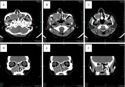
Axial and coronal computed tomographic scan of the paranasal sinuses demonstrated lamel-lar high-density shadow in both sides of the maxillary sinus, ethmoid sinus, frontal sinus and sphenoid sinus, as well as an heteroge-neous soft-tissue mass in the right maxillary sinus and causing sinus wall swelling bony destruction. There was bone destruction
involv-ing the orbital floor, medial, anterior, inferior
and posterior maxillary walls. The mass extend-ed into the right nasal cavity and the ipsilateral pterygopalatine fossa (Figure 1). Magnetic res-onance imaging (MRI) of the paranasal sinuses showed the right maxillary sinus component of
the mass to have heterogenous hypointensity on T1-weighted images (T1WI) and heteroge-nous hyperintensity with a peripheral hypoin-tense rim on T2-weighted images (T2WI). T2WI
showed the mass was well-defined and the
hyperintensity in the ipsilateral maxillary sinus indicated obstructive sinusitis. Diffusion wei- ghted magnetic resonance imaging (DWI) showed the mass of slightly high signal intensi-ty with a peripheral hypointense rim (Figure 2). The mass showed heterogeneous nodular and patchy enhancement with the non-enhanced peripheral hypointense rim after gadolinium contrast injection (Figure 3).
The patient was taken to the operating room with general anaesthesia and underwent surgi-cal excision of the mass by endoscopic sinus surgery. At surgery, we found that there were yellow soft masses alternating with black necrotic areas occupying the entire right maxil-lary sinus. A biopsy was performed, the
intraop-erative frozen section diagnosis was inflamma
[image:2.612.91.523.71.372.2]-tory infiltrates and necrosis. Based on the
Figure 1. Axial (A-C) and coronal (D-F) CT scan demonstrated an heterogeneous soft-tissue mass occupying the
entire right maxillary sinus with erosion of orbital floor, medial, anterior, inferior and posterior walls. The mass ex
Clinical and pathological findings in angiomatous polyp
whole findings, the diagnosis of an ANP was
made. The ANP was completely excised with no
significant hemorrhaging by intranasal endo -scopic approach.
Light microscopy was used after staining sec-tions with hematoxylin and eosin. The surface
of the mass was covered by pseudostratified
ciliated columnar epithelium with areas of squamous metaplasia, edematous
subepitheli-al stroma with infiltration by many inflamed
cells, The lesions showed numerous
extrava-sated red cells, fibrin, abundance of irregularly
shaped, thin-walled blood vessels, many show-ing intraluminal thrombus formation, which was associated with wide ares of ischemic necrosis (Figure 4). The patient remained asymptomatic and disease-free at follow-up 13 months later.
Discussion
There are many descriptions of ANP in the Eng- lish literature, names include cavernous
hem-angioma, pseudotumor, inflammatory granulo -ma telangiectaticum, pseudoangio-ma, orga-nized or organizing hematoma, vascular granu-loma, hemorrhage necrotic polyp, and angioec-tatic or angiomatous polyp. In this paper, we use the term angiomatous polyp, because it
reflects the fact that the mass is not a real
tumor and that the lesion is clinically character-ized by extensive vascular proliferation and hemorrhage.
There are many hypotheses of the pathogene-sis of ANP have been proposed, however, the
definite pathogenesis of ANP is still unclear.
One hypothesis is based on the presence of the maxillary sinus and/or nasal cavity polyp, the
polyp pedicle is suffer from significant vascular
[image:3.612.89.522.70.395.2]the ostial exit site, the poste-rior end of the infeposte-rior turbi-nate, the posterior choana and the most dependent part within the nasopharynx [7]. Compression of the feeder vessels in these areas is con-sidered to result in initial hemangiectasis and stasis, and edema. This leads to venous infarction followed by neovascularization of the polyp, then setting the stage for repeat vascular occlusion and infarction [7]. Another is that based on the formation of hematoma in the sinus antrum [1]. Many factors such
[image:4.612.91.523.71.383.2]as trauma, surgery, inflamma -tion and/or allergy of the max-illary sinus and nasal cavity, bleeding diatheses, and rup-Figure 3. Postcontrast Axial (A-C) and coronal (D-F), T1-weighted images showed heterogeneous nodular and patchy enhancement with the unenhanced peripheral hypointense rim.
Figure 4. Photomicrograph showed that the surface of the mass was covered by pseudostratified ciliated columnar epithelium with areas of squamous metaplasia, edematous
subepithelial stroma with infiltra
-tion by many inflamed cells (A), in
[image:4.612.91.376.434.693.2]Clinical and pathological findings in angiomatous polyp
tured aneurysm, which resulting in hematoma [1, 2, 8]. Reactive and reparative changes with
neovascularization and fibrosis lead to the
eventual formation of ANP. To our point of view, the two hypotheses are both reasonable. The most remarkable pathological features of ANP were the large numbers of dilated blood vessels, many with proof of intraluminal throm-bosis, and the sizable extent of necrosis of the lesion, with extravasation of blood components into the surrounding stroma [2-5]. The features of ANP under light microscopy are as follows [5]: (i) racemose aggregates of irregularly shaped blood vessels resembling dilated capil-laries, no elastic or muscular layers; (ii) acute
and chronic inflammation common; hemosider -in-laden macrophages; (iii) heterogeneity from
field to field; patchy areas with features of typi
-cal inflammatory polyps; (iv) paucicellular stro
-ma with scattered fibroblasts and myofibro -blasts; marked nuclear enlargement; large nucleoli; no mitoses. The features of ANP under electron microscopy are as follows [5]: (i)
typi-cal fibroblasts and myofibroblasts with indis
-tinct nuclear fibrous lamina; endothelial cell; (ii) amorphous extracellular matrix (fibrin, plasma,
cellular debris). To sum up, the characteristic pathological features of ANP are extensive vas-cular proliferation, accumulation of extracellu-lar amorphous eosinophilic material, and atypi-cal stromal cells [2-5].
Clinically, the symptoms of ANP were varied
and nonspecific, including nasal obstruction,
epistaxis, ophthalmoptosis, facial swelling and pain, nasal discharge, snoring, headaches, hyposmia, epiphora, and visual disturbances [1-5, 8-13]. Baumgarten et al [13] reported that the most common symptoms associated with ANP are nasal obstruction and an alteration in olfaction. However, other studies including our previous study found that the most common symptoms associated with ANP are nasal obstruction and epistaxis [1, 2]. In our case, the patient presented with an 2-month history of right-sided nasal obstruction, fulvous rhinor-rhea, headaches, right-sided tooth root pain under right face pressure, slight hyposmia and right-sided epiphora.
Radiologic examination is very important in the diagnosis of ANP. Previous reports of ANP
clari-fied that CT findings lacked specificity for ANP identification. The typical appearance of ANP
on CT are as follows [1, 2, 4, 7, 12, 14, 15]: (i)
an expansile mass filles the paranasal sinus
and/or nasal cavities and/or choana/naso-pharynx, causing bulge or destruction of the bony wall and heterogeneous isoattenuation on pcontrast CT scans; (ii) on contrast-enhanced CT, the center of the lesions is non-enhanced
with peripheral intensification; (iii) the mass on
CT shows clear and smooth edges and does not invade the peripheral soft tissue; (iv) does not usually invade the pterygopalatine fossa or sphenoid sinus. However, the imaging features of ANP on conventional MRI are quite charac-teristic. Due to the high soft-tissue resolution,
MRI has remarkable superiority to CT in reflect -ing the internal structures of ANP and the involved extent. The typical imaging features of ANP on MRI are as follows [1, 3, 4, 12]: (i) an expansive soft tissue mass; (ii) the margin of
the mass is well-defined; (iii) blockage of osti -um and secondary obstruction; (iv) extension into the nasal cavity, choana and nasopharynx; (v) heterogeneous hypointensity or isointense signal intensity on T1WI; (vi) heterogeneous hyperintense with hypointense linear septum internally and with a peripheral hypointense rim surrounding the lesion on T2WI; (vii) heteroge-neous nodular and patchy enhancement with the non-enhanced peripheral hypointense rim on postcontrast MRI. Most prominently, the hypointense peripheral rim of the mass is a
very specific contribution to the correct diagno -sis of ANP.
In conclusion, ANP of the maxillary sinus in- vades the pterygopalatine fossa is extremely rare. We believe that CT shows bone changes associate with ANP can provide useful informa-tion for the determinainforma-tion of surgical planning, while MRI that shows the typical imaging fea-tures in signal intensity can provide an accu-rate diagnosis of ANP. Therefore, we propose that combined application of CT and MRI is quite necessary for patients with suspicious of ANP. Moreover, endoscopic sinus surgery is considered the best approach for the treat-ment of ANP.
Acknowledgements
ce Foundation of China (grant nos. 81172562 and 81372903).
Disclosure of conflict of interest
None.
Address correspondence to: Dr. Shuihong Zhou, Department of Otolaryngology, The First Affiliated Hospital, College of Medicine, Zhejiang University, Hangzhou 310003, Zhejiang Province, China. Tel: 86-571-87236894; Fax: 86-571-87236895; E-mail: zhoushuihongzsh@sina.com
References
[1] Zou J, Man F, Deng K, Zheng Y, Hao D, Xu W. CT
and MR imaging findings of sinonasal angio
-matous polyps. Eur J Radiol 2014; 83: 545-551.
[2] Dai LB, Zhou SH, Ruan LX, Zheng ZJ. Correla- tion of computed tomography with pathologi-cal features in angiomatous nasal polyps. PLoS One 2012; 7: e53306.
[3] Wang YZ, Yang BT, Wang ZC, Song L, Xian JF. MR evaluation of sinonasal angiomatous pol-yp. AJNR Am J Neuroradiol 2012; 33: 767-772. [4] Sheahan P, Crotty PL, Hamilton S, Colreavy M, McShane D. Infarcted angiomatous nasal pol-yps. Eur Arch Otorhinolaryngol 2005; 262: 225-230.
[5] Yfantis HG, Drachenberg CB, Gray W, Papa- dimitriou JC. Angiectatic nasal polyps that clin-ically simulate a malignant process: report of 2 cases and review of the literature. Arch Pathol Lab Med 2000; 124: 406-410.
[6] Sinha SN. Observations on histology of nasal polypi. Indian J Otol 1967; 19: 164-168. [7] Batsakis JG, Sneige N. Choanal and
angioma-tous polyps of the sinonasal tract. Ann Otol Rhinol Laryngol 1992; 101: 623-625.
[8] Lee HK, Smoker WR, Lee BJ, Kim SJ, Cho KJ. Organized hematoma of the maxillary sinus: CT findings. AJR Am J Roentgenol 2007; 188: W370-W373.
[9] Lee BJ, Park HJ, Heo SC. Organized hematoma of the maxillary sinus. Acta Otolaryngol 2003; 123: 869-872.
[10] Song HM, Jang YJ, Chung YS, Lee BJ. Organizing hematoma of the maxillary sinus. Otolaryngol Head Neck Surg2007; 136: 616-620.
[11] Yagisawa M, Ishitoya J, Tsukuda M. Hemato- ma-like mass of the maxillary sinus. Acta Otolaryngol 2006; 126: 277-281.
[12] De Vuysere S, Hermans R, Marchal G. Sinochoanal polyp and its variant, the
angio-matous polyp: MRI findings. Eur Radiol2001;
11: 55-58.
[13] Baumgarten C, Kunkel G, Rudolph R, Staud RD, Sperner I, Gelderblom H. Histopathological examinations of nasal polyps of different etiol-ogy. Arch Otorhinolaryngol 1980; 226: 187-197.
[14] Sayed RH, Abu-Dief EE. Does antrochoanal polyp present with epistaxis? J Laryngol Otol 2010; 124: 505-509.
[15] Som PM, Cohen BA, Sacher M, Choi IS, Bryan
NR. The angiomatous polyp and the angiofi