Original Article
Malignant ovarian germ cell tumors in adolescents: 18
years of experience at a single institution
Ting Zhao1*, Yan Liu1*, Hao Zhang2, Xiao Wang1, Hongyuan Jiang1, Yuan Lu1
1Department of Gynecology, Obstetrics and Gynecology Hospital of Fudan University, Shanghai, China;
2Department of Pathology, Obstetrics and Gynecology Hospital of Fudan University, Shanghai, China. *Equal
con-tributors.
Received January 24, 2016; Accepted May 3, 2016; Epub June 15, 2016; Published June 30, 2016
Abstract: Malignant ovarian germ cell tumors (MOGCTs) in adolescents are rare. Although fertility-sparing surgery for such patients is suggested unequivocally, the necessity of staging surgery remains the subject of intense debate. Here, we evaluate the role of staging surgery in adolescents affected by MOGCTs and review the outcomes of these patients. We performed a retrospective study of patients aged 10-19 years who received surgery and were patho-logically diagnosed with MOGCTs at OB/GYN Hospital of Fudan University between January 1997 and October 2014. Forty-two patients diagnosed with immature teratomas, dysgerminomas, endodermal sinus tumors and mixed germ cell tumors were included in this study. The majority (90.5%) of them were of apparent early-stage with lesion
con-fined to the ovary grossly, and the majority (90.5%) received adjuvant chemotherapy. Although comprehensive stag -ing protocols were followed in only a small number of patients (28.6%) in our cohort, the recurrence rate was low at 4.8%. Five-year overall survival and disease-free survival were 97.2% and 94.8%, respectively. Comprehensive staging or individual factors such as peritoneal washings cytology, lymph nodes dissection and omentectomy were not independent risk factors for disease-free survival (DFS). Positive peritoneal washing cytology or lymph nodes
were not correlated with recurrence. Our findings show that there is a good prognosis for adolescents affected by MOGCTs and for adolescent patients receiving adjuvant chemotherapy, staging procedures may not provide any
additional benefits.
Keywords: Malignant ovarian germ cell tumors, adolescents, survival, comprehensive staging, chemotherapy, fertility-sparing
Introduction
Malignant ovarian germ cell tumors (MOGCTs) are rare and account for around 5% of all ovar-ian malignancies [1]. These tumors consist of several histological variations and exhibit sev-eral grades of differentiation. Dysgerminomas, immature teratomas and endodermal sinus tumors (EST) are the most common types and comprise over 90% of MOGCTs [2]. Approxima- tely 70% of patients are diagnosed during early stages [3].
MOGCTs predominate in pediatric and adoles-cent patients, unlike epithelial ovarian carcino-mas which are more commonly seen in women of more than 40 years of age. Most of the
patients afflicted with MOGCTs are nulliparous.
Therefore, therapeutic modalities should be
adjusted accordingly as fertility-sparing sur- gery is vital in the management of such cases. The National Comprehensive Cancer Network (NCCN) and the European Society for Medical Oncology (ESMO) both suggest in their guide-lines that fertility-sparing can be considered for patients with MOGCTs, even in advanced stag-es, on the premise that comprehensive staging procedures are also implemented at the same time [4].
The NCCN recommended that comprehensive or complete staging procedures for MOGCTs should be carried out in a protocol that is the same as the procedures used for treating
epi-thelial ovarian cancer. Specifically, the following
and excision of any nodules, (3) Infra-colic omentectomy, and (4) Sampling or excision of the retro-peritoneum lymph nodes [5]. Compre-hensive staging procedures are believed by some to be very important, as staging has been associated with lower recurrence rates [6]. However, a different point of view was put for-ward by the Children’s Oncology Group (COG). Based on their own results, COG suggested that when practicing fertility-sparing surgery for
children and adolescents, lymph node dissec-tion, biopsy of the peritoneal surfaces, and omentectomy are not indispensable if grossly absent of lesions. Omitting these steps had no obvious adverse impact upon survival [5]. Even if abnormalities of the omentum or lymph nodes was suspected, biopsy is preferred instead of total excision [7].
However, apart from the guidance from COG published in 2004, there have been very few studies relating to the role of staging surgery for adolescent patients in the past decade. Three studies concluded that comprehensive
staging surgery had no significant impact upon
prognosis for MOGCT patients [8-10]. However, these studies included both adolescents and adults in their survey population; and one arti-cle was written in a non-English language. More importantly, a common consensus has yet not to be reached on this topic, as the NCCN still approves of comprehensive staging surgery in their latest guidelines. We therefore conducted a retrospective study in order to review the treatment, survival, and more importantly, the role of staging surgery for adolescent patients diagnosed with MOGCTs at our hospital. Patients and methods
Forty-two patients aged 10-19 years under-went surgery and were pathologically diag-nosed with MOGCTs between January 1997 and October 2014 at the OB/GYN Hospital of Fudan University, one of the largest tertiary gynecological centers in China. No patient younger than 10 years of age is treated in this hospital as they are routinely transferred to specialized pediatric centers in China. Patient data was collected retrospectively from hospi-tal medical records, including age at diagnosis,
chief complaint, ultrasonogram (USG) findings,
serum tumor markers such as cancer anti-gen-125 (CA-125), alpha-fetal protein (AFP), and carcinoembryonic antigen (CEA) before and after surgery if available, surgical details, pathological reports, adjuvant chemotherapy, and clinical outcomes such as recurrence, death or survival.
All histological slides were reviewed by two independent pathologists. Histological type
was defined according to established classifi -cations by the World Health Organization
[image:2.612.91.289.96.563.2](WHO). Stage was verified according to the
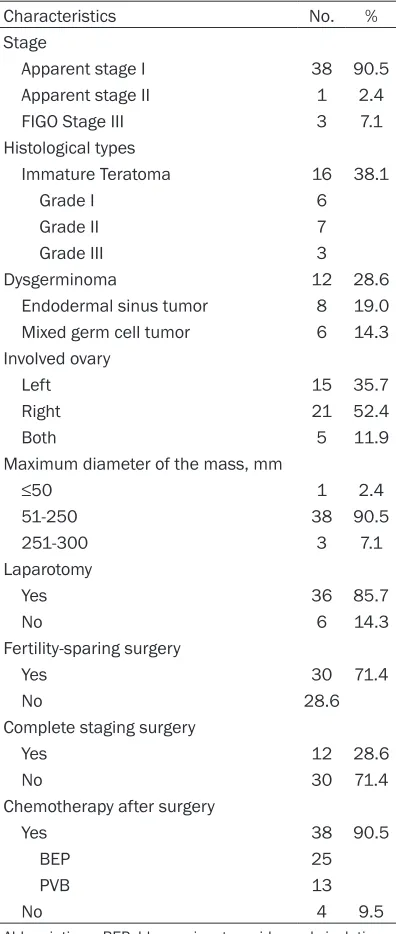
Table 1. Demographic information of the study population
Characteristics No. %
Stage
Apparent stage I 38 90.5
Apparent stage II 1 2.4
FIGO Stage III 3 7.1
Histological types
Immature Teratoma 16 38.1
Grade I 6
Grade II 7
Grade III 3
Dysgerminoma 12 28.6
Endodermal sinus tumor 8 19.0 Mixed germ cell tumor 6 14.3 Involved ovary
Left 15 35.7
Right 21 52.4
Both 5 11.9
Maximum diameter of the mass, mm
≤50 1 2.4
51-250 38 90.5
251-300 3 7.1
Laparotomy
Yes 36 85.7
No 6 14.3
Fertility-sparing surgery
Yes 30 71.4
No 28.6
Complete staging surgery
Yes 12 28.6
No 30 71.4
Chemotherapy after surgery
Yes 38 90.5
BEP 25
PVB 13
No 4 9.5
International Federation of Gynecology and
Obstetrics (FIGO) classification of ovarian
tu-mors (2015 version). Immature teratomas were graded according to the criteria developed by Norris et al [11].
Comprehensive staging surgery is defined ear -lier in this article, while fertility-sparing surgery
is defined as preservation of the uterus and at least part of an ovary. Radical surgery is defined
as bilateral salpingo-oophorectomy (BSO), or unilateral salpingo-oophorectomy (USO) when the contralateral ovary is congenitally dysgene-sis, with or without hysterectomy (TAH).
Patients were followed-up by periodic clinical, serological, and USG examinations via the out-patient service. A telephone interview was also performed to ascertain the latest status. Disease-free survival (DFS) period and overall
survival (OS) periods were defined as time inter -vals from the date of primary surgery to recur-rence or last disease-free visit (months), and to death or last visit (months), respectively. Kaplan-Meier analysis was utilized and univari-ate analysis was completed with the log-rank test. Fisher’s exact test was used to evaluate for correlation between recurrence and
perito-neal fluid or lymph node positivity. Statistical
analysis was performed by SPSS software (ver-sion 16.0, Chicago, IL, USA). A p-value <0.05
was defined as being statistically significant.
The study was approved by the Hospital Ethics Committee.
Results
Between January 1997 and October 2014, 42 adolescent patients were pathologically diag-nosed with MOGCTs; their demographic and clinico-pathological information is provided in Table 1. The median age was 16 years and none of our patients was sexually active. The most common histology was an immature teratoma (38.1%), followed by dysgerminoma (28.6%), EST (19.0%) and mixed germ cell tumor (14.3%). There were three dysgerminoma cases presented with gonadoblastoma in the same tumor. Twenty-two cases (52.4%) presented with a tumor in the right ovary while 15 cases (35.7%) presented a tumor in the left ovary.
Both ovaries were involved in five patients
(11.9%). However, although considered as uni-laterally involved, one patient with a dysgemi-noma had a concurrent gonadoblastoma in the contralateral ovary; in another two patients, the
contralateral ovary was congenitally dysgene-sis. The most common symptoms were (in descending order): pelvic mass (38.1%), pelvic pain (28.6%), abdominal distention (23.8%), dysmenorrhea (9.5%), absence of menarche (9.5%), and fever (7.1%). Four patients consult-ed clinicians due to the absence of menarche at the age of 16 years. Three patients com-plaining of pelvic pain were subsequently shown to have ovarian tumor torsion.
All patients were given a USG examination before surgery to estimate the size and gross nature of the tumor (e.g., solid, cystic, or com-plex). Thirty-seven (88.1%) cases were reported as complex while the others (11.9%) were solid. Maximum mass diameter ranged from 4.1 to 30 cm, with most (90.5%) measuring between
5 to 25 cm. There was no significant difference
in tumor size when compared between differ-ent histological types (p>0.05). There were 22/33 (66.7%), 8/20 (40%), 0/8, 3/22 (13.6%) and 21/28 (75.0%) patients presenting with elevation of CA-125 (>35 U/ml), CA-199 (>35 U/ ml), CA-153 (>35 U/ml), CEA (>5 ng/ml), and AFP (>10 ng/ml), respectively. Elevation of CA-125 was present in all the involved
patho-logical types identified of our study. This value
was positively correlated with stage (p<0.05). Elevation of CA-199 was neither correlated with stage nor type of tumor, nor CA-125 value. All patients exhibiting tumors containing an EST component showed elevation in AFP. To our sur-prise, 10 out of 12 patients with immature tera-tomas also showed elevation; and the AFP level positively correlated with tumor grade (p<0.05).
After surgery, there was a significant decline in
these tumor biomarkers in the majority of patients.
Laparotomy was carried out in 36 (85.7%) patients, while laparoscopy was performed in 6 (14.3%) patients. Most patients (30/42, 71.4%) received fertility-sparing surgery. Twelve patients (28.6%) did not receive fertility-sparing surgery: the reasons for this included advanced stage of disease in three patients, the
involve-ment of both ovaries in five patients, tumor or
dysgenesis of the contralateral ovary in three patients, and due to a decision made by the guardian of one patient.
There were 38 patients (90.5%) with lesions
confined to the ovary (or ovaries) grossly. Nine
stage IA/IB, and three patients were confirmed
to be FIGO stage I although incompletely staged because lymph nodes and omentum were both negative. The precise stage for the remaining 26 patients could not be determined due to the absence of lymph node dissection, or omentec-tomy, or both. There was one patient diagnosed with apparent stage II disease according to
sur-gical findings with involvement of the pelvic
peritoneum. Three patients had peritoneum lesions outside the pelvis were comprehensive-ly staged as FIGO stage III disease. In total, comprehensive staging procedures were fol-lowed in 12 (28.6%) patients. Peritoneal wash-ing cytology, omentectomy and lymph node dis-section were carried out in 22, 21 and 19 patients, respectively. There were 17 patients in whom none of the above staging items was implemented. Two patients (one of apparent early-stage and one of FIGO stage III disease) were positive for peritoneal washing cytology. One patient (of FIGO stage III disease) was posi-tive for lymph node involvement. No residual disease was left macroscopically in any of the patients.
Thirty-eight (90.5%) patients received post-operative chemotherapy for a median of 6 cycles (range, 1 to 8). A BEP (bleomycin, etopo-side, and cisplatin) regimen was adopted in 25 patients while a PVB regimen (vincristine, car-boplatin, and bleomycin) was adopted in 13 patients.
During the median follow-up of 61.3 months (range, 5 to 207 months), 2 patients were lost during follow-up, and 2 patients developed recurrences. One 13-year-old girl, with a 20 cm
mass in the right ovary with an intact surface, died. Earlier, USO and contralateral ovary par-tial excision had been carried out in this patient. As incompletely staged, this patient was appar-ent stage I with a mixed germ cell tumor con-taining EST and dysgerminoma. Seven courses of PVB regimen were given following surgery. Twelve months after the initial surgery a 5 cm mass was found in the left pelvis by USG while CA-125 was 450 U/ml and AFP was 836 ng/ml. AFP continued increasing to 1898 ng/ml before secondary debulking surgery, and only slightly reduced to 1406 ng/ml post-operatively. The patient survived for less than 6 months there-after and ultimately died.
The other recurrent case was a 16-year-old girl affected by a grade III immature teratoma in the right ovary. The CA-125 and AFP before sur-gery was 227.3 U/ml and >3000 ng/ml, respec-tively. We performed USO and comprehensive staging surgery and she was staged as FIGO stage IA. Six courses of BEP regimen were pre-scribed for this patient after surgery. After the third cycle both CA-125 and AFP fell to normal levels. However, after the sixth cycle (around four months after surgery), a small suspicious shadow was detected in Douglas’ pouch on USG and MRI. Chemotherapy was resumed but the mass increased to more than 3 cm in diam-eter six months following primary surgery. A secondary debulking surgery was carried out.
Pathology confirmed mature teratoma and neu -ronal tissue in Douglas’ pouch and on the sur-face of the peritoneum. This patient survived without this disease during the 41-month fol-low-up thereafter.
[image:4.612.92.305.108.254.2]Five-year OS and DFS for our patients cohort were 97.2% and 94.8%, respectively. None of the clinical factors such as histological type, laterality, fertility-sparing, comprehensive stag-ing, peritoneal washings cytology, lymph node dissection, omentectomy, or types of chemo-therapy regimen was shown to be an indepen-dent risk factor for DFS (Table 2). Further more, Fisher’s exact test showed that the positivity of peritoneal washing cytology or lymph nodes was not correlated with recurrence. With regard to the reproductive outcomes, we report that among the 30 patients who received fertility-sparing surgery, 25 cases (83.3%) reported regular menses. One patient successfully gave birth to a full-term baby while the other patients were either unmarried or taking contraception during the follow-up period.
Table 2. Univariate analysis: Chi-square and
p-value for various factors (log-rank test) for disease-free survival
Factor Chi-square p
Histological type 3.149 0.533
AFP level higher than 500 ng/ml 2.783 0.095
Laterality 1.703 0.427
Fertility-preserving 0.869 0.351 Comprehensive staging surgery 0.583 0.445
Peritoneal fluid cytology 0.857 0.355
Omentectomy 0.000 1.000
Discussion
Malignant tumors of the ovary are rare in chil-dren and adolescents [12], representing approximately 1% of all childhood malignant tumors [13]. Most ovarian malignant tumors occurring during childhood are germ cell tumors [14]. USG is considered to be a highly valuable technique for detecting the mass and distin-guishing between benign and malignant
tumors, with solid nature considered a definite
feature predicting malignancy [15]. However, it has been reported that cystic components are common in malignant ovarian tumors in child-hood with an incidence of 57% [5]. Tumor mark-ers are helpful but not always reliable for MOGCT screening. CA-125 levels were elevated in all of the histological types observed in our cohort and increased with stage. CA-199 was elevated in 40% of the patients but no correla-tion was found with stage. CA-153 and CEA seemed of limited importance as few patients showed elevation of these markers.
Elevated AFP levels usually suggest tumors containing an EST component. In our cohort, 10 out of 12 patients with an immature terato-ma also showed AFP elevation and the values were positively correlated with tumor grade. The COG and Pediatric Oncology Group (POG) have reported that 29.5% to 31.5% patients diagnosed with immature teratomas were
con-firmed to have co-existing microscopic foci of
EST, especially in grade III tumors. Furthermore, t hese organizations concluded that co-existing EST is the only valid risk factor for recurrence [16]. However, no concurrent microscopic foci of EST were reported in our study upon patho-logical examination. We speculate that a large
mass volume may increase the difficulty in
detecting tiny EST foci and more careful inspec-tion should be executed. In fact, one of the two patients experiencing recurrence in our cohort was a patient with a grade III immature terato-ma with terato-markedly elevated AFP levels (>3000 ng/ml) before surgery, which strongly suggest-ed the existence of EST.
For the treatment of MOGCTs in adolescents, surgical staging guidelines have been arbitrari-ly adopted from that of ovarian epithelial can-cers [5]. However, it has been reported that these guidelines were seldom followed com-pletely [5]. As described earlier in this article, the COG does not support staging procedures such as lymph node dissection and omentec-tomy for pediatric patients. Instead, the COG
developed another staging system specifically
for children and adolescents (listed in Table 3). For apparent early-stage patients with lesions
confined to the ovary, only peritoneal fluid cytol -ogy is suggested and if negative the guidelines would designate this as COG stage I. If positive, patients would be designated as COG stage III. In our study, we saw considerable variability in surgical procedures and only 28.6% (12/42) of patients received comprehensive staging according to the criteria described earlier. We found that neither comprehensive staging sur-gery, nor individual factors, including not only lymph node dissection or omentectomy, but
also peritoneal fluid cytology, was an indepen -dent impact factor for DFS. However, it should be mentioned that the majority (90.5%) of our patients were of apparent stage I with lesions
grossly confined to the ovary. More importantly,
most of these patients (90.5%) received adju-vant chemotherapy, which may have had
pro-found influence upon outcomes.
Prior to the 1980s, the 5-year overall survival of MOGCT patients was only 20%, even for early stage tumors [17]. The introduction of plati-num-based chemotherapy has resulted in
significant improvements in long-term survival,
even for patients of advanced stages. However, chemotherapy may cause severe latent effects.
Hearing loss was the most significant morbidity
[18]; renal impairment and neurotoxicity were also reported. Considering such risks, the COG and POG have made great efforts over the past decade to investigate the necessity for adju-vant therapy in early stage patients. The COG divided pediatric patients into 3 categories based on prognosis: low, intermediate and high Table 3. Pediatric staging system for ovarian cancer by the Children’s Oncology Group (COG)
Stage I Limited to ovary (ovaries); peritoneal washings negative; tumor markers normal after appropriate half-life decline (AFP, 5 days; β-HCG, 16 hours).
Stage II Microscopic residual disease or disease in lymph nodes <2 cm; peritoneal washings normal; tumor markers positive or negative. Stage III Gross residual disease or biopsy only; lymph nodes >2 cm; contiguous spread to other organs (omentum, intestine, bladder);
risk groups [18]. COG stage I MOGCT patients belong to the low risk group, and are prescribed with surveillance instead of chemotherapy after surgery [5, 18]. COG stage III patients belong to the intermediate risk group and rec-ommendations indicate that such patients receive 3 cycles of PEB regimen following sur-gery. Further more, the PEB regimen is also dif-ferent from the BEP regimen recommended by the NCCN as bleomycin is administered once per cycle instead of once a week [18].
However, a recent report showed that when prescribing COG stage I patients with surveil-lance after surgery, 48% (12/25) patients expe-rienced persistence or recurrence of disease within 8 months although they were all ulti-mately salvaged after chemotherapy [7]. These results were consistent with a French study which found that when surveillance was the only approach after surgery, a success rate of only 50% could be reached in COG stage I MOGCT patients [19]. The above results indi-cate that for COG stage I, girls who were incom-pletely staged according to the FIGO criteria are at risk omitting chemotherapy. Based on the FIGO staging system, one research study has shown that for FIGO stage I patients, the risk of
relapse was significantly increased (HR = 4.5)
with a recurrence rate of 33.3% if observation was chosen instead of chemotherapy after sur-gery; and relapse occurred only in patients of stage IC or IX (who were not completely staged according to FIGO protocols) [20]. Collectively, these results supported the fact that it takes great risks to omit adjuvant chemotherapy for incompletely staged early stage patients. In another respect, it was suggested in a recent article that for early stage patients not receiv-ing adjuvant chemotherapy, complete stagreceiv-ing is crucial because incomplete staging may lead to down-staging of tumors [21]. In fact, the NCCN guidelines for ovarian cancer (version 2.2015) suggest that only on the basis of com-prehensive staging surgery may pediatric and young adult patients of the following situations be considered for observation as a treatment option as well as chemotherapy: stage IA, IB dysgerminoma; stage IA, grade 1 immature terotomas; stage IA embryonal tumors; or stage IA ESTs. Taking these statements together, for early stage patients it is not rational to omit complete staging surgery and adjuvant chemo-therapy at the same time.
In our study, the majority (29/38, 76.3%) of the apparent early-stage patients did not receive comprehensive staging surgery, but only 2 (5.3%) patients relapsed. The recurrence rate is therefore much lower than reported results mentioned above. This may be attributed to the fact that 92.1% (35/38) of apparent early-stage patients received adjuvant chemotherapy. We speculate that for apparent early-stage adoles-cent patients who receive adjuvant
chemother-apy, staging surgery may add no further benefit.
The results of the log-rank and Fisher’s exact test in our cohort both supported this conclu-sion. We theorize that it might be rational there-fore to treat apparent early-stage adolescent patients with fertility-sparing surgery and adju-vant chemotherapy but without staging procedures.
Three patients with FIGO stage III disease in our study were precluded from fertility-sparing sur-gery. However, it has been suggested that fertil-ity-sparing procedures are safe for children and adolescents with advanced stages due to the high sensitivity to chemotherapy [21]. For the
five bilaterally involved patients and the three
unilaterally involved patients with the contralat-eral ovary absent or dysgenesis, whether it is safe to spare fertility, that is, to preserve part of the involved ovary, warrants further study. Conservative surgery and chemotherapy was reported to have minimal impact on fertility in young adult women [22, 23]. Other results have suggested that the adverse effect was closely related to the number of chemotherapy cycles [24]. The optimal duration of chemotherapy is still controversial. Three cycles in a completely resected disease and four cycles for patients with residual disease is generally accepted [4]. Patients receiving fertility-sparing treatment should undergo regular and long-term follow-up in order to evaluate their ovarian reserve. We acknowledge that our study has limitations as it was retrospective in nature and of limited size. However, this study represents data from a single institution over a long time period, and that treatment protocols were kept consistent throughout this prolonged interval.
Conclusion
current study were of apparent early-stage, and most (90.5%) received adjuvant chemotherapy. Although comprehensive staging protocols were followed in only a small number of patients (28.6%) in our cohort, the recurrence rate was as low as 4.8%. Comprehensive staging or indi-vidual factors such as peritoneal washing cytol-ogy, lymph node dissection and omentectomy were not independent risk factors for DFS. The positivity of peritoneal washing cytology or lymph node dissection did not correlate with recurrence. Based upon these results, we con-clude that for adolescent patients who have received adjuvant chemotherapy, staging
pro-cedures may not provide additional benefit.
Acknowledgements
Supported by Shanghai Science and Technology Commission grant: 15140903000 and Shang- hai Municipal health and family planning Com- mission grant: 201540224.
Disclosure of conflict of interest
None.
Address correspondence to: Dr. Yuan Lu, Shanghai OB/GYN Hospital, Fudan University, 419 Fangxie Road, Shanghai 200011, China. Tel: 86-21-3318- 9900; Fax: 86-21-5302-8000; E-mail: pipiluyuan@ 163.com
References
[1] Gadducci A, Lanfredini N and Tana R. Menstrual function and childbearing potential after fertility-sparing surgery and platinum-based chemotherapy for malignant ovarian germ cell tumours. Gynecol Endocrinol 2014; 30: 467-471.
[2] Vazquez I and Rustin GJ. Current controversies in the management of germ cell ovarian tu-mours. Curr Opin Oncol 2013; 25: 539-545. [3] Brown J, Friedlander M, Backes FJ, Harter P,
O’Connor DM, de la Motte Rouge T, Lorusso D, Maenpaa J, Kim JW, Tenney ME and Seckl MJ. Gynecologic Cancer Intergroup (GCIG) consen-sus review for ovarian germ cell tumors. Int J Gynecol Cancer 2014; 24: S48-54.
[4] Colombo N, Peiretti M, Garbi A, Carinelli S, Marini C and Sessa C. Non-epithelial ovarian cancer: ESMO Clinical Practice Guidelines for diagnosis, treatment and follow-up. Ann Oncol 2012; 23 Suppl 7: vii20-26.
[5] Billmire D, Vinocur C, Rescorla F, Cushing B, London W, Schlatter M, Davis M, Giller R, Lauer S and Olson T; Children’s Oncology Group
lignant germ cell tumors of the ovary in chil-dren and adolescents: an intergroup study. J Pediatr Surg 2004; 39: 424-429.
[6] Lin KY, Bryant S, Miller DS, Kehoe SM, Richardson DL and Lea JS. Malignant ovarian germ cell tumor - role of surgical staging and gonadal dysgenesis. Gynecol Oncol 2014; 134: 84-89.
[7] Billmire DF, Cullen JW, Rescorla FJ, Davis M, Schlatter MG, Olson TA, Malogolowkin MH, Pashankar F, Villaluna D, Krailo M, Egler RA, Rodriguez-Galindo C and Frazier AL. Surveil- lance After Initial Surgery for Pediatric and Adolescent Girls With Stage I Ovarian Germ Cell Tumors: Report From the Children’s Oncology Group. J Clin Oncol 2014; 32: 465-470.
[8] Jin Y, Pan LY, Huang HF, Shen K, Wu M, Yang JX and Lang JH. [Comprehensive staging surgery in treatment of malignant ovarian germ cell tu-mor]. Zhonghua Fu Chan Ke Za Zhi 2005; 40: 826-830.
[9] Yang ZJ, Liu ZC, Wei RJ and Li L. An Analysis of Prognostic Factors in Patients with Ovarian Malignant Germ Cell Tumors Who Are Treated with Fertility-Preserving Surgery. Gynecol Obstet Invest 2016; 81: 1-9.
[10] Liu Q, Ding X, Yang J, Cao D, Shen K, Lang J,
Zhang G, Xin X, Xie X and Wu Y. The signifi -cance of comprehensive staging surgery in malignant ovarian germ cell tumors. Gynecol Oncol 2013; 131: 551-554.
[11] Norris HJ, Zirkin HJ and Benson WL. Immature (malignant) teratoma of the ovary: a clinical and pathologic study of 58 cases. Cancer 1976; 37: 2359-2372.
[12] Schultz KA, Sencer SF, Messinger Y, Neglia JP and Steiner ME. Pediatric ovarian tumors: a review of 67 cases. Pediatr Blood Cancer 2005; 44: 167-173.
[13] von Allmen D. Malignant lesions of the ovary in childhood. Semin Pediatr Surg 2005; 14: 100-105.
[14] Zhang M, Jiang W, Li G and Xu C. Ovarian masses in children and adolescents - an analy-sis of 521 clinical cases. J Pediatr Adolesc Gynecol 2014; 27: e73-77.
[15] Al Jama FE, Al Ghamdi AA, Gasim T, Al Dakhiel SA, Rahman J and Rahman MS. Ovarian Tumors in Children and Adolescents--A Clinical Study of 52 Patients in a University Hospital. J Pediatr Adolesc Gynecol 2011; 24: 25-28. [16] Heifetz SA, Cushing B, Giller R, Shuster JJ,
Stolar CJ, Vinocur CD and Hawkins EP. Immature teratomas in children: pathologic considerations: a report from the combined Pediatric Oncology Group/Children’s Cancer Group. Am J Surg Pathol 1998; 22: 1115-1124.
analysis of 71 cases. Cancer 1976; 38: 2404-2419.
[18] Cushing B, Giller R, Cullen JW, Marina NM, Lauer SJ, Olson TA, Rogers PC, Colombani P, Rescorla F, Billmire DF, Vinocur CD, Hawkins EP, Davis MM, Perlman EJ, London WB and Castleberry RP. Randomized comparison of combination chemotherapy with etoposide, bleomycin, and either high-dose or standard-dose cisplatin in children and adolescents with high-risk malignant germ cell tumors: a pediat-ric intergroup study--Pediatpediat-ric Oncology Group 9049 and Children’s Cancer Group 8882. J Clin Oncol 2004; 22: 2691-2700.
[19] Baranzelli MC, Bouffet E, Quintana E, Portas M, Thyss A and Patte C. Non-seminomatous ovarian germ cell tumours in children. Eur J Cancer 2000; 36: 376-383.
[20] Palenzuela G, Martin E, Meunier A, Beuzeboc P, Laurence V, Orbach D and Frappaz D. Comprehensive staging allows for excellent outcome in patients with localized malignant germ cell tumor of the ovary. Ann Surg 2008; 248: 836-841.
[21] Park JY, Kim DY, Suh DS, Kim JH, Kim YM, Kim YT and Nam JH. Outcomes of pediatric and adolescent girls with malignant ovarian germ cell tumors. Gynecologic Oncology 2015; 137: 418-22.
[22] Zanetta G, Bonazzi C, Cantu M, Binidagger S, Locatelli A, Bratina G and Mangioni C. Survival and reproductive function after treatment of malignant germ cell ovarian tumors. J Clin Oncol 2001; 19: 1015-1020.
[23] Kanazawa K, Suzuki T and Sakumoto K. Treatment of malignant ovarian germ cell tu-mors with preservation of fertility: reproductive performance after persistent remission. Am J Clin Oncol 2000; 23: 244-248.