R E S E A R C H
Open Access
Early brain injury after aneurysmal subarachnoid
hemorrhage: a multimodal neuromonitoring study
Raimund Helbok
1*, Alois Josef Schiefecker
1, Ronny Beer
1, Anelia Dietmann
1, Ana Patrícia Antunes
1,2, Florian Sohm
3,
Marlene Fischer
1, Werner Oskar Hackl
4, Paul Rhomberg
5, Peter Lackner
1, Bettina Pfausler
1, Claudius Thomé
3,
Christian Humpel
6and Erich Schmutzhard
1Abstract
Introduction:There is a substantial amount of evidence from animal models that early brain injury (EBI) may play an important role for secondary brain injury after aneurysmal subarachnoid hemorrhage (aSAH). Cerebral microdialysis (CMD) allows online measurement of brain metabolites, including the pro-inflammatory cytokine interleukin-6 (IL-6) and matrix metalloproteinase-9 (MMP-9), which is indicative for disruption of the blood-brain barrier.
Methods:Twenty-six consecutive poor-grade aSAH patients with multimodal neuromonitoring were analyzed for brain hemodynamic and metabolic changes, including CMD-IL-6 and CMD-MMP-9 levels. Statistical analysis was performed by using a generalized estimating equation with an autoregressive function.
Results:The baseline cerebral metabolic profile revealed brain metabolic distress and an excitatory response which improved over the following 5 days (P<0.001). Brain tissue hypoxia (brain tissue oxygen tension of less than 20 mm Hg) was common (more than 60% of patients) in the first 24 hours of neuromonitoring and improved thereafter (P<0.05). Baseline CMD-IL-6 and CMD-MMP-9 levels were elevated in all patients (median = 4,059 pg/mL, interquartile range (IQR) = 1,316 to 12,456 pg/mL and median = 851 pg/mL, IQR = 98 to 25,860 pg/mL) and significantly decreased over days (P<0.05). A higher pro-inflammatory response was associated with the development of delayed cerebral ischemia (P= 0.04), whereas admission disease severity and early brain tissue hypoxia were associated with higher CMD-MMP-9 levels (P<0.03). Brain metabolic distress and increased IL-6 levels were associated with poor functional outcome (modified Rankin Scale of more than 3,P≤0.01). All models were adjusted for probe location, aneurysm securing procedure, and disease severity as appropriate.
Conclusions:Multimodal neuromonitoring techniques allow insight into pathophysiologic changes in the early phase after aSAH. The results may be used as endpoints for future interventions targeting EBI in poor-grade aSAH patients.
Introduction
Aneurysmal subarachnoid hemorrhage (aSAH) is a med-ical emergency with high mortality and morbidity [1,2]. The contribution of delayed cerebral ischemia (DCI) on outcome is undisputed, although the relief of cerebral vasospasm in the subacute phase after aSAH failed to improve functional outcome [3]. Despite advances in neurointensive care, the underlying mechanisms of sec-ondary brain injury remain incompletely understood. Animal data support the importance of pathophysiologic mechanisms in the very early phase after SAH with
changes including early vasospasm, inflammation, and global cerebral edema (GCE) [4]. Early brain injury (EBI) is now being recognized as an important cause of mortal-ity and disabilmortal-ity after SAH in humans and may be associ-ated with DCI [5]. So far, pathophysiologic mechanisms related to EBI are under-investigated in humans, and no treatment is available to adequately address these pro-cesses. Although difficulties exist in translating findings from the experimental setting to the patients’ bedside, animal data convincingly provide evidence of neuronal damage within minutes after SAH triggered by brain tis-sue hypoxia, cerebral inflammation, blood-brain barrier (BBB) breakdown, and others [4]. Monitoring of such events in the very early phase in humans is challenging; however, invasive multimodal neuromonitoring devices
* Correspondence:raimund.helbok@uki.at
1
Neurological Intensive Care Unit, Department of Neurology, Medical University of Innsbruck, Anichstreet 35, 6020 Innsbruck, Austria Full list of author information is available at the end of the article
allow continuous data acquisition for intracranial pres-sure (ICP), brain tissue oxygen tension (PbtO2), cerebral
blood flow, and at least hourly information on brain me-tabolism already within the first 24 hours after aneurysm bleeding [6]. Using multimodal neuromonitoring data, we previously showed derangement in cerebral metabol-ism and increased episodes of brain tissue hypoxia in the first days after aSAH in patients with radiologic evidence of GCE compared with those without GCE [7]. The pro-inflammatory cytokine interleukin-6 (IL-6) in the cere-bral microdialysate as a marker for neuroinflammation has been shown to be associated with DCI and unfavor-able outcome following aSAH [8-10]. Matrix metallopro-teinases (MMPs) are involved in vascular remodeling, neuroinflammation, BBB breakdown, and neuronal apop-tosis [11-13]. In the experimental setting, MMP-9 potenti-ates EBI and was associated with apoptosis of hippocampal neurons of rats [11]. In patients with SAH, MMP-9 was associated with disease severity and the development of cerebral vasospasm [14,15].
The goal of the current study was to study patho-physiological events involved in the development of EBI in poor-grade aSAH patients by investigating brain hemodynamics—ICP, cerebral perfusion pressure (CPP), and PbtO2—and brain metabolic changes in combination
with the local inflammatory response by cerebral micro-dialysis (CMD)-IL-6 and the function of the BBB by CMD-MMP-9 in the brain extracellular fluid. We intended to focus on the early phase after aSAH and re-late these findings to clinical course and outcome.
Methods
Patient selection and care
Between 2010 and 2012, 26 consecutive poor-grade aSAH patients admitted to the Neurological Intensive Care Unit at Innsbruck Medical University requiring multimodal neuromonitoring (Glasgow Coma Scale Score of not more than 8) were studied. One third of our patients presented with Hunt and Hess (H&H) grade 1 to 3 at hospital admission and were eligible for neuro-monitoring secondary to early neurological worsening (n = 4/26, 15%) or secondary brain swelling (n = 4/26, 15%). The clinical care of aSAH patients conforms to guidelines set forth by the American Heart Association [16]. All patients were followed with transcranial doppler sonography (TCD) (DWL Doppler-Box system; Compu-medics, Singen, Germany) and received continuous intra-venous nimodipine. All patients were comatose and treated with continuous sufentanil or ketamine and mid-azolam drips (or both) to facilitate mechanical ventilation. Acceleration of TCD mean blood flow velocity (mBFV) of more than 120 cm/s in the middle or anterior cerebral ar-tery or daily change in mean TCD velocities greater than 50 cm/s was suggestive of cerebral vasospasm. A catheter
cerebral angiogram was performed in patients with severe vasospasm (TCD-mBFV of more than 200 cm/s) refrac-tory to hypertensive therapy (CPP target of more than 80 mm Hg) and treated with intra-arterial nimodipine. Cerebral infarction from DCI was defined as appearance of new infarction on head computed tomography (CT) that was judged by an independent radiologist (PR) to be not attributed to other causes [17]. GCE was defined by an independent neuroradiologist (PR) on the basis of the initial head CT scan as previously described: (1) complete or near-complete effacement of the hemispheric sulci and basal cisterns and (2) bilateral and extensive disruption of the hemispheric gray-white matter junction at the level of the centrum semiovale, which was due to either blurring or diffuse peripheral‘finger-like’extension of the transition zone between gray and white matter [18].
Data collection, neuromonitoring, and ethical approval All admission variables and hospital complications were prospectively recorded in our institutional SAH outcome database, as approved by the local ethics committee (Medical University Innsbruck, AN3898 285/4.8, AM4091-292/4.6). Functional outcome was assessed at 3 months post-bleeding by using the modified Rankin Scale (mRS), and poor outcome was defined as mRS of more than 3. Based on clinical and imaging criteria, patients underwent monitoring of cerebral metabolism, PbtO2, and ICP
‘perilesional’ (less than 1 cm from the lesion). Brain metabolic distress was defined as lactate-to-pyruvate ratio (LPR) of more than 40, and brain tissue hypoxia as PbtO2of less than 20 mm Hg [19]. All continuously
[image:3.595.58.541.90.242.2]measured parameters were saved on a 3-minute average interval by using our patient data management system (Centricity* Critical Care 7.0 SP2; GE Healthcare Informa-tion Technologies, Dornstadt, Germany).
Figure 1Mean intracranial pressure (ICP), cerebral perfusion pressure (CPP), and brain tissue oxygen tension (PbtO2) of 26 aneurysmal
subarachnoid hemorrhage patients at given time points after neuromonitoring was started. (A)Mean (standard error of the mean) ICP over the first 144 hours of neuromonitoring. Significant increase of CPP(B)and PbtO2(C)over time. ***P<0.001. Dashed lines indicate commonly
used cutoffs (CPP at 70 mm Hg and PbtO2at 20 mm Hg). Values are presented as mean ± standard error of the mean.
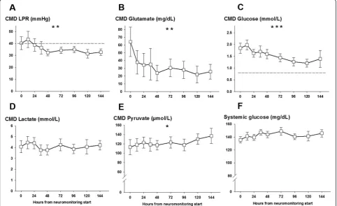
Figure 2Mean lactate-to-pyruvate ratio (LPR), glutamate, glucose, lactate, and pyruvate levels in the cerebral microdialysate and systemic glucose levels of 26 aneurysmal subarachnoid hemorrhage patients at given time points after neuromonitoring was started.
[image:3.595.57.540.369.664.2]Analytical methods
In all patients, IL-6 and MMP-9 levels could be mea-sured in a single microdialysis sample collected over a period of one hour. Analysis of IL-6 and CMD-MMP-9 was performed by enzyme-linked immunosorb-ent assays as described by the manufacturer (Aushon Custom Chemiluminescent Array Kit: 2-plex; Aushon Bio-Systems, Billerica, MA, USA). Calibrated protein stan-dards (50μL) and cerebral microdialysate (6μL) diluted in 50μL of buffer were added to pre-coated wells and incu-bated for 150 minutes. The wells were incuincu-bated for 30 minutes with biotinylated antibodies and then 30 mi-nutes with streptavidin-horseradish peroxidase conjugate. Finally, the SuperSignal Chemiluminescent Substrate was
added. All incubation steps were performed on a shaker at room temperature, and all wells were washed after every incubation step. The luminescent signal was detected by using a CCD (charge-coupled device) imaging and analysis system. The concentration of each sample was quantified by comparing the spot intensities to the corresponding standard curves calculated from the standard sample re-sults by using SearchLight® Analyst Software (Aushon Bio-Systems). CMD-IL-6 detection limit was 0.4 pg/mL.
Statistical analysis
[image:4.595.61.540.264.666.2]Continuous variables were assessed for normality. Nor-mally distributed data were reported as mean and standard error of the mean, and non-parametric data were reported
as median and interquartile range (IQR). Categorical vari-ables were reported as count and proportions in each group. Hourly recorded concentrations in the cerebral microdialysate were matched to continuously recorded pa-rameters (ICP, CPP, and PbtO2) averaged over the sampling
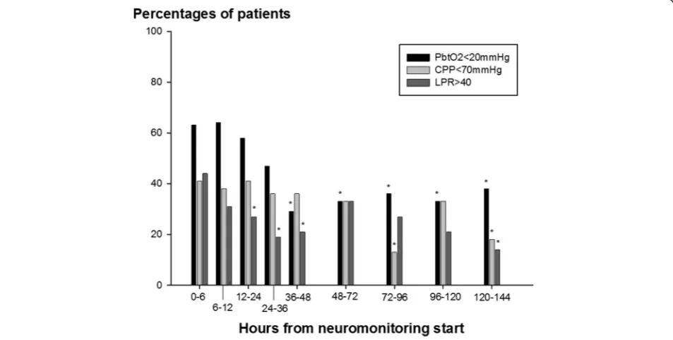
period (as shown in Figures 1, 2, and 3). Figure 4 displays the percentage of patients with at least one episode (hourly averaged data matched to microdialysis sampling time) in the abnormal range. CMD-derived metabolic parameters and PbtO2were categorized as previously defined
accord-ing to international accepted definitions to associate with CMD-IL-6 and CMD-MMP-9 levels. Time series data were analyzed by using a generalized linear model using a normal distribution and identity-link function and were extended by generalized estimating equations (GEEs) with an autoregressive process of the first order to handle re-peated observations within a subject [20]. Data were trans-formed (log for CMD-IL-6 and CMD-MMP-9) to meet assumptions of normality. In these GEE models, outcome was the dependent variable and important covariates were included (age and admission disease severity). For all tests, significance level was set at aPvalue of less than 0.05. All analyses were performed with IBM-SPSS V20.0 (SPSS Inc., Chicago, IL, USA).
Results
General characteristics
Clinical characteristics, hospital complications, and out-come data are summarized in Table 1. Aneurysm was
secured within the first 36 hours in all patients by endo-vascular coiling (n = 8, 31%) or surgical clipping (n = 18, 69%). In half of the patients (n = 13, 50%), CMD cathe-ters were located perilesional; in all other patients, cath-eters were located in normal appearing brain tissue. Six patients (23%) developed DCI and four patients died during hospitalization (15%).
Cerebrovascular hemodynamics and brain metabolism Neuromonitoring started at a median of 22 hours after ictus. Mean ICP, CPP, and PbtO2were 8 ± 1 mm Hg, 73 ±
2 mm Hg, and 16 ± 3 mm Hg, respectively. ICP remained less than 20 mm Hg and CPP significantly increased from neuromonitoring start to a maximum of 80 ± 2 mm Hg 6 days after ictus (P<0.001) (Figure 1A and B) in parallel to mean arterial pressure (P <0.001, data not shown). PbtO2significantly increased from baseline over the
moni-toring time (P <0.001) (Figure 1C) with at least one epi-sode of brain tissue hypoxia occurring in 63% of patients when neuromonitoring was initiated and decreasing to 29% and 12%, 48 and 96 hours later (Figure 4).
Brain metabolism and CMD-IL-6 and CMD-MMP-9
[image:5.595.61.538.446.687.2]Cerebral metabolism revealed a high LPR (42 ± 6) sig-nificantly improving over the following days (P= 0.002, Figure 2A) secondary to an increase in CMD-pyruvate from initially 113 ± 16 μmol/L to 136 ± 16 μmol/L on day 6 (P= 0.04, Figure 2E). CMD-lactate levels remained stable over the neuromonitoring time (Figure 2D).
Figure 4Percentage of 26 aneurysmal subarachnoid hemorrhage patients with at least one episode (mean hourly value) of brain tissue oxygen tension (PbtO2) below 20 mm Hg, cerebral perfusion pressure (CPP) below 70 mm Hg, and lactate-to-pyruvate ratio (LPR)
Initially upregulated CMD-glutamate (64 ± 18 mg/dL) decreased over days (P= 0.005, Figure 2B). CMD-glucose was normal at the start of neuromonitoring (1.8 ± 0.2 mmol/L) and significantly decreased during hospitaliza-tion (P= 0.001, Figure 2C) with 53% developing episodes
of cerebral glucose of less than 0.7 mmol/L on day 6. Sys-temic glucose levels were stable over the neuromonitoring time (Figure 2E). CMD-IL-6 levels were above the detec-tion limit in all patients with the highest levels at the start of neuromonitoring (7,600 ± 1,592 pg/mL) and decreased thereafter (P<0.001, Figure 3A). Initial highest levels were recorded in patients with aneurysm rebleeding (n = 4, 15%, range 10,500 to 25,300 pg/mL). CMD-MMP-9 was highly elevated (4,238 ± 1,737 pg/mL) at the start of neu-romonitoring and significantly decreased within 12 hours to levels below 1,000 pg/mL (P<0.001, Figure 3C). There was a correlation between CMD-IL-6 and CMD-lactate (r= 0.33, P<0.001) and LPR (r= 0.18, P= 0.01); however, no correlation was found between IL-6 and CMD-MMP-9 and systemic markers of inflammation (C-reactive protein, absolute leukocyte count, and body temperature) and hemoglobin levels.
Factors associated with CMD-IL-6 and CMD-MMP-9 Higher CMD-IL-6 levels were recorded initially (first 12 hours of neuromonitoring) in patients with admission GCE (4,440 pg/mL, IQR 805 to 16,194 pg/mL versus 1,542 pg/mL, IQR 155 to 4,059 pg/mL,P= 0.02) and pa-tients with a CPP of less than 70 mm Hg (n = 9/26, 35%, 16,281 pg/mL, IQR 6,501 to 21,929 pg/mL versus n = 17/26, 65%, 3,266 pg/mL, IQR 872 to 8,020 pg/mL;P= 0.03, Figure 3D) and overall in patients developing DCI independent of disease severity, aneurysm securing method, and perilesional probe location (Wald statistic = 5,4, degrees of freedom (df ) = 1, P= 0.02, Figure 3B). All other admission variables and hospital complications, including admission H&H grade, aneurysm size of more than 1 cm, the presence of aneurysm rupture-associated intraparenchymal hematoma on admission head CT scan, probe location, hydrocephalus requiring cerebro-spinal fluid diversion, and the aneurysm securing method, did not reveal significant differences in CMD-IL-6 levels.
CMD-MMP-9 levels were significantly elevated in the first hours of neuromonitoring in patients who lost consciousness at ictus (P= 0.005), H&H grade 5 patients (P= 0.002), and patients with initial brain tissue hypoxia (P= 0.03) after adjusting for probe location aneurysm re-pair method and disease severity as appropriate. All other admission and hospital complication were not as-sociated with higher CMD-MMP-9 levels.
Outcome
[image:6.595.56.289.109.614.2]CMD-IL-6 levels and LPR were higher in patients with poor 3-month mRS (Wald statistic = 5.7, df = 1, P =0.01 and Wald statistic = 10.1, df = 1,P =0.01). The relationship remained significant after adjusting for age, admission H&H grade, and probe location (Wald statistic = 6.5, df = 1, P =0.01 and Wald statistic = 19.5, df = 1, P <0.001).
Table 1 Baseline characteristics, complications, and outcome
Clinical characteristics N = 26
Age, years 55 (47-67)
Gender, female 15 (58%)
Admission H&H grade 2 2 (7.7%)
3 6 (23.1%)
4 2 (7.7%)
5 16 (61.5%)
Loss of consciousness 15 (58%)
Admission APACHE II score 17 (13-19)
Admission radiological characteristics
mFisher scale 1 3 (11.5%)
2 3 (11.5%)
3 9 (34.6%)
4 11 (42.3%)
SAH sum score 23 (15-27)
IVH Sum score 5 (0-8)
Aneurysm size above 10 mm 7 (27%)
Generalized cerebral edema 11 (42%)
Intracerebral hematoma 12 (46%)
Surgical procedures
Hydrocephalus requiring EVD/Shunt 20 (77%)
Clipping 18 (69%)
Hemicraniectomy 9 (35%)
Complications
Pneumonia 19 (73%)
Delayed cerebral infarction 6 (23%)
Anemia requiring transfusion 16 (62%)
Aneurysm rebleeding 4 (15%)
Hyperosmolar therapy 14 (54%)
Outcome characteristics
Length of hospital stay, days 40 (30-55)
3-month mRS 0-1 5 (19.2%)
2-3 5 (19.2%)
4 5 (19.2%)
5 6 (23.1%)
6 5 (19.2%)
CMD-MMP-9 levels were not associated with poor func-tional outcome after SAH.
Discussion
EBI is increasingly recognized to play a key role in path-ophysiologic changes contributing to poor functional outcome and mortality after aSAH. Here, we report evi-dence of brain metabolic derangement, brain tissue hyp-oxia, neuroinflammation, and BBB disruption in the first 72 hours of neuromonitoring in patients with poor-grade aSAH. Discovering mechanisms of EBI in humans may open the opportunity to target specific treatment endpoints in the early phase after SAH.
Neuroinflammation is increasingly recognized as an in-nate cerebral response to primary brain injury [21]. In the present study, we did not find an association between higher CMD-IL-6 levels and systemic inflammation, sup-porting the idea of compartmentalization of the central nervous system.
Pro-inflammatory cytokines may enhance brain edema through disruption of the BBB and induce neuronal apoptosis and therefore directly contribute to early brain damage [22,23]. Cerebral IL-6 has an estimated half-life of several hours and is produced by microglia, astro-cytes, and neurons [24]. In previous studies using cere-bral microdialysis, the pro-inflammatory cytokine IL-6 was associated with SAH disease severity, the develop-ment of DCI, and poor outcome [8-10]. In the present study, we furthermore found an association with admis-sion GCE, metabolic derangement, and a CPP of less than 70 mm Hg. The association between high CMD-LPR and a CPP<70mmHg has been previously reported in SAH patients with admission GCE [7]. Defining the optimal CPP in the early phase after SAH remains a challenge without having predefined brain physiologic endpoints even after aneurysm securing. Brain multi-modal monitoring data may be used to target endpoints on the cellular level. In a series of 30 patients with poor-grade SAH, a CPP of less than 70 mm Hg was associated with metabolic distress and brain tissue hypoxia; how-ever, these data cannot be extrapolated to the first 72 hours after SAH [25]. A higher CPP was associated with improved brain metabolism reflected by a lower LPR in a retrospective analysis of aSAH patients with admission GCE [7]. Improving substrate delivery espe-cially in the early phase after SAH may be beneficial in patients with increased need. As shown in patients with traumatic brain injury, CPP augmentation may translate into increased PbtO2 and a reduction in oxygen
extrac-tion fracextrac-tion [26]. However, a beneficial effect on brain metabolism was not observed. Defining the optimal CPP in the early phase after SAH and identifying patients who may benefit from early augmentation of CPP re-main important issues for future research and should
include multimodal neuromonitoring data as treatment endpoints.
Another potential treatment target in the early phase after aSAH is to suppress neuroinflammation by the ap-plication of systemic anti-inflammatory drugs. Potential benefits in patients with SAH have been postulated [27-29] and are furthermore supported by the improve-ment of cerebral edema and decreasing neuronal cell apoptosis in experimental SAH models [30]. With the limitation of associated hemodynamic side effects [31] when applied as a rapid infusion, a continuous low-dose infusion may be considered [32].
We found an early upregulation of CMD-MMP-9 in our study population, and higher levels were associated with disease severity, loss of consciousness at ictus, and early brain tissue hypoxia. Loss of consciousness at ictus is highly correlated with poor clinical grade and the de-velopment of early or delayed brain edema [18]. MMP-9 contributes to endothelial basal membrane damage, neu-roinflammation, and apoptosis and therefore plays a piv-otal role in EBI [11-13]. Serum-MMP-9 levels were elevated in patients who developed cerebral vasospasm, although both an initial upregulation and a sustained prolonged increase have been described [15,33]. This again supports the importance of local measurements in the brain as serum markers may reflect a dilution of the innate cerebral response or exaggerated systemic levels originating from multiple organ systems [14,21]. Antag-onizing MMP-9 diminished cortical apoptosis, was asso-ciated with improved outcome after experimental SAH [34,35], and was recently postulated as potential therapy in ischemic stroke [36].
episodes of mitochondrial dysfunction compared with epi-sodes of cerebral ischemia as cause for disturbed cerebral energy metabolism in patients with SAH [38]. Although no specific treatment to improve mitochondrial dysfunc-tion is currently available, further research is warranted as mitochondrial dysfunction may increase tissue sensitivity to secondary adverse events such as vasospasm and de-creased cerebral blood flow.
We observed improvement in brain metabolism and PbtO2over the monitoring time most likely secondary to
the parallel increase in CPP. Brain extracellular glucose concentrations significantly decreased to a critical level in a substantial amount of patients, whereas systemic glucose levels remained constant and this is suggestive of increased cerebral glucose consumption. Achieving normal cerebral glucose levels should be recommended as neuroglucopenia is associated with metabolic distress and poor outcome after SAH [39].
Quantifying brain metabolism and neuroinflammation may be of importance as both were associated with poor functional outcome. All statistical models were corrected for important covariates, including probe location, as in half of our patients the microdialysis catheter was within 1 cm from the lesion.‘Perilesional’probe positioning im-plies that the microdialysate was collected adjacent to radiological damaged brain tissue, where cell necrosis, blood compounds, autophagy, and apoptosis may alter brain metabolism and ameliorate cytokine release into the extracellular compartment.
Our study was designed as a pilot study and included only a small number of patients and this is a potentially limiting factor. Moreover, early pathophysiologic changes described in the present study may be relevant for patients with poor-grade aSAH and not be generalizable to all clin-ical grades. We were not able to define specific treatment targets based on the following limitations: (1) a localized metabolic information using cerebral microdialysis tech-nique (2) the small sample size and (3) local treatment strategies which may differ from other institutional proto-cols and substantially influence longitudinal brain physio-logic data. Importantly, patient- and disease-specific data were prospectively documented, and statistical models were corrected for important covariates.
Conclusions
EBI is believed to substantially contribute to secondary brain injury and to cause significant morbidity and mor-tality following aSAH. The present study proves that multimodal neuromonitoring techniques can provide insight into pathophysiologic changes in the early phase after aSAH. In our series of 26 patients, catheters were placed within the first 36 hours, revealing metabolic de-rangement and (to a certain degree) hemodynamic in-stability, a pro-inflammatory cerebral response, and BBB
breakdown. Multimodal neuromonitoring data may as-sist the neurointensivist in defining treatment targets on the cellular level, eventually opening the door for spe-cific treatment options to minimize early brain injury in patients with aSAH.
Key messages
Early brain injury (EBI) is common after
subarachnoid hemorrhage (SAH) and is associated with poor outcome.
Pathophysiologic mechanisms of EBI include blood-brain barrier breakdown, blood-brain tissue hypoxia, neuroinflammation, and excitotoxicity leading to brain edema and metabolic derangement.
Neuromonitoring techniques may identify underlying pathophysiologic mechanisms occuring in the early phase after aSAH and therefore help to understand mechanisms of EBI.
Multimodal neuromonitoring data may assist the neurointensivist in defining treatment targets on the cellular level, eventually opening the door for specific treatment options to minimize early brain injury in patients with aneurysmal SAH.
Abbreviations
aSAH:aneurysmal subarachnoid hemorrhage; BBB: blood-brain barrier; CMD: cerebral microdialysis; CPP: cerebral perfusion pressure; CT: computed tomography; DCI: delayed cerebral ischemia; df: degrees of freedom; EBI: early brain injury; GCE: global cerebral edema; GEE: generalized estimating equation; H&H: Hunt & Hess; ICP: intracranial pressure; IL-6: interleukin-6; IQR: interquartile range; LPR: lactate-to-pyruvate ratio; mBFV: mean blood flow velocity; MMP: matrix metalloproteinase; mRS: modified Rankin Scale; PbtO2: brain tissue oxygen tension;
SAH: subarachnoid hemorrhage; TCD: transcranial Doppler sonography.
Competing interests
The authors declare that they have no competing interests.
Authors’contributions
RH was involved in the idea, study design, interpretation of data, statistical analysis, and writing of the manuscript and final revision of the manuscript. AS made substantial contributions to the design and data acquisition, analysis, and interpretation and performed laboratory analysis of brain-derived biomarkers. RB and BP made substantial contributions to the idea, study design, data analysis and interpretation, and final revision of the manuscript. ES made substantial contributions to the idea, study design, data interpretation, and final revision of the manuscript. AD, APA, FS, MF, and CH contributed to the design, data acquisition, and interdisciplinary data interpretation and performed laboratory analysis of brain-derived biomarkers. WOH made substantial contributions to the design, data acquisition (multimodal neuromonitoring high-frequency data), and data interpretation. PL substantially contributed to the study design, statistical analysis, and data interpretation. PR performed radiographic analysis as independent radiologist and substantially contributed to the data interpretation. CT substantially contributed to the design and data interpretation and performed placement of multimodal neuromonitoring devices. All authors critically reviewed, drafted, and approved the final version of the manuscript and agree to be accountable for all aspects concerning the work.
Acknowledgments
Author details
1
Neurological Intensive Care Unit, Department of Neurology, Medical University of Innsbruck, Anichstreet 35, 6020 Innsbruck, Austria.2Department
of Neurosciences, Santa Maria Hospital, Hospital de Santa Maria, 1649-028 Lisbon, Portugal.3Department of Neurosurgery, Innsbruck Medical University,
Anichstreet 35, 6020 Innsbruck, Austria.4Institute of Biomedical Informatics, UMIT-University for Health Sciences, Medical Informatics and Technology, Eduard Wallnöfer-Zentrum I, 6060 Hall in Tirol, Austria.5Department of Radiology, Innsbruck Medical University, Anichstreet 35, 6020 Innsbruck, Austria.6Department of Psychiatry and Psychotherapy, Medical University Innsbruck, Anichstreet 35, 6020 Innsbruck, Austria.
Received: 8 November 2014 Accepted: 12 February 2015
References
1. Suarez JI, Tarr RW, Selman WR. Aneurysmal subarachnoid hemorrhage. N Engl J Med. 2006;354:387–96.
2. Broessner G, Lackner P, Hoefer C, Beer R, Helbok R, Grabmer C, et al. Influence of red blood cell transfusion on mortality and long-term functional outcome in 292 patients with spontaneous subarachnoid hemorrhage. Crit Care Med. 2009;37:1886–92.
3. Etminan N, Vergouwen MD, Ilodigwe D, Macdonald RL. Effect of
pharmaceutical treatment on vasospasm, delayed cerebral ischemia, and clinical outcome in patients with aneurysmal subarachnoid hemorrhage: a systematic review and meta-analysis. J Cereb Blood Flow Metab. 2011;31:1443–51. 4. Sehba FA, Hou J, Pluta RM, Zhang JH. The importance of early brain injury
after subarachnoid hemorrhage. Prog Neurobiol. 2012;97:14–37. 5. Sehba FA, Pluta RM, Zhang JH. Metamorphosis of subarachnoid
hemorrhage research: from delayed vasospasm to early brain injury. Mol Neurobiol. 2011;43:27–40.
6. Stuart RM, Schmidt M, Kurtz P, Waziri A, Helbok R, Mayer SA, et al. Intracranial multimodal monitoring for acute brain injury: a single institution review of current practices. Neurocrit Care. 2010;12:188–98.
7. Helbok R, Ko SB, Schmidt JM, Kurtz P, Fernandez L, Choi HA, et al. Global cerebral edema and brain metabolism after subarachnoid hemorrhage. Stroke. 2011;42:1534–9.
8. Sarrafzadeh A, Schlenk F, Gericke C, Vajkoczy P. Relevance of cerebral interleukin-6 after aneurysmal subarachnoid hemorrhage. Neurocrit Care. 2010;13:339–46.
9. Hillman J, Aneman O, Persson M, Andersson C, Dabrosin C, Mellergard P. Variations in the response of interleukins in neurosurgical intensive care patients monitored using intracerebral microdialysis. J Neurosurg. 2007;106:820–5.
10. Mellergard P, Aneman O, Sjogren F, Saberg C, Hillman J. Differences in cerebral extracellular response of interleukin-1beta, interleukin-6, and interleukin-10 after subarachnoid hemorrhage or severe head trauma in humans. Neurosurgery. 2011;68:12–9. discussion 19.
11. Guo Z, Sun X, He Z, Jiang Y, Zhang X. Role of matrix metalloproteinase-9 in apoptosis of hippocampal neurons in rats during early brain injury after subarachnoid hemorrhage. Neurol Sci. 2010;31:143–9.
12. Wang Z, Meng CJ, Shen XM, Shu Z, Ma C, Zhu GQ, et al. Potential contribution of hypoxia-inducible factor-1alpha, aquaporin-4, and matrix metalloproteinase-9 to blood–brain barrier disruption and brain edema after experimental subarachnoid hemorrhage. J Mol Neurosci. 2012;48:273–80. 13. Candelario-Jalil E, Yang Y, Rosenberg GA. Diverse roles of matrix
metalloproteinases and tissue inhibitors of metalloproteinases in neuroinflammation and cerebral ischemia. Neuroscience. 2009;158:983–94. 14. Sarrafzadeh A, Copin JC, Bengualid DJ, Turck N, Vajkoczy P, Bijlenga P, et al.
Matrix metalloproteinase-9 concentration in the cerebral extracellular fluid of patients during the acute phase of aneurysmal subarachnoid hemorrhage. Neurol Res. 2012;34:455–61.
15. Fischer M, Dietmann A, Beer R, Broessner G, Helbok R, Pfausler B, et al. Differential regulation of matrix-metalloproteinases and their tissue inhibitors in patients with aneurysmal subarachnoid hemorrhage. PLoS One. 2013;8:e59952. 16. Bederson JB, Connolly Jr ES, Batjer HH, Dacey RG, Dion JE, Diringer MN, et al.
Guidelines for the management of aneurysmal subarachnoid hemorrhage: a statement for healthcare professionals from a special writing group of the stroke council, American heart association. Stroke. 2009;40:994–1025. 17. Vergouwen MD, Participants in the International Multi-Disciplinary Consensus
Conference on the Critical Care Management of Subarachnoid H. Vasospasm
versus delayed cerebral ischemia as an outcome event in clinical trials and observational studies. Neurocrit Care. 2011;15:308–11.
18. Claassen J, Carhuapoma JR, Kreiter KT, Du EY, Connolly ES, Mayer SA. Global cerebral edema after subarachnoid hemorrhage: frequency, predictors, and impact on outcome. Stroke. 2002;33:1225–32.
19. Schmidt JM, Ko SB, Helbok R, Kurtz P, Stuart RM, Presciutti M, et al. Cerebral perfusion pressure thresholds for brain tissue hypoxia and metabolic crisis after poor-grade subarachnoid hemorrhage. Stroke. 2011;42:1351–6. 20. Zeger SL, Liang KY. Longitudinal data analysis for discrete and continuous
outcomes. Biometrics. 1986;42:121–30.
21. Helmy A, Carpenter KL, Menon DK, Pickard JD, Hutchinson PJ. The cytokine response to human traumatic brain injury: temporal profiles and evidence for cerebral parenchymal production. J Cereb Blood Flow Metab. 2011;31:658–70. 22. Ostrowski RP, Colohan AR, Zhang JH. Molecular mechanisms of early brain
injury after subarachnoid hemorrhage. Neurol Res. 2006;28:399–414. 23. Sozen T, Tsuchiyama R, Hasegawa Y, Suzuki H, Jadhav V, Nishizawa S, et al.
Role of interleukin-1beta in early brain injury after subarachnoid hemorrhage in mice. Stroke. 2009;40:2519–25.
24. Spooren A, Kolmus K, Laureys G, Clinckers R, De Keyser J, Haegeman G, et al. Interleukin-6, a mental cytokine. Brain Res Rev. 2011;67:157–83. 25. Ko SB, Choi HA, Parikh G, Helbok R, Schmidt JM, Lee K, et al. Multimodality
monitoring for cerebral perfusion pressure optimization in comatose patients with intracerebral hemorrhage. Stroke. 2011;42:3087–92. 26. Johnston AJ, Steiner LA, Coles JP, Chatfield DA, Fryer TD, Smielewski P, et al.
Effect of cerebral perfusion pressure augmentation on regional oxygenation and metabolism after head injury. Crit Care Med. 2005;33:189–95. 27. Muroi C, Hugelshofer M, Seule M, Keller E. The impact of nonsteroidal
anti-inflammatory drugs on anti-inflammatory response after aneurysmal subarachnoid hemorrhage. Neurocrit Care. 2014;20:240–6.
28. Broessner G, Lackner P, Fischer M, Beer R, Helbok R, Pfausler B, et al. Influence of prophylactic, endovascularly based normothermia on inflammation in patients with severe cerebrovascular disease: a prospective, randomized trial. Stroke. 2010;41:2969–72.
29. Simard JM, Tosun C, Ivanova S, Kurland DB, Hong C, Radecki L, et al. Heparin reduces neuroinflammation and transsynaptic neuronal apoptosis in a model of subarachnoid hemorrhage. Transl Stroke Res. 2012;3:155–65. 30. Zhang XS, Zhang X, Wu Q, Li W, Wang CX, Xie GB, et al. Astaxanthin offers
neuroprotection and reduces neuroinflammation in experimental subarachnoid hemorrhage. J Surg Res. 2014;192:206–13.
31. Schiefecker AJ, Pfausler B, Beer R, Sohm F, Sabo J, Knauseder V, et al. Parenteral diclofenac infusion significantly decreases brain-tissue oxygen tension in patients with poor-grade aneurysmal subarachnoid hemorrhage. Crit Care. 2013;17:R88. 32. Cormio M, Citerio G. Continuous low dose diclofenac sodium infusion to
control fever in neurosurgical critical care. Neurocrit Care. 2007;6:82–9. 33. Chou SH, Feske SK, Simmons SL, Konigsberg RG, Orzell SC, Marckmann A,
et al. Elevated peripheral neutrophils and matrix metalloproteinase 9 as biomarkers of functional outcome following subarachnoid hemorrhage. Transl Stroke Res. 2011;2:600–7.
34. Sherchan P, Lekic T, Suzuki H, Hasegawa Y, Rolland W, Duris K, et al. Minocycline improves functional outcomes, memory deficits, and histopathology after endovascular perforation-induced subarachnoid hemorrhage in rats. J Neurotrauma. 2011;28:2503–12.
35. Guo ZD, Wu HT, Sun XC, Zhang XD, Zhang JH. Protection of minocycline on early brain injury after subarachnoid hemorrhage in rats. Acta Neurochir Suppl. 2011;110:71–4.
36. Chaturvedi M, Kaczmarek L. Mmp-9 inhibition: a therapeutic strategy in ischemic stroke. Mol Neurobiol. 2014;49:563–73.
37. Westermaier T, Jauss A, Eriskat J, Kunze E, Roosen K. The temporal profile of cerebral blood flow and tissue metabolites indicates sustained metabolic depression after experimental subarachnoid hemorrhage in rats. Neurosurgery. 2011;68:223–9. discussion 229–30.
38. Jacobsen A, Nielsen TH, Nilsson O, Schalen W, Nordstrom CH. Bedside diagnosis of mitochondrial dysfunction in aneurysmal subarachnoid hemorrhage. Acta Neurol Scand. 2014;130:156–63.