Prismatic adaptation effects on non-spatial and spatial
processes in the healthy adult.
By
Lorraine Boran
A dissertation submitted for the degree of Doctor of Philosophy of the University of Dublin, Trinity College Dublin, Ireland.
Declaration
This thesis has not been submitted as an exercise for a degree at this or any other University and it is entirely my own work. I agree that Trinity College Dublin Library may lend or copy this thesis upon request.
Signed
Acknowledgements
This thesis has been a major personal undertaking and there have been times in the past few years when I felt like the plaintiff in Hamer v Sidway (1891) who forbore from drinking, swearing, smoking and gambling after his uncle promised him a large sum of money. The withdrawal from normal life toward the end of the write-up phase was eased by a number of people who have provided assistance to me. It is really a pleasure to have the opportunity of thanking them.
I am a self-confessed tree-lover. I focus on small details and worry about them until everything makes sense! My many thanks to Ian Robertson for his ability to get me to see the wood for the trees. I’d like to thank him for his continued support throughout the thesis and for taking the time to read draft chapters. I’d also like to take the opportunity of thanking Stuart Smith. Stuart gave me invaluable assistance in the optic flow study. I enjoyed our chats and you encouraged me to think of research as exciting and personally challenging.
My attention now swings to the people I met everyday in the department, who are always so professional and helpful. Thank you June for your advice, smiles and repeated offers of assistance. My many thanks to Rose and Marcella, who were always so kind and helpful. Thanks also to Dave, Lisa and Eddie for technical support. I’d also like to thank Tony in the library for his help. My work in the department was funded by IRCHSS and TCIN, which gave me the opportunity to work with and meet wonderful people. This research would not have been possible without willing participants, who individually took up to 7 hours or more out of their busy schedules to be tested on numerous occasions.
I met the most wonderful and talented people during my time in Trinity. I will never forget the times spent with Mary, Laura, Aisling and Celine.
Finally, I cast my attention homeward. I have an incredibly loving and supportive family; mum, dad and Marie. I won’t even try to communicate how thankful I am for their love and support. Mid-way through my thesis I met you, Dave. You have been so loving, strong and encouraging. Your Renaissance-intellect amazes me!
Brief Summary
The structure of the thesis is broadly based on the hierarchical division of attention into two domains – intensity and selectivity components of attention (Sturm, 1996; Van Zomeren & Brouwer, 1994). The intensity components of attention (arousal and vigilance) are a requisite for the more complex aspects of attention selectivity (Sturm et al., 2004). Spatial neglect is characterized by impairment in both the intensity (Heilman et al., 1978; Husain & Rorden, 2003; Robertson et al., 1996; Samuelsson et al., 1998) and the selectivity components of attention (Bartolomeo & Chokron, 2002; Posner & Peterson, 1990). In the context of understanding how prism adaptation rehabilitates spatial neglect, researchers have attempted to produce (Michel et al., 2003; Redding & Wallace, 2005) mild neglect-like patterns of behaviour in healthy individuals following prism adaptation. The aim of this thesis was to induce ‘neglect-like’ patterns of performance in both non-spatial and spatial attentional tasks following left prism adaptation (Berberovic & Mattingley, 2003; Colent et al., 2000; Michel et al., 2003).
Chapter 2 examined the adaptation process and aftereffects in sensory motor function. A prismatic aftereffect in both visual and proprioceptive spatial maps was observed post adaptation to either left or right shifting prisms. Adaptation caused re-alignment in the visual (eye) and proprioceptive (hand) systems. Aftereffects were direction specific. Left shifting prisms produced a leftward shift in a visual indication of subjective straight-ahead (SSA) and an opposite rightward shift in a proprioceptive indication of subjective straight-ahead. Right shifting prisms produced a rightward shift in visual SSA and a leftward shift in proprioceptive SSA. The mechanisms underpinning these prismatic effects are unclear. The thesis investigated chapter by chapter, the effects of adapting to either prism shift on non-spatial and spatial function.
advanced that LPA impaired arousal and vigilance by depressing right parietal function, and hence ‘simulating’ neglect-like impairment in non-spatial attention.
Chapter 5 examined the effects of adaptation on non-spatial selective attention within hierarchical stimuli. Adaptation to right shifting prisms (RPA) facilitated processing on the global level, indicative of left parietal suppression. LPA did not have any effect on local global processing. Chapters 6 and 7 examined the spatial effects of adaptation – attentional orienting and reorienting across space. Both prism shifts produced a rightward shift in lateral bias as measured by the greyscales task. This finding was interpreted in terms of right parietal suppression and prismatic effects in the allocentric spatial frame of reference. However, both prism shifts differentially influenced spatial orienting. LPA made it easier to reorient from right space, and RPA made it easier to reorient from left space. The spatial effects of adaptation to either prism shift was interpreted as depression of function in the right and left parietal area following left and right adaptation respectively.
List of Publications
Published Abstract
Boran L. Robertson I (2004) Prismatic effects on arousal and lateral bias. Cognitive Neuroscience Society Meeting, San Francisco, USA
Presentation
Boran L. Robertson I (2005) Mechanisms of prism adaptation: a model proposing the involvement of a cerebello-parietal-frontal network International Neuroscience Society Meeting, Dublin, Ireland
Boran L. Robertson I (2005) ‘How might rehabilitation work?’ Possible mechanisms underpinning the rehabilitative effects of prism adaptation. International Neuroscience Society Meeting, Dublin, Ireland
Boran L. Robertson I (2004) Prism Adaptation: Effects and the Mechanisms
List of Abbreviations
Ach Acetycholine
ADHA Attention Deficit and Hyperactivity Disorder ANOVA Analysis of Variance
DART Dual sustained attention to response task DLPFC Dorsolateral prefrontal cortex
EEG Electroencephalogram ERP Event-related Potential
FMRI Functional Magnetic Resonance Imaging IPL Inferior parietal lobule
IPS Inferior parietal sulcus LPA Left Prism Adaptation
MACCL-R Mood and Affect Questionnaire NE Norepinephrine
PET Positron Emission Tomography RPA Right Prism Adaptation
PFC Pre Frontal Cortex
RCBF Regional Cerebral Blood Flow RT Reaction Time
SART Sustained Attention to Response Task SD Standard Deviation
SPL Superior Parietal Lobe STG Superior Temporal Gyrus TBI Traumatic Brain Injury
List of Figures
Figure 1.1:Illustration of hemispatial Neglect 2 Figure 2.1: Illustration of first pointing performance with LPA 25
Figure 2.2: Process of Adaptation 27
Figure 2.3: Illustration of Adaptation process 39
Figure 2.4: Direct Effect 43
Figure 2.5: Adaptation Curve 44
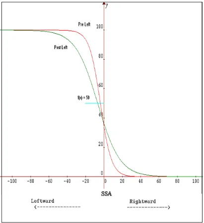
Figure 2.6: Prismatic Aftereffects (PSSA) 46
Figure 2.7: Heading Estimation 52
Figure 2.8: Continuous Flow with FOE 53
Figure 2.9: Mean Visual SSA 55
Figure 2.10: Sample data (visual SSA LPA) 56 Figure 2.11: Sample data (visual SSA RPA) 57 Figure 2.12: Continuous Flow with eye fixation 60
Figure 3.1: Sympathetic Arousal 85
Figure 3.2: Brain Areas involved in Arousal 87
Figure 3.3: Illustration of EDA response 93
Figure 3.4: Mean Arousal Responding level 101
Figure 3.5: Mean Phasic Arousal 103
Figure 4.1: Illustration of the vigilance system 126
Figure 4.2: SART sequence 132
Figure 4.3: Mean EoC in SART 135
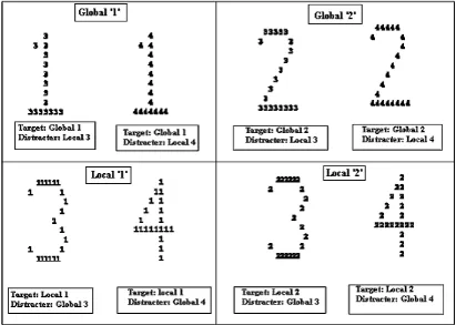
Figure 5.1: Illustration of a Navon Figure 154 Figure 5.2: Typical performance on Local Global Processing 160 Figure 5.3: Illustration of hierarchical stimulus 170
Figure 5.4: Exogenous cues 171
Figure 5.5 Cue Timing 172
Figure 5.6: Endogenous cues 173
Figure 5.7: Cue Timing 173
Figure 5.8: Target and Distracter 174
Figure 5.9: RPA effect on LG 180
Figure 5.10: RPA effect on CES 181
Figure 6.1: Greyscales task 202
Figure 6.2: Pointing Aftereffects (reproduced from chapter 2) 204 Figure 6.3: Directional shift in Lateral Asymmetry 207 Figure 6.4: Lateral Shifts post adaptation 208 Figure 6.5: Accuracy in Lateral Asymmetry 210 Figure 6.6: Lateral Asymmetry and hand-used-to-adapt 213 Figure 7.1: Illustration of exogenous Attentional Control 231 Figure 7.2: Illustration of endogenous Attentional Control 233
Figure 7.3: Exogenous task 243
Figure 7.4: Timing in exogenous task 244
Figure 7.5: Endogenous task 244
Figure 7.6: Timing in endogenous task 245
List of Tables
Table 4.1: Self-report measures 133
Table 4.2: Mean EoC in SART 136
Table 4.3: Total Error in SART 137
Table 4.4: Mean RT in SART 138
Table 4.5: Mean EoC in DART 139
Table 4.6: Total Error in DART 141
Chapter 1 Spatial Neglect: A General Introduction ... 1
1.1 Spatial Neglect ... 1
1.2 Rehabilitation of Spatial Neglect – Prism Adaptation ... 7
1.3 Spatial and non-spatial mechanisms underpinning prismatic aftereffects in the healthy brain ... 12
1.4 Thesis Outline ... 12
1.5 Thesis Extended Summary ... 13
Chapter 2: The Adaptation Process ... 13
Chapter 3: Prism Adaptation affects non-spatial lateralized alertness (arousal) ... 14
Chapter 4: Prism Adaptation affects non-spatial lateralized vigilance network ... 16
Chapter 5: Prism Adaptation affects non-spatial selective attention ... 18
Chapter 6: Prism Adaptation affects lateral asymmetry ... 18
Chapter 7: Prism Adaptation affects spatial attentional reorienting ... 19
Chapter 2 Model of Prism Adaptation: Visual & Proprioceptive Aftereffect ... 22
2.1 Summary ... 22
2.2 Aims ... 23
2.3 Introduction to Prism Adaptation ... 23
2.3.1 Models of Prism Adaptation ... 28
2.4 Measuring prismatic effects – Experiment 1 ... 36
2.5 Predictions ... 36
2.5.1 Left Prism Predictions ... 36
2.5.2 Right Prism Predictions ... 37
2.6 Method – Experiment I ... 37
2.6.1 Participants ... 37
2.6.2 Apparatus and Stimuli ... 38
2.6.3 Procedure ... 39
2.7 Results – Experiment I ... 42
2.8 Discussion – Experiment I ... 47
2.9 Optic flow – Experiment II ... 47
2.9.1 Visual Straight-ahead – optic flow ... 48
2.10 Predictions ... 50
2.10.1 Left Prism Predictions ... 50
2.10.2 Right Prism Predictions ... 50
2.11 Method- Experiment IIa Optic flow with free movement of eyes ... 51
2.12 Results – Experiment IIa ... 54
2.13 Discussion – Experiment IIa ... 58
2.14.1 Participants ... 59
2.14.2 Stimuli and Procedure ... 60
2.15 Results- Experiment IIb ... 61
2.16 Discussion – Experiment II ... 61
2.17 General Discussion ... 61
2.18 Future Research ... 65
2.19 Conclusion ... 66
Chapter 3 Effects of prismatic adaptation on arousal. ... 67
3.1 Summary ... 67
3.2 Aim ... 68
3.3 General Introduction ... 69
3.3.1 Attention ... 69
3.3.2 Arousal ... 70
3.3.3 Neglect as an attentional deficit ... 70
3.3.4 Spatial Lateralized Attentional Deficits ... 71
3.3.5 Non Spatial Lateralized Attentional Deficits ... 73
3.3.6 Neuropsychological Models of Attention ... 76
3.3.7 Sympathetic Arousal ... 83
3.4 Measures ... 92
3.5 Predictions ... 94
3.5.1 Arousal Responding Level (tonic and phasic components) ... 94
3.5.2 Phasic Arousal (phasic component only) ... 95
3.6 Method ... 95
3.6.1 Participants ... 95
3.6.2 Apparatus and Stimuli ... 97
3.6.3 Procedure ... 99
3.7 Results Experiment ... 99
3.7.1 Summary of Mean EDA responding level ... 99
3.7.2 Summary of Phasic EDA responses ... 102
3.8 Discussion of Arousal Findings ... 105
3.9 Introduction to Mood and Arousal ... 109
3.9.1 Theories of Emotional responding & Mood states ... 109
3.10 Measures ... 112
3.11 Predictions ... 113
3.12 Method ... 113
3.12.2 Stimuli/Procedure ... 113
3.13 Results ... 114
3.14 Criticisms & Study Limitations ... 115
3.15 Conclusion ... 115
Chapter 4 Prism Adaptation and Vigilance ... 116
4.1 Summary ... 116
4.2 General Introduction ... 116
4.2.1 Vigilance and Spatial Attention ... 117
4.2.2 The relationship between Arousal and Vigilance ... 119
4.2.3 Vigilance ... 121
4.3 Predictions ... 127
4.4 Method ... 127
4.4.1 Participants ... 127
4.4.2 Apparatus/ Stimuli ... 128
4.4.3 Procedure ... 131
4.5 Results ... 133
4.5.1 Behavioural Results ... 133
4.5.2 CFQ ... 133
4.5.3 MACCLR ... 133
4.5.4 SART ... 134
4.5.5 DART - Behavioural Results ... 139
4.5.6 EDA analysis – SART random ... 143
4.5.7 EDA analysis – DART ... 143
4.6 Discussion ... 144
4.7 Study Limitations ... 149
4.8 Future Research ... 149
4.9 Conclusions ... 150
Chapter 5. Prism Adaptation and Spatial Attention: Effects on global & local deployment of attention ... 151
5.1 Summary ... 151
5.2 Aims ... 152
5.3 General Introduction - Local-Global Processing ... 153
5.3.1 Lateralization – Perceptual phase of processing ... 155
5.3.2 Counter Evidence ... 157
5.3.4 ‘Global’ neglect: a case of biasing attention toward local detail in spatial neglect
... 162
5.3.5 Attentional Mechanism underpinning Local-Global Processing ... 163
5.4 Measures ... 166
5.4.1 Local-Global Covert Orienting of Attention ... 166
5.5 Predictions ... 167
5.6 Method ... 168
5.6.1 Participants ... 168
5.6.2 Stimuli ... 168
5.6.3 Prism Adaptation Procedure ... 174
5.6.4 Data Analysis ... 174
5.7 Results ... 176
5.7.1 LG exogenous ... 176
5.7.2 LG endogenous ... 183
5.8 Discussion ... 184
5.9 Study Limitations ... 187
5.10 Further Research ... 187
5.11 Summary ... 188
Chapter 6: Effects of prismatic adaptation on free-viewing perceptual asymmetries in the greyscales task. ... 189
6.1 Summary ... 189
6.2 Aims of Study ... 190
6.3 General Introduction ... 190
6.3.1 Pseudoneglect and Perceptual Asymmetry ... 190
6.3.2 Spatial neglect & Perceptual Asymmetry ... 193
6.3.3 Neuropsychological Models of Performance on the Greyscales Task ... 194
6.3.4 Prism Adaptation and lateral asymmetry ... 196
6.4 Measures ... 197
6.4.1 Attentional Asymmetry ... 197
6.4.2 Arousal ... 198
6.5 Predictions ... 198
6.6 Method ... 199
6.6.1 Participants ... 199
6.6.2 Apparatus and Stimuli ... 199
6.6.3 Procedure ... 200
6.7 Results ... 204
6.7.2 Prismatic Effects on the greyscale task ... 205
6.8 Discussion ... 215
6.9 Study Limitations ... 224
6.10 Further Research ... 224
6.11 Conclusions ... 225
Chapter 7: Spatial Orienting ... 227
7.1 Summary ... 227
7.2 General Introduction to Spatial Orienting ... 228
7.2.1 The Posner Paradigm ... 229
7.2.2 Spatial mechanism of Neglect ... 230
7.2.3 Attentional Models of impaired orienting behaviour in Spatial Neglect ... 233
7.3 Measures of Attentional Orienting ... 237
7.3.1 COVAT exogenous ... 238
7.3.2 COVAT endogenous ... 239
7.4 Predictions ... 239
7.5 Method ... 240
7.5.1 Participants ... 240
7.5.2 Stimuli and Procedure ... 241
7.5.3 Prism Adaptation Procedure ... 246
7.5.4 Data Analysis ... 246
7.6 Results ... 249
7.6.1 Mean RT exogenous ... 249
7.6.2 Mean RT endogenous ... 250
7.6.3 Left Prism Goggles ... 251
7.6.4 Right Prism Goggles ... 255
7.6.5 Sham Prism Goggles ... 258
7.7 Discussion ... 261
8 General Discussion ... 268
8.1 Research Aims ... 268
8.2 The adaptation process ... 269
8.3 Neural Bases of Adaptation ... 272
8.4 Non-spatial Mechanisms underpinning LPA induced ‘neglect-like’ cognitive impairment ... 277
8.5 Spatial Mechanisms underpinning LPA induced ‘neglect-like’ cognitive impairment ... 280
8.6 Summary ... 287
Chapter 1 Spatial Neglect: A General Introduction
“…My experience is what I agree to attend to … without selective interest, experience is an utter chaos” (James, 1890, p. 402).
1.1 Spatial Neglect
Edoardo Bisiach and Claudio Luzzati (1978) “studied two patients with damage to their right parietal lobes that left them with a visual neglect syndrome. [Their]…eyes register the whole visual field, but they attend only to the right half” (Pinker, 1997, p. 286). Patients with spatial neglect are typically unaware of the extent of their disorder, which manifests in a number of profound spatial difficulties in everyday encounters. “They ignore the cutlery to the left of the plate, draw a face with no left eye or nostril, and when describing a room, ignore large details [such as] a piano on their left” (p. 289). As noted by Driver and Vuilleumier (2006) unilateral spatial neglect is the failure to represent and respond to events occuring in the side of space opposite a unilateral brain lesion (Heilman, 1979; 1993). Low level primary sensory or motor loss deficits are not at issue, but deficits in higher-order spatial representation and attention (Halligan & Marshall, 1994).
home country (Bartolomeo et al., 1994; Rode & Perenin, 1994) or familiar details from the left side of their room (Denny-Brown & Banker, 1954).
Fig.1.1 Adapted illustration of left hemispatial neglect based on Bisiach & Luzzati (1978). A patient was asked to imagine and then name as many buildings as possible when facing (green arrow) the cathedral (cross figure on map) in the Piazza del Duomo in Milan. Buildings located to the right (in green) were recalled, but buildings on the left were ignored. When the patient was asked to turn around and imagine facing the opposite end of the piazza (red arrow), buildings previously ignored were then named (in red).
There is debate in the literature regarding the neural bases of spatial neglect. This controversy has served to highlight the heterogeneity of the behavioural deficit.
Behrmann et al. (2004) credits Karnath et al., (2001) with identifying the superior temporal gyrus (STG) and not the inferior parietal lobule (IPL) as the candidate site for cortical damage associated with spatial neglect. In a related study, Karnath et al. (2002) identified subcortical sites associated with spatial neglect. These sites included the putamen, the caudate nucleus within the basal ganglia, and the pulvinar within the thalamus. The identified regions have reciprocal connections with the STG. This result was interpreted by the authors to be consistent with their prior argument that the STG is a critical region in spatial neglect.
In contrast to the STG hypothesis, others have argued that the inferior parietal lobule (IPL) is the crucial anatomical analogue of spatial neglect (Mort et al., 2003). Using structural imaging, the authors found that all of their neglect patients had damage to the angular gyrus, and 62% of their sample (9 out of 14 neglect patients) had damage to the TPJ and the intraparietal sulcus (IPS divides the parietal lobe into the superior and inferior regions). A further 57% (8 patients) had damage to the supramarginal gyrus and the inferior frontal gyrus, whereas just 50% (7 patients) sustained damage to the STG. Damage to the superior parietal lobe (SPL) and middle frontal gyrus (MFG) was present in 29% (4 patients) of their neglect sample. Crucially, none of the control patients had damage in either the TPJ or the STG, whereas two or more had damage in other areas common to the neglect patients.
these types of neglect, and previous studies have failed to correlate neglect type as measured on line bisection with lesion location (Hillis et al., 2005). Patients with allocentric neglect may process the page containing the line as one stimulus and neglect the left side of the line, whereas patients with egocentric neglect may neglect the end of the line located in left space (left of patient’s midline). The authors determined that hypoperfusion rather than the cortical infarct per se predicted neglect type. “Reperfusion of the cortex resulted in recovery of spatial neglect despite the continued presence of subcortical infarcts” (Hillis et al., 2005, p. 3161). The authors reported a correlation between frontal and dorsal hypoperfusion in the right posterior inferior frontal gyrus, angular gyrus and the supramarginal gyrus with neglect in egocentric space. Allocentric neglect was predicted by ventral hypoperfusion in the right superior temporal gyrus and the posterior inferior temporal gyrus. Thus, egocentric neglect was predicted by hypoperfusion in right parietal areas, and allocentric neglect was predicted by hypoperfusion in right superior temporal areas.
As will be discussed shortly, spatial neglect is generally argued to be an attentional disorder (Driver & Mattingley, 1997; Husain & Rorden, 2003). As noted by many including George et al. (2005, p. 265), it is a disorder associated with “slowed recovery, difficulties in many activities in everyday life and a high degree of dependence on others” (see Paolucci et al., 2001).
1.1.1 Varieties of Attention
As noted by Bartolomeo et al. (2001), Parasuraman (1998) identified at least three independent but interconnected components of attention: (1) selective components that bias processing; (2) vigilance, or temporal attentional maintenance; and (3) control, regulation and coordination. Other authors have proposed broadly similar frameworks when investigating attention. Attention may be subdivided into two subsystems with a hierarchical organization (Sturm et al., 1999), one representing the selective or spatial aspects of focused and divided attention; and the other representing the non-spatial intensity aspects of arousal and vigilance (Sturm, 1996; Van Zomeren & Brouwer, 1994). Taken together, the mechanisms defining and underpinning attention are discussed in terms of spatial and non-spatial functions. Impairment in the systems subserving either spatial or non-spatial mechanisms, and in the interaction between these systems underpins the aetiology of spatial neglect (Husain & Rorden, 2003).
1.1.2 Spatial lateralized mechanisms in spatial neglect
The visual system is bombarded with information from the environment, so the advantage of efficient selection of goal-relevant stimuli is clear (Johnston & Dark, 1986). Chronic forms of unilateral neglect are usually seen following right hemisphere damage, which results in impaired attentional selectivity and lack of awareness of left space (Stone et al, 1992). Neglect may be the result of a spatially lateralized gradient of attention that has become biased toward the right side (Kinsbourne, 1970; 1993; Smania et al., 1998). It is also characterized by another particular attentional deficit, and that is the inability to disengage attention and shift it in a leftward direction (Posner et al., 1984).
1.1.3 Non-spatial lateralized mechanisms in spatial neglect
The most commonly argued viewpoint accounting for higher incidents of spatial neglect following right hemisphere stroke relative to left hemisphere stroke has been the suggestion that the right hemisphere is dominant for a number of spatial functions (Heilman et al., 2002). However, spatial neglect can occur following stroke on the left side of the brain, so a number of authors have advanced the view that the right hemisphere is dominant for a number of non-spatial functions which impede recovery following right hemisphere damage (Heilman et al., 1979; see Husain & Rorden, 2003; Manly et al., 2005; Posner, 1993; Robertson & Manly, 1999; Samuelsson et al., 1998). The candidate functions are known collectively as the “intensity” aspect of attention (Posner & Boies, 1971; Posner & Rafal, 1987; Van Zomeren & Brouwer, 1994), including arousal and vigilance.
As already introduced, clinical and experimental evidence has been garnered suggesting that spatial neglect is a direction-specific disorder in attention. These deficits include a pathological rightward bias in attentional orienting (Kinsbourne, 1970), a reduced ability to disengage the focus of attention from “sticky” right space when attention is to be shifted in a leftward direction (Posner et al., 1984), and finally a non-spatial deficit. Patients with spatial neglect show low arousal levels (Heilman et al., 1978) and impaired ability to endogenously maintain an alert and vigilant state (Robertson et al., 1997). Robertson & Frasca (1992) and Robertson (1993) argued that a general right hemisphere attentional deficit is partially responsible for the persistence of spatial neglect. Disruption to the arousal and vigilance systems combine with impaired spatial lateralized mechanisms to exacerbate the neglect syndrome (Husain & Rorden, 2003; Robertson et al., 1997; Samuelsson et al., 1998).
2005; Robertson, 2001; Sturm & Willmes, 2001). In fact, an extensive right hemisphere attentional network including the right anterior cingulate, the right frontal dorsolateral cortex, the right inferior parietal lobule as well as thalamic and brainstem structures underpin the voluntary control of arousal. Vigilance tasks robustly activate the right fronto-parietal network (Lewin et al., 1996; Manly et al., 2003; Mottaghy et al., 2005; Pardo et al., 1991; Paus et al., 1996; Sturm et al., 1999; 2004), while thalamic and brainstem structures regulate arousal (Berridge et al., 1993; Foote et al., 1980; Kinomura et al., 1996; Mottaghy et al., 2005; Paus et al., 1997; Sturm et al., 1999; 2004). The frontal cortex modulates arousal via the thalamus, and co-activates parietal regions known to be involved in both spatial and non-spatial attention (Sturm & Willmes, 2001). Structures such as the parietal cortex within the right hemisphere lateralized attentional network mediating spatial and non-spatial attentional processes may be implicated in the aetiology of spatial neglect (Husain & Rorden, 2003; Mort et al., 2003; Vallar & Perani, 1986).
1.2 Rehabilitation of Spatial Neglect – Prism Adaptation
The effects of neglect treatment have been evanescent (Pierce et al., 2002). Most interventions have yielded relatively short-lived benefits (e.g. vestibular stimulation; Rubens, 1985) and are appropriate for certain patient types (e.g. limb activation therapy; Robertson et al., 1992), or require prolonged training (e.g. attention training; Làdavas et al., 1994). Unfortunately, it is beyond the scope of this chapter to introduce the many cognitive and sensory treatments of neglect (e.g. but see Pierce et al., 2002; Rossetti & Rode, 2002 for review). The discovery of long lasting (e.g. anecdotal evidence suggests up to 17 weeks; Frassinetti et al., 2002), efficient and non-invasive treatment of neglect following adaptation to right-shifting prisms (see Rossetti et al., 1998) offers an exciting and hopeful new avenue of research into spatial neglect, visuo-motor adaptation and rehabilitation.
1.2.1 Brief Introduction to Prism Adaptation
world appear upside down). Stratton gradually adapted to this new visual world, and was able to complete tasks as accurately as before he donned the prisms (e.g. writing etc.). He even noted that subjectively the world as seen through the prisms was the right side up when he didn’t concentrate on the image.
Adaptation to prism lenses induces a plastic change in the brain (Fernàndez-Ruiz & Díaz, 1999). “Plasticity is a neural property, which allows sensorimotor systems to adapt to novel situations and maintain that adaptive change” (Roller et al., 2001, p. 341). Visuomotor Adaptation leads to measurable changes at the level of the synaptic connections in the central nervous system (Rosenzweig & Bennett, 1996). This phenomenon has been studied since Stratton’s time. During the mid 1960s, Held (1965) demonstrated that visuo-motor adaptation induces experience-dependent change in the brain. Most prism research aims to identify the neural locus of such plasticity, whether it occurs in the low level motor and sensory systems or both (Harris, 1965;Kornheiser, 1976; Sugita, 1996; Welch, 1974).
A more recent study by Sekiyama et al., (2000) required participants to adapt to right-left reversing prisms for 35-39 days. Although visually guided behaviour was disrupted at the beginning of prism wearing, all the participants could ride a bicycle after one month of adaptation with prisms on! It took the participants close to a month to be able to accurately report if a target was located to the right or the left of the midline. At around this time, participants reported that they could visualize their hands in the old way (veridical left and right hand) and in the new reversed way (veridical left hand is new right hand and vica versa). Activation in the inferior part of the posterior frontal cortex, intraparietal sulcus (IPS) and prefrontal cortex was unique to the ‘new’ representation of both hands. Activity in the frontal region was limited to the left hemisphere regardless of the hand imagined (left or right). The neural locus of adaptivity was located within a fronto-parietal system.
adaptive change has not yet been identified and attempts to unveil the mechanisms underpinning this adaptive change have been frustrating (Rossetti & Rode, 2002). However, it is becoming clear that the answers may lie within the fronto-parietal-cerebellar system involved in the adaptation process (Rossetti et al., 2005).
1.2.2 The prism adaptation paradigm & the rehabilitation of spatial neglect
Prism glasses were initially used to shift images from neglected left space into attended right space. This method of bringing objects into attended right space improved performance on some neuropsychological tests such as line bisection or cancellation and on some measures of attention, but activities of daily living did not seem to improve dramatically (Rossi et al., 1990). However, Rossetti et al. (1998) provided the first evidence that adaptation (as opposed to mere exposure) to right shifting prisms ameliorated spatial neglect for six patients as measured by classic manual line bisection, target cancellation, copying drawings, drawing from memory and text reading. The reduction of the pathological rightward bias that characterizes spatial neglect lasted for 2 hours following a brief adaptation period of 5 minutes. The critical feature of the Rossetti et al. (1998) study was the demonstration of a leftward shift in manual pointing toward subjective straight-ahead (proprioceptive straight-ahead or PSSA). Patients indicated SSA to be located about 9º to the right of veridical straight-ahead prior to adaptation. Following a short burst of right prism adaptation (RPA), patients had significantly reduced their pathological rightward bias. Left prism adaptation (LPA) did not significantly influence any of the measures.
The effectiveness of prism adaptation as a possible rehabilitation method was undoubtedly confirmed in a study by Frassinetti et al. (2002). A battery of clinical measures of spatial neglect was administered pre and post adaptation. Clinical measures included cancellation, line bisection and drawing; behavioural measures included interacting with everyday items such as using the phone, reading a menu and a clock. Tests of near and far space (object reaching and room description respectively) as well as tests of personal space (removing fluff from the body) were also administered. A measure of PSSA was taken pre and post adaptation. Patients showed little or no bias in pointing straight-ahead (PSSA) pre adaptation. Following RPA, a leftward shift was noted in pointing (about 2.7º), which persisted for 6-12 hours post adaptation (1.4º and 1.3º respectively).
This study also revealed increasing amelioration following prism adaptation as time lengthened from the prism-training period. Take for example the average correct response in the clinical and behavioural measures of spatial neglect; this average correct response actually increased from pre adaptation measures of 64%, to 88% one-week post adaptation and reaching a plateau of 92% five weeks post adaptation. Anecdotal evidence suggests that some patients showed ameliorative effects up to 17 weeks post adaptation. Neglect was also ameliorated for measures in far and near space, but not statistically for personal neglect. As with average correct responses, omission errors on the left side significantly decreased post adaptation.
Colent et al. (2000) reported a cognitive effect following adaptation to left shifting prisms for the first time in healthy adults. The authors had healthy participants perform the line bisection and landmark task. The line bisection task requires the participant to manually indicate the centre of a line and the landmark task requires the participant to make a judgment about a pre-bisected line. Before adaptation, participants made leftward errors indicative of pseudoneglect. That is, they bisected lines to the left of veridical centre. Following adaptation to left prisms; mean error in the landmark task was shifted rightward, so that the subjective centre of the line was shifted to the right of veridical centre.
This finding was later replicated and extended to the manual line bisection task (Michel et al., 2003). Left prism adaptation (LPA) significantly shifted bisection error rightward in a sample of healthy participants. In fact, the authors showed that the mild ‘neglect-like’ rightward shift was observed in different amounts for lines located in left and central space, and for long lines. The rightward bias was not observed for lines located in right space, or for short lines. These effects cannot be explained by the aftereffects of prism adaptation, which generalize homogenously across space (Bedford, 1989). Adaptation to right shifting prisms had no effect.
Berberovic & Mattingley (2003) replicated earlier reports that LPA induces a ‘neglect-like’ rightward shift in perceptual judgments. LPA produced a rightward shift for lines presented in near reaching space (50cm from participant) and far space (116cm from participant). Thus, the authors concluded that LPA produces a rightward shift in both spatial reference frames.
1.3 Spatial and non-spatial mechanisms underpinning prismatic aftereffects in the healthy brain
The thesis presents evidence that a mainly right lateralized fronto-parietal system is disrupted following a short period of adaptation to left shifting prisms in the healthy brain. It is argued throughout the thesis that non-spatial and spatial functions lateralized to the right fronto-parietal system underpin adaptation to left shifting prisms in the healthy brain. One method of understanding how prism adaptation works is to study its effect in the healthy adult (Clower & Boussaoud, 2000; Mattingley, 2002; Redding & Wallace, 1996). Left prism adaptation (LPA) produces a mild ‘neglect-like’ rightward shift in perceptual asymmetry, weight-bearing and tactile exploration of right space (Berberovic & Mattingley, 2003; Colent et al., 2000; Michel et al., 2003) in the healthy adult. In the context of understanding how prism adaptation rehabilitates spatial neglect, this approach attempts to ‘induce’ (Redding & Wallace, 2005) or ‘simulate’ (Michel et al., 2003) neglect-like patterns of behaviour in healthy individuals following prism adaptation (Girardi et al., 2004).
1.4 Thesis Outline
performance in both non-spatial and spatial attentional tasks following left prism adaptation (Berberovic & Mattingley, 2003; Colent et al., 2000; Michel et al., 2003).
Chapter 2 examines the adaptation process and aftereffects in sensory motor function. Chapters 3 and 4 explore the effects of left prism adaptation (LPA) on the intensity aspects of attention – arousal and vigilance respectively. Chapter 5 examines the effects of LPA on non-spatial selective attention within hierarchical stimuli. Then chapters 6 and 7 examine the spatial effects of LPA – attentional orienting and reorienting across space. Chapter 8 looks back on the main findings in this thesis and critically examines the extent to which the results support the original argument that LPA depresses right parietal function. The discussion will also focus on whether we are any closer to understanding the mechanisms that underpin prism adaptation in the rehabilitation of spatial neglect.
1.5 Thesis Extended Summary
Chapter 2: The Adaptation Process
increase our understanding of spatial neglect, and the mechanisms, which underpin the rehabilitation process. However, recent translational applications have typically taken into account the long history (e.g. Held & Hein, 1958; Kohler, 1951; Stratton, 1896) or even the complexity of the adaptation process (see Redding & Wallace, 2005).
Prism adaptation evokes two kinds of adaptive processes: strategic control (recalibration) and spatial realignment of sensory-motor reference frames. Calibration is the reduction of prism-induced pointing error. Alignment refers to the repositioning of spatial maps. Both processes are involved in reducing pointing error caused by the prism shift, and both produce adaptive aftereffects in behaviour following prism removal. Aftereffects occur in the eye (visual) and the hand (proprioceptive) systems. Recent application of prism adaptation in both neglect rehabilitation and production of ‘neglect-like’ biases have neglected to measure both aftereffects (but see Girardi et al., 2004).
The present study measured the (a) direct effect of calibration and (b) the indirect effects of alignment on the visual and proprioceptive systems. Left prism adaptation (LPA) produced a leftward shift in initial pointing error with visual guidance (direct effect) and a leftward shift in the visual system following adaptation (indirect visual aftereffect). An opposite rightward shift was observed in the proprioceptive system following adaptation (indirect proprioceptive aftereffect). Right prism adaptation (RPA) produced a rightward shift in the direct effect and the visual aftereffect, and an opposite leftward shift in the proprioceptive aftereffect. The implications for direct and indirect effects upon the eye-head system are discussed.
Chapter 3: Prism Adaptation affects non-spatial lateralized alertness (arousal)
2000). This right hemisphere fronto-parietal attentional network subserves both spatial and non-spatial functions, and includes regions anatomically linked to the aetiology of spatial neglect (Husain & Rorden, 2003).
Spatial neglect patients with damage to the right hemisphere are generally hypo-aroused and display reduced arousal or emotional responses to significant and novel stimuli (Heilman et al., 1978). The right inferior parietal region is specialized for mediating the arousal response (Tranel, 2000; Tranel & Damasio, 1994) and damage to frontal and parietal areas can attenuate the sympathetic arousal response as measured by electrodermal activity (EDA) (Critchley et al., 2002). Frontal and parietal areas exert top-down control on the noradrenergic brainstem via the thalamus, which regulates the arousal response (Sturm et al., 1999).
Impaired arousal has been associated with a pathological rightward bias in spatial attention. It has also been shown that altering arousal levels in the healthy brain can affect spatial attention (Manly et al., 2005). Sleep deprived participants display a mild ‘neglect-like’ rightward shift in perceptual judgment (Dobler et al., 2003; Manly et al., 2005), while exogenous alerting has been shown to attenuate spatial neglect (Robertson et al., 1995). Thus, altering arousal levels in the brain can influence spatial attention.
The mechanisms that underpin adaptation in both spatial neglect and the healthy brain remain a subject of academic debate. Uncovering these mechanisms has important implications for the rehabilitation of spatial neglect and for understanding attentional processes in the normal brain.
Given that EDA provides an index of the socio-emotional response to significance, it was hypothesized that the mean EDA response following a pointing movement to target stimuli while wearing left shifting prisms would be significantly reduced throughout adaptation. Visual feedback of pointing error following each pointing response made each target psychologically significant.
During the early phase of adaptation, participants misreach and make more pointing errors as they adapt to the prism goggles. This phase of adaptation is typically characterized by a rapid learning curve as participants strategically adapt to the prisms (Fernandez-Ruiz et al., 2001; Redding & Wallace, 1996). With time, participants finesse their pointing movements so that they point accurately to targets while still wearing the prisms. Previous studies have demonstrated that this period of adaptation is critical in establishing a lasting aftereffect in the healthy participant as measured in spatial attention tasks (Boran & Robertson, 2004 unpublished manuscript; Berberovic et al., 2004; Ferber & Murray, 2005).
EDA recordings were made after each pointing movement. Results confirmed the predictions that left prism adaptation significantly reduces the mean arousal response to pointing error compared to the control condition due to possible depressed right parietal function.
Chapter 4: Prism Adaptation affects non-spatial lateralized vigilance network
predicted that left prism adaptation (LPA) would significantly impair sustained attention, and significantly reduce the arousal response to psychologically significant no-go targets. It is argued that LPA operates by depressing right parietal function as measured by arousal and vigilance.
Sustained attention (vigilance) refers to the endogenous maintenance of an alert state in the absence of exogenous input (Robertson et al., 1997). Vigilance is achieved by a predominantly right hemisphere fronto-parietal attentional network (Sturm & Willmes, 2001). Impaired vigilance has been shown to be a better predictor of the severity of spatial neglect compared to spatial tests such as line bisection (Robertson et al., 1995). Vigilance ability is also associated with spatial bias in healthy normals (Bellgrove et al., 2004). Participants who were good at sustaining attention over a short period displayed a normal leftward spatial bias (pseudoneglect), compared to participants who were impaired at the vigilance task. Those who were impaired at maintaining vigilance displayed a mild “neglect-like” rightward shift in asymmetry scores in a chimera task.
In the present study, vigilance performance was measured using two separate tasks – the SART and the Dual task SART (DART). The simplicity of both tasks tend to encourage a routine response set, taxing the individual’s ability to maintain an alert state and to keep in mind the overall goal of withholding a routine response to a no-go target. The SART is a sensitive measure of everyday attentional failures (Robertson et al., 1997) and robustly activates the right hemisphere alertness network (Manly et al., 2003).
Chapter 5: Prism Adaptation affects non-spatial selective attention
The right TPJ is a possible candidate site of LPA modulation (Berberovic & Mattingley, 2003; Colent et al., 2000; Connolly & Robertson, 2004 unpublished thesis; Girardi et al., 2004). It is also involved in processing global features in a visual scene. Patients with right temporal parietal damage presenting with spatial neglect (Lux et al., 2005) and without neglect (Robertson et al., 1988) ignore global levels of a hierarchical stimulus. These patients bias attentional processing toward local levels (Lux et al., 2005). The present chapter extends previous results by attempting to induce a mild “local” processing bias in the healthy brain following left prism adaptation. Processing the local level in a hierarchical stimulus and “ignoring” the global level is a behavioural impairment common to right parietal damage presenting with spatial neglect (Lux et al., 2005).
The present study failed to demonstrate a significant modulation of local global processing following left prism adaptation. However, evidence was garnered suggesting RPA (right prism adaptation) facilitated global processing.
Chapter 6: Prism Adaptation affects lateral asymmetry
Patients with spatial neglect typically over-attend to features on the right side of a target stimulus. In contrast, healthy adults choose the left half of a stimulus as more salient in forced choice lateral asymmetry tasks. The greyscales task was developed to quantify the early and automatic rightward orienting of visual attention in spatial-neglect patients and the early leftward orienting in healthy adults (Mattingley et al., 1994; 2003). Explanations for this attentional imbalance have been couched in terms of differential hemispheric contralateral control of spatial attention.
Adaptation to right shifting prisms reduces the pathological rightward attentional bias in spatial neglect patients. In contrast, adaptation to left shifting prisms produces a mild “neglect-like” rightward shift in perceptual asymmetry in healthy participants. The present chapter investigates whether adaptive after-effects extend to free-viewing perceptual asymmetries. Before and after adaptation, participants performed a visual greyscales task that examined attentional biases by forcing participants to judge the darker of two left-right mirror-reversed brightness gradients under free viewing conditions. It was predicted that left prism adaptation (LPA) would shift attentional bias rightward due to depressed right parietal function. This prediction was confirmed. However contrary to prediction, right prism adaptation (RPA) also significantly shifted spatial attention rightward. These results are discussed in terms of depressed right parietal function and prismatic effects in the allocentric spatial frame of reference.
Chapter 7: Prism Adaptation affects spatial attentional reorienting
The clinical syndrome of spatial neglect may be modelled on a series of attentional events beginning with an early orienting and lateral preference to the right side of space (ipsilesional). This early and automatic orienting of attention rightward is then followed by a deficit in disengaging attention from “sticky” right side of space in order to reorient it toward left space (contralesional). The Posner task was developed to quantify the pathological rightward orienting and the subsequent deficit in disengaging attention from right space in order to reorient it leftward in spatial-neglect patients (Morrow & Ratcliffe, 1988; Posner et al., 1984).
(Losier & Klein, 2001). Patients with spatial neglect show impairment in reflexively orienting to invalidly cued targets in left space. The disruption of reflexive alerting mechanisms in spatial neglect (within the right hemisphere attentional system) may lead to decreased alerting input to the right hemisphere dorsal IPS-FEF system. This may in turn, lead to decreased spatial orienting and ineffective selection of left-sided contralesional stimuli.
As already introduced, prism adaptation to a lateral shift of the visual field produces significant perceptual aftereffects in right hemisphere hemi-neglect patients and in normal participants. The mechanisms underpinning these effects are unclear. It is possible that prism adaptation alters the way attention is deployed across space. Chapter 7 investigates whether adaptive after-effects extend to spatial orienting.
Before and after adaptation, participants performed two Posner-type tasks. One task presented exogenous cues (peripheral flashes) equally divided between valid, invalid and neutral predictors of target location. Patients with spatial neglect typically respond more quickly to validly cued targets in right space (orienting bias) and more slowly to invalidly cued targets in left space (disengage deficit as reviewed by Losier & Klein, 2001). Disordered orienting and attentional disengagement is less pronounced in a Posner task with endogenous and informative cues (majority of cues validly predict target location).
It was predicted that left prism adaptation (LPA) would simulate mild neglect-like spatial orienting behaviour by (1) reducing response time (RT) to validly cued targets in right space and (2) increasing RT to invalidly cued targets in left space. This pattern of responding was predicted in the exogenous version of the task. Orienting and disengage deficits are not robustly observed in the endogenous version (Losier & Klein, 2001).
Chapter 2 Model of Prism Adaptation: Visual & Proprioceptive Aftereffect
2.1 Summary
The mechanisms underpinning visuo-motor adaptation to prism shifts are not well known. Investigating the methodology employed in adaptation is likely to increase our understanding of normal visuo-motor control (e.g. Fernandez-Ruiz et al., 2001) and neuropathology (e.g. rehabilitation of spatial neglect; Berberovic et al., 2004; Frassinetti et al., 2002; Pisella et al., 2002; Rode et al., 1999; Rossetti et al., 1998; Tilikette et al., 2001). Clinical studies within the last decade have advanced the argument that adaptation to shifting prisms rehabilitates neglect behaviour. Adaptation to right-shifting prisms (RPA) affects low and high levels of spatial representation (Rossetti & Rode, 2002). RPA produces a leftward shift in subjective straight-ahead (Rossetti et al., 1998), posture (Tilikette et al., 2001), perceptual judgments (Rode et al., 1999) and exploration (Ferber et al., 2002; McIntosh et al., 2002). A mild neglect-like rightward bias can be produced in perceptual judgments (e.g. line bisection), tactile exploration (Girardi et al., 2004) and posture (e.g. weight-bearing) following adaptation to the opposite left-shifting prisms in the healthy brain (e.g. Berberovic & Mattingley, 2003; Colent et al., 2000; Michel et al., 2003). These lines of research promise to increase our understanding of spatial neglect, and the mechanisms, which underpin the rehabilitation process.
see Girardi et al., 2004). The present study measured the (a) direct effect of calibration and (b) the indirect effects of alignment on the visual and proprioceptive systems. Left prism adaptation (LPA) produced a leftward shift in initial pointing error with visual guidance (direct effect) and a leftward shift in the visual system following adaptation (indirect visual aftereffect). An opposite rightward shift was observed in the proprioceptive system following adaptation (indirect proprioceptive aftereffect). Right prism adaptation (RPA) produced a rightward shift in a measure of the direct effect and in the visual aftereffect, and an opposite leftward shift in the proprioceptive aftereffect. The implications for direct and indirect effects upon the eye-head system are discussed.
2.2 Aims
The present study aimed to measure the (a) direct effect of a prism shift on pointing accuracy, and the (b) indirect effects of adaptation to the prism shift within the visual and proprioceptive systems. A direct effect of prism exposure is the initial shift in pointing accuracy with visual guidance in the direction of the prism shift (e.g. leftward pointing error while wearing left-shifting prisms). Indirect effects of prism exposure are measured in two ways. A participant is required to point straight-ahead without any visual guidance (proprioceptive straight-ahead or PSSA). A participant is also required to judge heading estimation with moving visual stimuli (optic flow) and this measure is called the visual subjective straight-ahead (VSSA). Measures of PSSA and VSSA were taken before and after prism adaptation. A change in SSA following adaptation is a measure of prism aftereffects in the proprioceptive (PSSA) and the visual (VSSA) systems.
2.3 Introduction to Prism Adaptation
Fig 2.1: Illustration of first pointing performance with left shifting prisms. Note that the participant points to the left (black cross) of the ‘real’ target location (red cross). Prism induced pointing error modulates both visual and proprioceptive systems. This calibration error initiates the adaptation process.
Possibly the most complex component of prism adaptation is the aftereffect. Aftereffects may be specific to tasks, which are similar to the adaptation process. For example, a common measure of prism adaptation is the ‘negative aftereffect’ in pointing error after the prism goggles are removed. The pointing task is similar in terms of motor and visual requirements to the prism exposure process. Aftereffect magnitude may show generalization if pointing movement speed is similar to the adaptation process (Kitazawa et al., 1997). Aftereffects may also show complete generalization for all points in the spatial frame of reference implicated in the prism adaptation process (Bedford, 1993; Guigon & Baraduc, 2002; Redding & Wallace, 1997).
aftereffect of a leftward prism shift is a negative rightward shift in the eye-hand system.
2.3.1 Models of Prism Adaptation
2.3.1.1 Motor Control Theory of Prism Adaptation
Redding & Wallace (1976-2005) have developed a comprehensive model of prism adaptation in the healthy brain over the past two decades. It is only very recently that their prism adaptation model has been mentioned in the spatial neglect literature (Redding et al., 2005). The authors propose that the adaptation process to a lateral shift “evokes all the mechanisms of adaptive perceptual-motor performance in all their complexity” (Redding & Wallace, 1997, as cited by Redding & Wallace, 2005, p. 433). For the purposes of this thesis, three adaptive processes are introduced and later discussed; (1) postural control, (2) strategic control and (3) spatial realignment.
Postural Control
Postural control requires information from visual, vestibular1, and proprioceptive systems. Information regarding the centre of gravity from all three systems is integrated centrally. This information elicits a coordinated motor response to maintain the body’s centre of gravity over the support surface of the feet. Prism adaptation produces a shift in the medial-lateral axis (weight bearing) in the direction opposite to the visual shift. LPA shifts weight bearing to the right in the healthy normal (Gizzi et al., 1997; Michel et al., 2003). Adaptation to right shifting prisms (RPA) shifts weight bearing to the left in the spatial neglect patient (Tilikette et al., 2001). The effects of prism adaptation on postural control are asymmetric e.g. RPA does not affect weight bearing in the healthy normal and LPA has no effect on balance for the spatial neglect patient. Michel et al. (2003) have argued that the prismatic effects on balance are not due to low-level sensorimotor aftereffects in the visual and proprioceptive systems, but due to a modification in the representation of the body.
Strategic Control
Error correction during prism adaptation is conscious (Welch, 1978). “Strategic control includes the selection, alteration and learning of movement plans appropriate to the task” (Redding et al., 2005, p. 434). A movement plan consists of both feedforward and feedbackward control. Feedforward control refers to the anticipation of errors either before they occur or before they become too large (Redding & Wallace, 1997). Error feedback permits the participant to compare current performance to a pre-specified goal state. When a movement plan fails to achieve its goal performance (e.g. direct effect of prism exposure), concurrent feedback control may help the participant to correct performance error if sufficient time is available. Knowledge of results (KOR) from early pointing trials can be used by the participant to reduce error on subsequent trials. KOR permits the recalibration of target position for the next movement plan. Recalibration is a form of associative learning (Bedford, 1993; Welch, 1978) specific to regions within the
adapted reference frame, but it may generalize to post prism adaptation performance as long as conditions are similar (Redding & Wallace, 2001; 2002; 2003).
The greatest carryover of prism effects is observed in conditions where the participant makes a pointing response to a target with her own hand and observes the pointing error (direct KOR). If a participant adapts to prism goggles but receives feedback by virtual means (real-time video broadcast of hand and target positions, or abstract symbols representing the location of the hand and target position), then the magnitude of the aftereffect is reduced (Norris et al., 2001). KOR must be on-line and direct in order to ensure successful adaptation. Previous studies suggest that performance feedback (regardless of mode) activates the right inferior parietal and anterior cingulate, part of a cortical network involved in multi-modal performance feedback during motor learning (Kawashima et al., 2000). Recalibration is one of three processes involved in prism adaptation. The third is called spatial realignment.
Spatial Realignment
A number of authors within the prism adaptation literature (Redding & Wallace, 1993; 1997, 2002, 2003, 2005a,b) and within the clinical literature (Rossetti et al., 1998; Rossetti & Rode, 2002; Pisella et al., 2004; 2005) have argued that spatial alignment is the source of prism aftereffects. The initial difference registered between expected goal performance (hit the target) and the performance achieved (overshoot due to prism shift) under feedforward control signals a spatial discordance between spatial maps. This registered discordance produces incremental realignment among the spatial maps that improves adaptation performance. If pointing movements are made so slowly as to permit limb movement to be entirely under feedback control, then no error signal is generated and no aftereffect is observed (Redding & Wallace, 1996; 1997).
Baraduc, 2002, p 545). Adopting a strategy (recalibration) in order to adapt to the prism shift can reduce the detected spatial discordance (e.g. deciding to point to the right of the target while wearing left shifting prisms will achieve accuracy but will eliminate the spatial discordance necessary to initiate realignment of the spatial maps). In contrast to strategic control, spatial alignment is a form of non-associative learning (Bedford, 1993; Redding & Wallace, 1997). The effects of spatial alignment generalize to actual reference frames (not just regions in space involved in adaptation), when task performance following adaptation involves the realigned spatial maps, thereby producing aftereffects.
Strategic control and spatial alignment
Strategic control (calibration) is “high level” while spatial alignment (alignment) is “low level”. Calibration is high level in the sense that attentional2 mechanisms are involved in the calibration process. Redding et al. (1992) tested this assumption by varying the cognitive load and measuring its effects on both calibration (direct effect) and alignment (aftereffects). Cognitive load was manipulated by requiring participants to complete mental arithmetic (Redding et al., 1992) or mental imagery tasks (Redding et al., 1985) while adapting to a prism shift compared to a control condition. Aftereffects were not affected by increasing cognitive load, but pointing error during exposure (direct effects) was higher when performing concurrent mental arithmetic. These findings provide evidence that calibration is “high level” and requires central processing capacity, but alignment is “low level” and does not require central processing capacity (it is an automatic unconscious process).
Calibration is an adaptive process and occurs countless times. Take the example of reaching for an object. The participant sees the object, represents its location in the visual-motor reference frame that can inform hand reaching commands. This reaching command specifies the object’s location and the region of extrapersonal space relevant to
the task. Using Redding & Wallace’s terminology, one would say that the focus of the proprioceptive-motor reference frame (hand) is shifted to the region of space, which contains the salient object – it is calibrated for the reaching task. A number of strategies may be used to reach for the object, such as feedforward and feedbackward strategies. Feedforward control refers to the anticipation of errors before they can occur or before they become too large (Redding & Wallace, 1997). For example, reaching for the alarm clock with eyes closed may be an automatic and highly practiced behaviour because of feedforward signals. In contrast, error feedback permits the participant to compare current performance to a pre-specified goal state. The participant may choose to visually guide the reaching behaviour, and use on-line feedback to finesse the movement or for future reaches.
Alignment is an independent and different process to calibration (Redding & Wallace, 2001). It involves the transformation of visual-motor coordinates into proprioceptive-motor coordinates (Redding et al., 2005). The visual-proprioceptive-motor reference frame is centered on the head, while the proprioceptive-motor reference frame is centered on the shoulder (of reaching hand). There is a constant difference between the centre of the head and the centre of the shoulder, which must be taken into account when reaching for an object. If reaching toward the clock must be accurate, head-centered location commands must be transformed into shoulder-centered locations for the reaching hand. The spatial relationship between the visual and proprioceptive spatial frames is determined by the constant difference between them. However, prism goggles displace the visual-motor reference frame (visual shift), and the difference between the spatial frames is changed (e.g. that is the reason why participants mispoint the first time they wear prism goggles). The process of ‘re-alignment’ is necessary to realign the two reference frames so that the world makes sense again (e.g. point accurately with visual guidance while wearing prism lenses).
sensory-motor systems…” (Redding & Wallace, 2005, unpublished manuscript). In Redding’s words, the noetic reference frame is a “fiction describing the connections that enable alignment among multiple [spatial] frames. It is not a reference frame per se but a switching point for coordinative linkage among sensory-motor systems (e.g. eye-head, hand-head)”. For example, coordinates (e.g. for alarm clock) from the eye-head sensory-motor system are transformed into corresponding proprioceptive for the reaching hand.
Calibration cannot ‘know’ the state of alignment. In fact, misalignment between the visual and proprioceptive spatial maps (prism shift) appears as a calibration error (direct effect) and initiates a fast phase of error reduction (re-calibration) and a slower phase of alignment between the visual and proprioceptive maps (re-alignment).
The effects of calibration and alignment are separable (Redding & Wallace, 2001). Re-calibration produces a shift in pointing accuracy in the direction opposite the prism shift, which is in the same direction as for the transfer of training. Take adaptation to left shifting prisms as an example (LPA). Left prisms shift the visual field to the left. Misalignment between the visual and proprioceptive maps is interpreted as a calibration error (e.g. the participant overshoots to the left for the first pointing response). The participant observes this error, and uses a number of strategies to reduce the error for the next pointing response. Reducing pointing error involves pointing to the right of the visual target. After prism adaptation, this training generalizes to pointing without prisms to a target with visual feedback. Participants are generally surprised to observe that they overshoot this time to the right of the visual target after taking off the prism goggles!
2.3.1.2 Berberovic & Mattingley (2003)
The Berberovic & Mattingley (2003) model advances the argument that the right hemisphere processes spatial errors (during adaptation) in both left and right space, whereas the left hemisphere processes errors in right space only. A corollary of this argument is that damage to the right hemisphere should interfere with adaptation to left-shifting prisms (as intact left hemisphere only engages in error processing in right space). This prediction has been confirmed a number of times: Lauté et al., 2002; Rossetti et al. (1998); Tilikette et al. (2001).
This model proposes that pointing errors arising from prism shifts are processed asymmetrically. The right hemisphere is sensitive to pointing errors induced by both prism shifts (left and right of fixation), whereas the left hemisphere is sensitive to pointing errors induced by right shifting prisms only (right of fixation). The error signal is critical for calibration (Redding & Wallace, 2005). The effect of calibration (and not alignment) is the “suppression of visuomotor processes in the hemisphere responsible for signaling the error” (Berberovic & Mattingley, 2003, p. 500). This model predicts that adaptation to either prism shift should suppress the right hemisphere more than the left. Suppression of the right hemisphere results in a rightward shift in perceptual bias (left hemisphere swings spatial attention rightward; Kinsbourne, 1970). This rightward shift is observed in the allocentric reference frame for both prism shifts.