ORIGINAL RESEARCH
FUNCTIONAL
Role of Semantic Paradigms for Optimization of Language
Mapping in Clinical fMRI Studies
D. Zaca`, S. Jarso, and J.J. Pillai
ABSTRACT
BACKGROUND AND PURPOSE: The optimal paradigm choice for language mapping in clinical fMRI studies is challenging due to the variability in activation among different paradigms, the contribution to activation of cognitive processes other than language, and the difficulties in monitoring patient performance. In this study, we compared language localization and lateralization between 2 commonly used clinical language paradigms and 3 newly designed dual-choice semantic paradigms to define a streamlined and adequate language-mapping protocol.
MATERIALS AND METHODS:Twelve healthy volunteers performed 5 language paradigms: Silent Word Generation, Sentence Comple-tion, Visual Antonym Pair, Auditory Antonym Pair, and Noun-Verb Association. Group analysis was performed to assess statistically significant differences in fMRI percentage signal change and lateralization index among these paradigms in 5 ROIs: inferior frontal gyrus, superior frontal gyrus, middle frontal gyrus for expressive language activation, middle temporal gyrus, and superior temporal gyrus for receptive language activation.
RESULTS:In the expressive ROIs, Silent Word Generation was the most robust and best lateralizing paradigm (greater percentage signal change and lateralization index than semantic paradigms atP⬍.01 andP⬍.05 levels, respectively). In the receptive region of interest, Sentence Completion and Noun-Verb Association were the most robust activators (greater percentage signal change than other paradigms,P⬍.01). All except Auditory Antonym Pair were good lateralizing tasks (the lateralization index was significantly lower than other paradigms,P⬍.05).
CONCLUSIONS: The combination of Silent Word Generation andⱖ1 visual semantic paradigm, such as Sentence Completion and Noun-Verb Association, is adequate to determine language localization and lateralization; Noun-Verb Association has the additional advantage of objective monitoring of patient performance.
ABBREVIATIONS:BOLD⫽blood oxygen level– dependent; IFG⫽inferior frontal gyrus; MFG⫽middle frontal gyrus; LI⫽lateralization index; MNI⫽Montreal Neurological Institute; MTG⫽middle temporal gyrus; PSC⫽percentage signal change; SFG⫽superior frontal gyrus; STG⫽superior temporal gyrus
T
he 2-fold aim of presurgical language mapping is to localize eloquent language cortical tissue and determine hemispheric language lateralization for surgical planning. To date, presurgicallanguage mapping is challenged by the inability to a priori define an individual’s language network.1The classically accepted model
of language representation, describing expressive language (speech production) in the left inferior frontal gyrus (IFG) (Broca area) and language comprehension (receptive language process-ing) in the Wernicke area, in the posterior aspect of the left supe-rior temporal gyrus (STG), is inadequate to fully describe the entire language network. A newer framework, validated by both functional and structural MR imaging, proposes that language processing occurs through a ventral and dorsal pathway.2
Other limitations of clinical language blood oxygen level– depen-dent (BOLD) fMRI include both variability in activation patterns among different language paradigms and the variable specificity of activation for the delineation of the essential language cortex. Cogni-tive processes such as decision-making, attention, and working memory are also involved in language processing, thereby making it more difficult to explicitly use language mapping to distinguish es-sential and noneses-sential regions of the language network.
Received November 21, 2012; accepted after revision January 26, 2013.
From the Division of Neuroradiology (D.Z., J.J.P.), Russell H. Morgan Department of Radiology and Radiological Science, Johns Hopkins University School of Medicine, Baltimore, Maryland; Center for Mind/Brain Sciences (D.Z.), University of Trento, Trento, Italy; and F.M. Kirby Research Center for Functional Brain Imaging (S.J.), The Kennedy-Krieger Institute, Baltimore, Maryland.
This work was supported in part by a research grant from Siemens Medical Solu-tions (Dr Pillai is the Principal Investigator). Siemens was not involved in the study design, data collection, analysis, or manuscript preparation but did provide salary support for Dr Zaca`.
Paper previously presented in part at: 50th Annual Meeting of the American Soci-ety of Neuroradiology, April 21–26, 2012; New York, New York.
Please address correspondence to Jay J. Pillai, MD, Division of Neuroradiology, Russell H. Morgan Department of Radiology and Radiological Science, The Johns Hopkins Hospital, Phipps B-100, 1800 Orleans St, Baltimore, MD 21287; e-mail: [email protected]
Additionally, monitoring patient compliance during task per-formance is important to assess the task-activation pattern. Co-vert verbal fluency tasks have been shown to be the best for deter-mination of expressive language regions and hemispheric lateralization, but they do not allow adequate monitoring of task performance inside the scanner.3
Determination of language lateralization is, in many cases, as im-portant as language localization for surgical planning. The risk of postoperative deficits is generally thought to be higher in surgical procedures in which the lesion is located in the language-dominant hemisphere. However, the assessment of language lateralization by fMRI can be affected by many factors, including task selection, ROIs used for lateralization index (LI) computation, and statistical thresholding.4,5
For all of the above-mentioned reasons, fMRI for language map-ping is still not universally accepted as a standard of care for presur-gical planning. In light of the recently developed language models and to contribute to the effort to establish effective protocols for language fMRI, we designed this study to determine which paradigm or combination of paradigms provides an adequate and streamlined protocol for comprehensive language mapping in clinical fMRI stud-ies. This was carried out by comparing patterns of activation between 2 commonly used clinical language fMRI paradigms and 3 newly designed semantic language tasks that allow objective monitoring of patient task performance.
MATERIALS AND METHODS
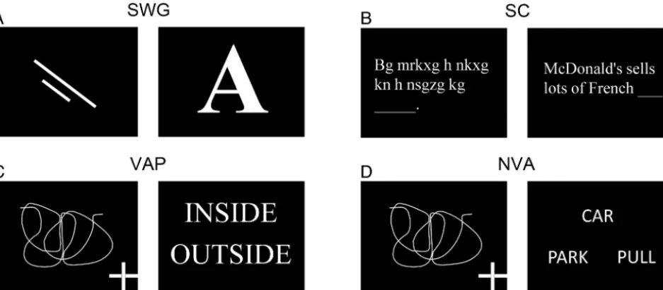
Study DesignTwelve right-handed (6 men/6 women; range, 21– 45 years of age) primarily English-speaking healthy volunteers participated in this study approved by the institutional review board. Two commonly used clinical language paradigms—Silent Word Generation and Sentence Completion—and 3 newly designed semantic para-digms—Visual Antonym Pair, Auditory Antonym Pair, and Noun-Verb Association—were performed by all the participating subjects (Fig 1). All paradigms were block design with alternating active and control blocks lasting 20 seconds each for a total of 4
minutes. The paradigms were implemented by using Prism Ac-quire (Prism Clinical Imaging Inc, Elm Grove, Wisconsin). See below for a description of each paradigm:
Silent Word Generation
Control Block. Visual fixation on 2 consecutive nonsense draw-ings, each for 10 seconds.
Active Block. Covert generation of words for 2 consecutively pre-sented letters, each for 10 seconds.
Sentence Completion
Control Block. Scan through 5 consecutive samples of scrambled letters arranged to resemble words in a sentence.
Active Block. Covert reading of 5 consecutive real sentences with the last word missing and covert generation of a word to complete each sentence.
Visual Antonym Pair
Control Block. Visual fixation of 5 consecutive drawings with a cross placed in 1 of the 4 corners of the screen. Keypad button press was re-quired if the cross location was in the upper or lower right corner.
Active Block. Reading of 5 consecutive pairs of words. Keypad button press was required if the 2 words were antonyms.
Auditory Antonym Pair
Control Block. Listening to 5 consecutive pairs of tones. Keypad button press was required if the 2 tones were identical.
Active Block. Listening to 5 consecutive pairs of words. Keypad button press was required if the 2 words were antonyms.
Noun-Verb Association
Control Block. Same as “Visual Antonym Pair.”
Active Block. Visual presentation of 5 samples of a noun on the top row and a pair of verbs on the bottom row. Keypad button
[image:2.594.63.532.48.253.2]press was required if the verb presented on the right of the bottom row was more closely semantically associated with the presented noun than the verb on the bottom left (eg, Fig 1D; the verb “park ” on the bottom left was more closely associated with the noun “car ” than the verb “pull ” located on the bot-tom right; therefore, a button response was not required in this case; vice versa, if the verb “park ” was on the bottom right and the verb “pull,” on the bottom left, a button response would have been required).
For the dual-choice tasks, the number of expected button presses in the control and active blocks was balanced. A train-ing and practice session was performed outside the MR imag-ing scanner with each participant to provide task instructions and opportunity to practice the tasks by using similar but dif-ferent stimuli from those that were included in the actual examination.
Imaging
Images were acquired by using a 3T Magnetom Trio scanner (Sie-mens, Erlangen, Germany) equipped with a 12-channel head ma-trix coil.
BOLD images were acquired by using a single-shot T2*WI gradient-echo EPI sequence. Imaging parameters were the follow-ing: TR⫽2000 ms; TE⫽30 ms; flip angle⫽90°; FOV⫽24 cm; 80⫻80 matrix acquisition; section thickness⫽3 mm with a 1-mm gap between sections.
Structural images for coregistration and overlay of functional activation maps were acquired by using a standard 3D T1WI
gra-dient-echo sequence (TR ⫽ 2300 ms; TI⫽900 ms; TE⫽3.5 ms; flip angle⫽ 9°; FOV⫽256 cm2; 256⫻256 matrix
acquisition; section thickness⫽1 mm).
Image Analysis
AFNI software (http://afni.nimh.nih. gov/afni) was used for image processing. Preprocessing included section timing, motion correction, spatial smoothing, and registration in a stereotactic space, the Montreal Neurological Institute (MNI)-152 atlas. Regression analysis was then performed by fitting each voxel time-se-ries, divided by the mean and multiplied by 100, to a theoretic expected time-series (ideal TS) generated by convoluting each paradigm timing with a hemody-namic impulse response function. Per-centage signal change (PSC) maps from the baseline were calculated for each paradigm as
PSC⫽100⫻a⫻PP共ideal TS兲 Baseline
where PP indicates peak to peak; PP (ideal TS), maximum (ideal TS) to min-imum (ideal TS); and Baseline⫽b0⫹
b1 ⫻(average polynomial grade 1) ⫹ a⫻minimum (ideal TS), where b0is the
baseline constant, b1a linear trend slope, andathe regression
coefficient.
Statistical Analysis
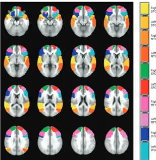
Language localization and lateralization were compared among the 5 paradigms in 5 ROIs (IFG, superior frontal gyrus [SFG], middle frontal gyrus [MFG], middle temporal gyrus [MTG] and STG) automatically defined for the left and right hemispheres on the MNI atlas available in AFNI (Fig 2). To assess statistically significant differences in language localization, we performed a nonparametric analysis with statistical significance considered at theP⬍.01 level. A Friedman test was run voxelwise in each region of interest followed by Wilcoxon signed rank tests between each pair of paradigms in the voxels where the Friedman test score achieved statistical significance. Multiple comparison correction was applied on the results of the Wilcoxon tests by using a com-bination of probability and clustering thresholding obtained by using the AlphaSim simulation program available in AFNI.
Lateralization was expressed by calculating, in each region of interest for each paradigm, the LI by using a threshold-indepen-dent method6:
LI⫽LH⫺RH LH⫹RH
where LH and RH are the weighted sum of all voxeltvalues in the left and right hemisphere portion, respectively, of each region of interest.
[image:3.594.56.366.45.363.2]The same nonparametric tests used for language localization analysis were performed to assess differences in LI among the paradigms in the 5 ROIs. Statistical significance was considered at theP⬍.05 level.
RESULTS
Language Localization
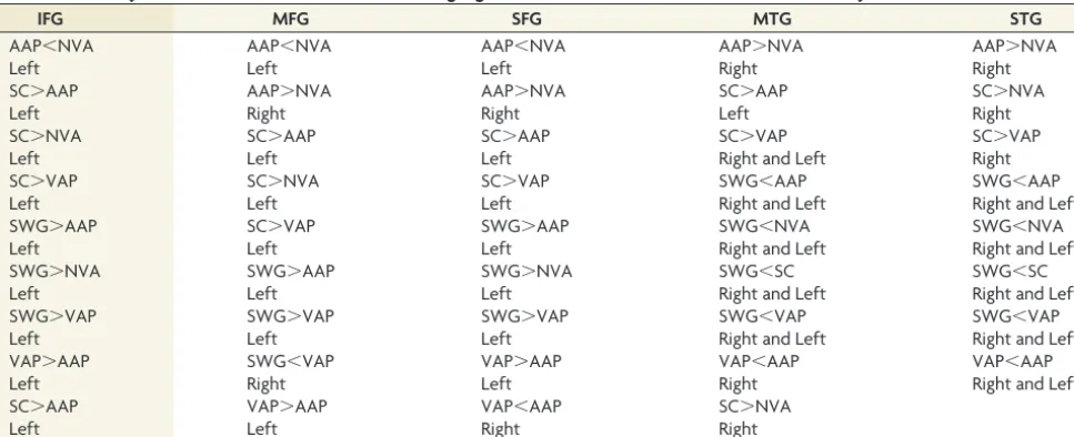
All 5 ROIs showed voxels withscores above the threshold asso-ciated with statistical significance (P⬍.01). Table 1 summarizes the significant results of the Wilcoxon tests comparing the PSC between pairs of paradigms in each region of interest. The results of the simulation run in AlphaSim determined a cluster size of 9 voxels to apply multiple comparison correction on the Wilcoxon testscore maps at theP⬍.01 level. The verbal fluency paradigm Silent Word Generation and Sentence Completion were demon-strated to be more robust activators than the semantic paradigms in frontal gyri ROIs (IFG, MFG, SFG) in the dominant left hemi-sphere, with Auditory Antonym Pair showing the weakest activation in the left IFG, MFG, and SFG but stronger than Visual Antonym Pair and Noun-Verb Association in the right IFG, MFG, and SFG. Silent Word Generation provided the weakest activation in both the right and left MTG and STG. Sentence Completion was more robust than Auditory Antonym Pair and Visual Antonym Pair in the left MTG and STG, but not more robust than Noun-Verb Association. Auditory Antonym Pair and Sentence Completion were the stron-gest activators of the right MTG and STG.
Language Lateralization
In Table 2, the results of the Wilcoxon tests demonstrating a sig-nificant difference atP⬍.05 in LI between each pair of paradigms
in each region of interest are reported. Silent Word Generation was the best lat-eralizing task in the expressive ROIs, whereas Sentence Completion did not outperform, in general, Noun-Verb As-sociation and Visual Antonym Pair as a lateralizing task, except for Noun-Verb Association in the SFG. Auditory Ant-onym Pair provided the most bilateral activation both in the frontal regions and in the MTG. The other 4 paradigms did not demonstrate statistically significant differ-ences in LI in the MTG. No significant differdiffer-ences in LI between the paradigms were found in the STG.
DISCUSSION
In this study, we compared the localization (as locally detectable statistically significant PSC) and lateralization (via the LI) pro-vided by 2 clinically used covert language tasks, Silent Word Gen-eration and Sentence Completion, with those provided by 3 newly designed dual-choice block-design semantic language paradigms, Visual Antonym Pair, Auditory Antonym Pair, and Noun-Verb Association in a group of right-handed healthy volunteers. We made this comparison to determine which paradigm or set of paradigms would be most useful for presurgical mapping of the eloquent language cortex. The analyzed ROIs included more than just the classic Broca and Wernicke areas (left IFG and left STG, respectively), because it is well-recognized that language activa-tion extends well beyond these 2 areas.2Language is not a unitary
[image:4.594.54.538.55.252.2]process but rather a collection of processes operating at distinct levels, such as phonetics, phonology, orthography, and semantics. Therefore, the representation areas related to these processes de-serve attention in presurgical planning because their inadvertent resection may also cause postoperative language deficits. We ad-opted a nonparametric statistical approach because the variables that we compared among the different tasks in the different ROIs for localization (PSC) and lateralization (LI) are defined as ratios of Gaussian variables, and in general, the distribution of a quo-tient of 2 normal variables can be multimodal.
Table 1: Summary of Wilcoxon test results in PSC for language localization in the 5 ROIs included in the analysis
IFG MFG SFG MTG STG
AAP⬍NVA AAP⬍NVA AAP⬍NVA AAP⬎NVA AAP⬎NVA
Left Left Left Right Right
SC⬎AAP AAP⬎NVA AAP⬎NVA SC⬎AAP SC⬎NVA
Left Right Right Left Right
SC⬎NVA SC⬎AAP SC⬎AAP SC⬎VAP SC⬎VAP
Left Left Left Right and Left Right
SC⬎VAP SC⬎NVA SC⬎VAP SWG⬍AAP SWG⬍AAP
Left Left Left Right and Left Right and Left
SWG⬎AAP SC⬎VAP SWG⬎AAP SWG⬍NVA SWG⬍NVA
Left Left Left Right and Left Right and Left
SWG⬎NVA SWG⬎AAP SWG⬎NVA SWG⬍SC SWG⬍SC
Left Left Left Right and Left Right and Left
SWG⬎VAP SWG⬎VAP SWG⬎VAP SWG⬍VAP SWG⬍VAP
Left Left Left Right and Left Right and Left
VAP⬎AAP SWG⬍VAP VAP⬎AAP VAP⬍AAP VAP⬍AAP
Left Right Left Right Right and Left
SC⬎AAP VAP⬎AAP VAP⬍AAP SC⬎NVA
Left Left Right Right
[image:4.594.53.379.279.347.2]Note:—AAP indicates Auditory Antonym Pair; NVA, Noun-Verb Association; SC, Sentence Completion; SWG, Silent Word Generation; VAP, Visual Antonym Pair.
Table 2: Summary of Wilcoxon test results in the LI for lateralization analysis
IFG MFG SFG MTG
SWG⬎SC SWG⬎SC SWG⬎AAP SWG⬎AAP
SWG⬎AAP SWG⬎VAP SWG⬎NVA SC⬎AAP
NVA⬎AAP SWG⬎NVA SC⬎NVA VAP⬎AAP
NVA⬎VAP SWG⬎AAP NVA⬎AAP
NVA⬎AAP (trend)
The results demonstrate that Silent Word Generation is the most robust task for language localization and the most effective for determining language lateralization in the frontal gyri (IFG, MFG, and SFG) of the dominant (left) language hemisphere (Ta-bles 1 and 2). A verbal fluency task, such as Silent Word Genera-tion, requires phonologic access, verbal working memory, and lexical search activity, and these functions are localized in the left inferior frontal gyrus as demonstrated in multiple studies.7,8In
addition, the MFG and SFG are regions of the brain involved in speech production because of the activation of the middle frontal cortex in word retrieval9and the presupplementary motor area
for initiation and execution.10Sentence Completion activates the
dominant hemisphere frontal gyri as robustly as Silent Word Generation because of the word-generation component present in this paradigm (a word required to complete each sentence). However, its pattern of activation is significantly less lateralized than Silent Word Generation in the IFG and MFG because of the contribution of the right hemisphere homologous areas in speech comprehension tasks involving executive processing.11Semantic
decision tasks also elicit activation in the IFG, MFG, and SFG,12
but the lack of word retrieval, initiation, and execution compo-nents of the Noun-Verb Association, Visual Antonym Pair, and Auditory Antonym Pair tasks explains their weaker activation in these gyri compared with our 2 tasks with a verbal fluency com-ponent. In addition, the auditory semantic task Auditory An-tonym Pair showed weaker activation than Visual AnAn-tonym Pair and Noun-Verb Association. This result is consistent with the findings of multiple studies reporting activation of the pars trian-gularis and pars opercularis both for speech and nonspeech sounds when they had to be held in auditory working memory, as in both the control and active blocks of the Auditory Antonym Pair task.13,14
The requirement of working memory and articulary recording activity both in the control and active tasks for Auditory Antonym Pair justifies the same findings in the MFG and SFG.15,16The
analysis of patterns of activation in the temporal gyri demon-strated a weaker BOLD PSC for the purely verbal fluency task Silent Word Generation both in the left and right hemispheres compared with the other 4 paradigms, all of which are associated with semantic processing localized in the middle temporal and angular gyri.17The relative strength of activation among Sentence
Completion, Noun-Verb Association, Visual Antonym Pair, and Auditory Antonym Pair was somewhat variable depending also on the considered hemisphere. In particular, as reported in Table 1, Auditory Antonym Pair was the most robust activator task in the right MTG and STG because its PSC in these 2 regions of interest was greater than Silent Word Generation, Noun-Verb Association, and Visual Antonym Pair in both regions. Auditory Antonym Pair activation results are also highly bilateral in the STG (average LI⫽0.20⫾0.20) and in the MTG (average LI⫽ 0.15⫾0.20), where it was also significantly lower in comparison with the other 4 paradigms (P⬍.05).
Results from the literature indicate a bilateral superior tempo-ral activation for both speech and nonspeech sounds.18However,
the reason for bilateral STG activation even after the subtraction of the speech (tones) control task from the speech (antonyms) active task may be that more demand is placed on short-term
auditory memory during the speech task than during the tone task. Furthermore, contrary to phonologic processing studies re-porting lateralized activation in receptive language areas, the se-mantic component of the active task (antonym versus nonan-tonym) explains the bilateral pattern of activation in our group of volunteers.4We found, in our study, greater activation for
Sen-tence Completion in the left MTG compared with Visual An-tonym Pair and Auditory AnAn-tonym Pair but not compared with Noun-Verb Association, and these findings could be attributed to the higher level of lexical-semantic processing required for Sen-tence Completion and Noun-Verb Association.19Instead
Sen-tence Completion activation was stronger than Noun-Verb Asso-ciation and Visual Antonym Pair in the right MTG and STG, confirming the role of the right hemisphere in semantic tasks involving executive processing.11
The Silent Word Generation group LI was not significantly different from either Sentence Completion or the semantic para-digms but was higher than that of Auditory Antonym Pair. There-fore one could consider using only Silent Word Generation in a clinical language fMRI examination because of its capability to determine language lateralization in both the frontal and tempo-ral lobes. However its PSC is weaker in both the left and right temporal gyri compared with the other 4 paradigms that include language comprehension in the active blocks of the paradigms; therefore, the semantic tasks are more adequate to map the tem-poral regions involved in the language network because they elicit a stronger BOLD response than a verbal fluency task. Further-more, it has been demonstrated that the combination of multiple tasks, a language-specific region of interest approach and imple-mentation of statistical threshold-independent approaches for determination of hemispheric lateralization, provides more reli-able lateralization that correlates better with the criterion stan-dard Wada test.20,21
One limitation of this study includes the exclusion of the cer-ebellum from region-of-interest analysis. The cercer-ebellum has demonstrated fMRI activation during silent articulation.22Cases
of cognitive deficits in association with cerebellar damage have been reported,23yet frank aphasic disturbances are rare. Multiple
cognitive studies performed on a group of healthy volunteers have demonstrated a further parceling out of language functions in each of the 5 ROIs considered in our work.24,25However, since
this study aimed to improve language presurgical mapping, where analysis is conducted at the single subject level, such parcellation would not be practical and it most likely would not add any crit-ical information for patient surgcrit-ical management. Finally, no Wada testing was performed to actually confirm language lateral-ization in these subjects, but this would not be ethical in a group of healthy volunteers.
CONCLUSIONS
advisable in clinical studies to have a variety of available para-digms because if a patient finds a particular task too challenging because of neurologic impairment, it will rarely produce mean-ingful fMRI results.
ACKNOWLEDGMENTS
The authors thank Gayane Yenokyan, MD, PhD, for assistance with data analysis.
Disclosures: Domenico Zaca`—RELATED:Grant: Siemens Medical Solutions,* Com-ments: Siemens provided support for my postdoctoral fellowship salary at Johns Hopkins University. Jay J. Pillai—RELATED:Grant: Siemens Medical Solutions,* Com-ments: This research grant paid for Dr Zaca`’s (my former postdoctoral fellow) salary. However, Siemens was not involved in the study design, analysis, reporting of results, or writing of the manuscript in any way,OTHER RELATIONSHIPS: I am an unpaid member of the Medical Advisory Board of Prism Clinical Imaging. *Money paid to the institution.
REFERENCES
1. Seghier ML, Lazeyras F, Pegna AJ, et al.Variability of fMRI activa-tion during a phonological and semantic language task in healthy subjects.Hum Brain Mapp2004;23:140 –55
2. Saur D, Kreher BW, Schnell S, et al.Ventral and dorsal pathways for language.Proc Natl Acad Sci U S A2008;105:18035– 40
3. Partovi S, Konrad F, Karimi S, et al.Effects of covert and overt par-adigms in clinical language fMRI.Acad Radiol2012;19:518 –25 4. Zaca` D, Nickerson JP, Deib G, et al.Effectiveness of four different
clinical fMRI paradigms for preoperative regional determination of language lateralization in patients with brain tumors. Neurora-diology2012;54:1015–25
5. Ruff IM, Petrovich Brennan NM, Peck KK, et al.Assessment of the language laterality index in patients with brain tumor using func-tional MR imaging: effects of thresholding, task selection, and prior surgery.AJNR Am J Neuroradiol2008;29:528 –35
6. Branco DM, Suarez RO, Whalen S, et al.Functional MRI of mem-ory in the hippocampus: laterality indices may be more mean-ingful if calculated from whole voxel distributions.Neuroimage 2006;32:592– 602
7. Palmer ED, Rosen HJ, Ojemann JG, et al.An event-related fMRI study of overt and covert word stem completion.Neuroimage2001; 14(1 pt 1):182–93
8. Eulitz C, Elbert T, Bartenstein P, et al.Comparison of magnetic and metabolic brain activity during a verb generation task.Neuroreport 1994;6:97–100
9. Paulesu E, Goldacre B, Scifo P, et al.Functional heterogeneity of left inferior frontal cortex as revealed by fMRI. Neuroreport 1997;8:2011–17
10. Kawashima R, Okuda J, Umetsu A, et al.Human cerebellum plays an important role in memory-timed finger movement: an fMRI study.
J Neurophysiol2000;83:1079 – 87
11. Vigneau M, Beaucousin V, Herve PY, et al.What is right-hemisphere contribution to phonological, lexico-semantic, and sentence pro-cessing? Insights from a meta-analysis.Neuroimage2011;54:577–93 12. Binder JR, Desai RH, Graves WW, et al.Where is the semantic sys-tem? A critical review and meta-analysis of 120 functional neuro-imaging studies.Cereb Cortex2009;19:2767–96
13. Burton MW, Small SL, Blumstein SE.The role of segmentation in phonological processing: an fMRI investigation.J Cogn Neurosci 2000;12:679 –90
14. Hsieh L, Gandour J, Wong D, et al.Functional heterogeneity of in-ferior frontal gyrus is shaped by linguistic experience.Brain Lang 2001;76:227–52
15. Wilson SM, Iacoboni M.Neural responses to non-native phonemes varying in producibility: evidence for the sensorimotor nature of speech perception.Neuroimage2006;33:316 –25
16. Wilson SM, Saygin AP, Sereno MI, et al.Listening to speech activates motor areas involved in speech production. Nat Neurosci 2004;7:701– 02
17. Vandenberghe R, Price C, Wise R, et al.Functional anatomy of a common semantic system for words and pictures. Nature 1996;383:254 –56
18. Booth JR, Burman DD, Meyer JR, et al.Functional anatomy of intra-and cross-modal lexical tasks.Neuroimage2002;16:7–22
19. De´monet JF, Chollet F, Ramsay S, et al.The anatomy of phonological and semantic processing in normal subjects. Brain1992;115(pt 6):1753– 68
20. Niskanen E, Kononen M, Villberg V, et al.The effect of fMRI task combinations on determining the hemispheric dominance of lan-guage functions.Neuroradiology2012;54:393– 405
21. Suarez RO, Whalen S, Nelson AP, et al.Threshold-independent functional MRI determination of language dominance: a valida-tion study against clinical gold standards. Epilepsy Behav 2009;16:288 –97
22. Ackermann H.Cerebellar contributions to speech production and speech perception: psycholinguistic and neurobiological perspec-tives.Trends Neurosci2008;31:265–72
23. Fiez JA, Petersen SE, Cheney MK, et al.Impaired non-motor learn-ing and error detection associated with cerebellar damage: a slearn-ingle case study.Brain1992;115(pt 1):155–78
24. Specht K, Reul J.Functional segregation of the temporal lobes into highly differentiated subsystems for auditory perception: an audi-tory rapid event-related fMRI-task.Neuroimage2003;20:1944 –54 25. Thierry G, Giraud AL, Price C.Hemispheric dissociation in access to