ISSN Online: 2475-7349 ISSN Print: 2475-7330
DOI: 10.4236/ym.2017.14021 Dec. 13, 2017 202 Yangtze Medicine
Molecular Motors—Self-Organization
of Cytoskeletal Network
Kishore Dutta
Department of Physics, Handique Girls’ College, Guwahati, India
Abstract
Molecular motors play an important role in the organization of cytoskeletal filament networks. These nanometer-sized natural molecular machines opened up a new frontier of nano-technology. This article describes biomole-cular nano-machines, their internal structures, and dynamical interactions between molecular motors and their molecular tracks which reorganize a network of long protein filaments, particularly during cell division to form cytoskeleton of daughter cells. Towards the end, the article also takes up some still-to-be resolved matters and prospects for future developments in this ex-citing multidisciplinary area of science.
Keywords
Cell Motility, Molecular Motors, Self-Organization
1. Introduction
Spontaneous, self-generated movement is a hallmark of almost all biological systems. Even cells that are incapable of active movement within their environ-ment perform essential intracellular motility processes. Cell motility is a com-plex and integrated process governed by the dynamics of cytoskeleton filaments and collective activities of a series of biological nanomachines in the cell
machi-nery [1] [2] [3] [4] [5]. Most forms of movement in the living world are powered
by these tiny protein machines known as molecular motors. Molecular motors are essential molecules of life which actively involve in diverse functions of cells such as muscle contraction, vesicle transport, chromosome separation, replica-tion, transcripreplica-tion, and translation. Although recent advent of highly sophisti-cated novel biophysical techniques, such as fluorescence microscopy, high speed atomic force microscopy, and optical tweezers [6] [7] [8] [9] [10], has
signifi-How to cite this paper: Dutta, K. (2017) Molecular Motors—Self-Organization of Cytoskeletal Network. Yangtze Medicine, 1, 202-215.
https://doi.org/10.4236/ym.2017.14021
Received: September 14, 2017 Accepted: December 10, 2017 Published: December 13, 2017
Copyright © 2017 by author and Scientific Research Publishing Inc. This work is licensed under the Creative Commons Attribution International License (CC BY 4.0).
http://creativecommons.org/licenses/by/4.0/
DOI: 10.4236/ym.2017.14021 203 Yangtze Medicine
cantly increased our understanding regarding the function and role of molecular motors in the cell, how Nature designed these tiny powerful ingenious na-no-scale devices to achieve coordination and collective action in all living organ-isms, is still remains a mystery.
Nature offers a wide variety of different motor families characterized by their chemical structures, environments, and direction of motions. From a broad theoretical perspective, a molecular motor can be imagined as a microscopic ob-ject that moves predominantly in one direction along the molecular track—a polarized one-dimensional periodic structure. Within the cell, there are several molecular tracks, namely, cytoskeletal filaments, DNA, RNA, and mitochondrial membranes. The movement of the molecular motors along these molecular tracks is characterized by unidirectionality (yielding non-equilibrium behavior), discrete steps, and intrinsic stochasticity. All these events that take place stochas-tically and characterized by the intrinsic rate are known as Poisson processes in statistical physics. Interestingly, the dynamic interactions of the motors and fi-laments exhibit cooperative collective behavior yielding a rich variety of stable self-organized structure [11] [12] [13]. In this review article, we shall first intro-duce and elucidate few common molecular motors and their dynamic characte-ristics. Focusing on fundamental physical principles underlying the collective phenomena resulting from the interaction of many molecular motors and linear protein filaments, we then describe the self-organization of long protein filament networks, particularly during cell division to form cytoskeleton of daughter cells. Our discussion suggests a new perspective to our understanding of collective motion, a topic of increasing interest among physicists, mathematicians, engi-neers and biologists.
2. Various Molecular Motors and Their Tracks
Based on the tracks on which they move, the motor proteins are classified as cy-toskeletal based, nucleic acid based, and membrane based motor proteins. Among various molecular motors, kinesin, dynein, and myosin are cytoskeleton- based motors which move in a directed manner along cytoskeletal filaments [1]. The majority of active transport in the cell is driven by these three classes of mo-lecular motors. The helicases, polymerases, and ribosomes are nucleic acid based motors which move in the track of single-stranded DNA or mRNA. On the oth-er hand, pumps, protein translocation motors, ATP synthase (commonly called ATPase), and flagellar motors are associated with membranes. Recent revolutio-nary advances in single molecule detection techniques enable to observe directly the activity and performance of individual molecular motors [9]. In Table 1, we summarize some common characteristics of these molecular motors. In what fol-lows, we shall briefly elucidate the dynamic behavior of these molecular motors.
2.1. Microtubule Based Motors
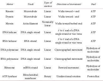
DOI: 10.4236/ym.2017.14021 204 Yangtze Medicine Table 1. Characteristics of some common molecular motors.
Motor Track Type of motion Direction of movement Fuel
Kinesin Microtubule Linear Walks toward + end ATP
Dynein Microtubule Linear Walks toward - end ATP
Myosin Actin filament Nominally linear Walks toward barbed end ATP
DNA helicase DNA single strand Linear single strand or vice versa 3' to 5' end of a DNA ATP
RNA helicase RNA strand Linear single strand or vice versa 3' to 5' end of a DNA ATP
DNA polymerase DNA single strand Linear Choreographed movement Hydrolysis of nucleotides
RNA polymerase DNA single strand Linear Choreographed movement Hydrolysis of nucleotides
Ribosome mRNA strand Linear Forward movement Hydrolysis of nucleotides
ATP Synthase Mitochondrial membrane Rotary Unidirectional rotation Proton flux
called tubulin. The α- and β-tubulins pairwise aggregate into tubulin hetero-dimers, which are longitudinally associated with one another to form protofila-ments. Microtubules are hollow fibers consist of typically 13 such parallel proto-filaments. As the heterodimers are polar in nature, the microtubules also possess a polarity, usually denoted as the (−) and the (+) end; plus-ends located in the cell periphery and minus-ends located at the microtubule organizing center near the nucleus. Due to this polarity, molecular motors such as kinesins and dyneins can walk unidirectionally on the microtubules and carry various cargo particles such as vesicles, mRNA, mitochondria, endosomes, virus particles, or other or-ganelles many times the size of the motor, to their required destination [4] [5].
Figure 1 depicts a schematic diagram for the unidirectional movement of a
ki-nesin motor over a microtuble.
One of the astonishing dynamic properties of kinesins and dyneins is their stepwise movement over long distances without detachment from the filament
[14] [15]. This movement is based on the alternated binding of the two motor
DOI: 10.4236/ym.2017.14021 205 Yangtze Medicine Figure 1. Schematic representation of the unidirectional movement of a kinesin motor over a mi-crotubule filament.
of nucleotide triphosphates. The Brownian ratchet or thermal ratchet was de-signed and analyzed by Feynman [16] simply as an illustration of Carnot's prin-ciple. This mechanism is strongly rely on nonequilibrium statistical physics and it not only account for the general principles behind molecular motors, but has also been successfully applied as a prototype in various fluctuation-driven transport occurring in many mesoscopic and microscopic systems. Numerous single molecule experimental investigations [14] [19] have shown that, about one hundred times per second, kinesin hydrolyzes one ATP molecule, generates a force of up to 7 pico-Newton and takes an 8 nanometer step towards the posi-tive end of a microtubule. These indigenous experiments suggested that the ATP hydrolysis is responsible for changing the local conformation of the microtu-bules that provides the information for the foot where to reattach.
2.2. Actin Based Motors
Actin filaments or F-actin are the most abundant proteins in a eukaryotic cell and mostly concentrated near the cell membrane. They are two-stranded helical polymers built from dimer pairs of globular-actin monomers (or, G-actin) which are polar in nature. The two halves of an actin monomer are separated by a cleft that can bind ATP or its hydrolyzed form ADP. This is responsible for the exis-tence of two distinct ends to the whole filament, namely a fast growing end, known as plus end or barbed end, where mostly ATP-bound monomers are lo-cated, and a slow growing end known as minus end or pointed end, which is rich in ADP-bound monomers.
DOI: 10.4236/ym.2017.14021 206 Yangtze Medicine Figure 2. Schematic sketch of a myosin motor.
with each other and form myosin filaments. On the other hand, the heads of the myosin molecules are bound to polymerized actin filaments. Actin and myosin together exhibit a periodic arrangement organized in a polarity-alternated fa-shion. The motion at the heart of myosin motors is a rotary motion which is transmitted to actin fiber as a linear motion through a long lever-arm. This lever- arm amplifies the motion of the molecular motor from a few nanometers to 10 nanometers. During muscular contraction, the free energy of ATP hydrolysis by myosin drives the myosin filaments to slide along the actin filaments.
2.3. Nucleic Acid Based Motors
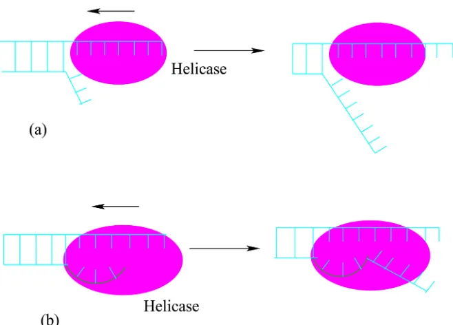
Motors that utilize nucleic acid strands (DNA and RNA) as their linear tracks are classified into two groups—helicases and polymerases. Helicase motors un-zip double-stranded nucleic acids (DNA or RNA) and translocate along one of the two strands either in 5′→3′ or in 3′→5′ direction. DNA helicase binds
DOI: 10.4236/ym.2017.14021 207 Yangtze Medicine Figure 3. Unwinding the double-stranded DNA by a DNA helicase motor via (a) passive and (b) active mechanism.
interacts directly with the double-stranded DNA and plays a direct role in desta-bilizing the duplex DNA. However, the exact mechanism by which helicases ac-complish unwinding the strands of double-stranded DNA and couple the NTPase activity to movement is still unclear and a subject of intense study [23]. Significant progress has been made and intense structural, biochemical, as well as genetic approaches are currently being pursued to gain insight into both the mechanism and role of helicases in various biological processes.
The helicases are involved in almost all DNA and/or RNA metabolic processes. They play a fundamental role in transcription, replication, DNA re-pair, and recombination [24]. The chromosome replication mechanism in all cells is solely dependent upon the action of DNA helicases. Malfunctioning of certain DNA helicases may lead to several severe human genetic diseases, such as Werner syndrome, Bloom syndrome or xeroderma pigmentosum [25].
inten-DOI: 10.4236/ym.2017.14021 208 Yangtze Medicine
sive research work over the years has been devoted, analysis of RNA polymerase as molecular motor is still in its infancy.
2.4. Membrane Based Motors
Besides the aforementioned molecular motors that move along a linear sub-strate, there is an another class of motor namely, rotary motors which plays a key role in many biological membranes [18] [26]. As depicted in Figure 4, the molecular motor F F0 1-adenosine triphosphatase (commonly called ATPase)
comprises of two rotary motors F0 and F1, where the F0 motor is driven
by proton flow and the other one by ATP hydrolysis. It has been recognized that the collective swimming movement of bacteria is due to the presence of rotary motors that rotate their flagellum. The bacteriophage—a virus that infects bacte-ria, possesses a rotary motor which packs DNA into the bacteriophage head. All known biological rotary motors are driven by ion-gradient-based sources of energy, particularly, the hydrogen-ion (or proton) gradients that provides the necessary electrochemical forces. These rotary machines usually converts such electrochemical energy stored in proton concentration gradients, first into me-chanical motion, and then back into chemical energy under the form of ATP. Surprisingly, rotary motors are reversible in the sense that they can harness the chemical energy of ATP to produce or maintain the transmembrane electro-chemical gradient of proton concentration.
[image:7.595.250.493.523.706.2]From the above discussions, we have seen that the hydrolysis of ATP with the release of ADP and inorganic phosphate is known to be the power source for many motor proteins which, in turn, powers cell motility by enabling the motors to bind to and move along their tracks in a stepwise linear or rotary motion. They are unique in their ability to transform the chemical energy stored in an energy-rich bond of ATP into mechanical work and thereby able to transport a wide variety of cargo, power cell locomotion, build or destroy other proteins, pump ions, drive cell division, and when combined in large ensembles, allow
DOI: 10.4236/ym.2017.14021 209 Yangtze Medicine
organisms to move.
In addition to transport vesicles and organelles, molecular motors actively participate in the process of self-organization of the cytoskeletal structures in the cell including physical separation of molecules or molecular aggregates [13] [27]
[28] [29]. In what follows we shall make a comprehensive analysis on how these
molecular motors actively take part in the self-organization of protein filaments.
3. Self-Organization of Protein Filaments
Self-organization is a fascinating physical process of many non-equilibrium sys-tems and is a manifestation of nonlinearity. It is the spontaneous emergence of order, regularity, coherence, and coordination within the system from numerous short-range interactions among the constituents [27] [30] [31]. The simple mi-croscopic local interaction rules are the key factors that give rise to a global self-organized collective state leading to various fascinating natural patterns. From a thermodynamic point of view, formation of such self-organized complex spatial patterns takes place due to constant work done against entropy increase. In order to maintain a high level of differentiation, all living organisms actively work against entropy increase by consuming energy from the environment. Liv-ing organisms are, therefore, far from thermal equilibrium. ApproachLiv-ing equili-brium would result in a decrease of the organisms internal structure and thereby a loss in its ability of performing basic tasks. This is akin to the possible heat death of the universe.
DOI: 10.4236/ym.2017.14021 210 Yangtze Medicine
Cross-linking with myosins, actin filaments organize into linear bundles, two- dimensional networks, and three-dimensional gels. Most notable examples of bundles are muscle sarcomere and stress-fiber of an adherent cell. Not only the
in vitro experiments and integrated modeling efforts, but also analytic theories, demonstrated how the interaction of motors and active micro-filaments lead to a complex self-organized structure. Depending on the interactions with actin fi-laments, myosins perform a wide variety of cellular tasks, from cellular transport to muscle contraction. Myosin I perform transport of endocytic vesicles, myosin II family powers muscle contraction and cytokinesis, myosin V phagocytosis and transport of cellular elements, myosins VI and VII provide hearing and balance cells. When myosins bind tightly to actin in the absence of ATP, temporary stiffness of joints and muscular rigidity occurs—a physiological condition after death [29].
From the perspective of statistical physics, the active filaments are example of a many-degrees of freedom nonlinear dynamical system that are far from equili-brium and exhibit self-organizing property at certain critical values of external parameters [13] [31] [34]. As such, the fascinating self-organizing behavior of micro filaments continues to attract enormous attention and inspired many theoretical efforts directed towards modeling active filament solutions [35]. Since capturing the dynamics of individual nanomotors and microscopic fila-ments is a difficult task, coarse-grained modeling achieves increasingly impor-tant to elucidate generic features and emergent behavior. In the following sec-tions, we shall discuss the role of motor proteins in chromosome segregation during mitosis and meiosis and their self organization process.
3.1. Self-Organization during Mitosis
The most-spectacular event of intracellular transport occurs during the essential process of eukaryotic mitosis, in which duplicated chromosomes are segregated from the mother cell and delivered to each of the nascent daughter cells. Under-standing the complex interplay between molecular motors and microtubules that carry out faithful segregation of genetic material in all eukaryotic cells, is a long standing goal among scientists to reveal the mechanism, structure, and organi-zation in biological systems.
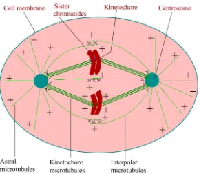
During mitosis, the accurate chromosome segregation is carried out by a complex and extraordinarily sophisticated macromolecular machine—the mi-totic spindle [36] [37] [38]. The mitotic spindle itself is a highly dynamic struc-ture comprises of two microtubule asters in which the minus ends are focused, and the plus ends extend outwards, overlapping into the characteristic bipolar shape as schematically depicted in Figure 5. Although the structure of the poles differs in different organisms, all spindles are bipolar in nature. Any defects in spindle bipolarity or chromosome bi-orientation lead to potentially lethal errors in chromosome segregation.
DOI: 10.4236/ym.2017.14021 211 Yangtze Medicine Figure 5. Schematic representation of mitotic spindle of somatic animal cells in meta-phase. The minus ends of the microtubules (green) are embedded in a spindle pole (the centrosome) and the plus ends are pointing outward from the pole. Numerous motors and other proteins (represented by the symbol ×) cross-link the minus ends of microtu-bules at the spindle poles and the plus ends of inter-polar microtumicrotu-bules in the spindle midzone.
DOI: 10.4236/ym.2017.14021 212 Yangtze Medicine
seems to be an essential and remarkable physical self-organization process.
3.2. Self-Organization during Meiosis
Sexual reproduction, which involves the successful mixing and recombination of genetic material during meiosis, requires concerted movement of the nucleus
[41]. This dynamic physical process is driven by molecular motors that move the nucleus back and forth inside the cell with the aid of microtubules. How motors and microtubules work together to produce these large-scale movements during meiosis, however, remains a mystery. Experiments on fission yeast revealed that asymmetric redistribution of motor proteins from microtubules behind the moving nucleus to those in front of the nucleus, creates nuclear oscillation. Re-cent highly sophisticated experiments demonstrated that this dynamic motor re-distribution occurs purely as a result of changes in the mechanical strain sensed by the motor proteins. However, a complete understanding of complex meiotic processes requires a great deal of multi-disciplinary efforts.
4. Discussion and Conclusion
Over the last couple of years, the rapid development of single-molecule tech-niques and structural studies leads to a considerable progress in understanding how the biological motors operate. How do they move? How do they generate force? How much fuel do they consume, and with what efficiency? How do they determine which cargo is to be transported and when to transport the cargo to its proper location within the cell? Although the key proteins involved in the processes have been identified and localized, the underlying physical mechanism is still remains unknown. The intriguing complex behavior of these natural biomolecular machines represents a formidable challenge for theoretical de-scriptions and numerical approaches that aim at a fundamental analysis of the underlying interaction mechanisms.
signifi-DOI: 10.4236/ym.2017.14021 213 Yangtze Medicine
cant step towards future bio-nanomachines and bio-nanorobots.
Perhaps the most exciting goal of these ingenious biological nanomotors is the molecular repair of the human body. Molecular motors might be applied as a drug delivery vehicle to the cell bodies of motor neurons by axonal transport. Using currently available biochemical methods, scientists designed and created synthetic motors to disrupt specific cellular functions and to transport drugs or other substances to specific cells or regions of cells. Motor defects lead to motili-ty defects of the animal cells which can, in turn, lead to severe diseases including male infertility, deafness, chronic inflammatory diseases, neurodegenerative diseases, or may even be lethal. Although Nature offers a maintaining and re-pairing mechanism for its damaged molecular systems, such complex repair mechanisms are beyond the capabilities of current nanotechnology. We hope that extensive experimental, theoretical and computational investigations in near future will lead to a deeper understanding of these collective physical phenome-na, which will possibly help us to cure and control life-threatening diseases and will provide possible design principles that can be utilized to synthesize artificial nanomachines. At last, I hope this introductory article will encourage students and particularly young researchers to become the active participants in this challenging endeavor.
References
[1] Howard, J. (2001) Mechanics of Motor Proteins and the Cytoskeleton. Sinauer As-sociates, Sunderland, Massachusetts.
[2] Kreis, T.E. and Vale, R.D. (1999) Guidebook to the Cytoskeletal and Motor Pro-teins. 2nd Edition, Oxford University Press, Oxford.
[3] Davis, A.P. (1999) Synthetic Molecular Motors. Nature, 401, 120.
[4] Engelke, M.F., et al. (2016) Engineered Kinesin Motor Proteins Amenable to Small-Molecule Inhibition. Nature Communications, 7, Article Number: 11159.
https://doi.org/10.1038/ncomms11159
[5] Ravichandran, A., et al. (2017) Enhanced Dynamics of Confined Cytoskeletal Fila-ments Driven by Asymmetric Motors. Biophysical Journal, 113, 1121-1132.
https://doi.org/10.1016/j.bpj.2017.07.016
[6] Svoboda, K., Schmidt, C.F., Schnapp, B.J. and Block, S.M. (1993) Direct Observa-tion of Kinesin Stepping by Optical Trapping Interferometry. Nature, 256, 721-727.
https://doi.org/10.1038/365721a0
[7] Finer, J.T., Simmons, R.M. and Spudich, J.A. (1994) Single Myosin Molecule Me-chanics: Piconewton Forces and Nanometer Steps. Nature, 368, 113-119.
https://doi.org/10.1038/368113a0
[8] Greenleaf, W.J., Woodside, M.T. and Block, S.M. (2007) High-Resolution, Sin-gle-Molecule Measurements of Biomolecular Motion. Annual Review of Biophysics and Biomolecular Structure, 36, 171-190.
https://doi.org/10.1146/annurev.biophys.36.101106.101451
[9] Kodera, N., et al. (2010) Video Imaging of Walking Myosin V by High-Speed Atomic Force Microscopy. Nature, 468, 72-77. https://doi.org/10.1038/nature09450
DOI: 10.4236/ym.2017.14021 214 Yangtze Medicine
Networks. Physical Biology, 9, Article ID: 026005.
https://doi.org/10.1088/1478-3975/9/2/026005
[11] Nedelec, F., et al. (1997) Self-Organization of Microtubules and Motors. Nature, 389, 305-308. https://doi.org/10.1038/38532
[12] Surrey, T., et al. (2001) Physical Properties Determining Self-Organization of Mo-tors and Microtubules. Science, 292, 1167-1171.
https://doi.org/10.1126/science.1059758
[13] Marchetti, M.C., Joanny, J.F., Ramaswamy, S., Liverpool, T.B., Prost, J., Rao, M. and Simha, R.A. (2013) Hydrodynamics of Soft Active Matter. Reviews of Modern Physics, 85, 1143-1189. https://doi.org/10.1103/RevModPhys.85.1143
[14] Schnitzer, M.J. and Block, S.M. (1997) Kinesin Hydrolyses One ATP Per 8-nm Step.
Nature, 388, 386-390. https://doi.org/10.1038/41111
[15] Fallesen, T.L., Macosko, J.C. and Holzwarth, G. (2011) Measuring the Number and Spacing of Molecular Motors Propelling a Gliding Microtubule. Physical Review E, 83, Article ID: 011918. https://doi.org/10.1103/PhysRevE.83.011918
[16] Feynman, R.P., Leighton, R. and Sands, M. (1963) The Feynman Lectures on Phys-ics. Vol. 1, Addison-Wesley, Reading.
[17] Astumian, R.D. (1997) Thermodynamics and Kinetics of a Brownian Motor.
Science, 276, 917-922.https://doi.org/10.1126/science.276.5314.917
[18] Hernández, J.V., Kay, E.R. and Leigh, D.A. (2004) A Reversible Synthetic Rotary Molecular Motor. Science, 306, 1532-1537.https://doi.org/10.1126/science.1103949
[19] Visscher, K., Schnitzer, M.J. and Block, S.M. (1999) Single Kinesin Molecules Stu-died with a Molecular Force Clamp. Nature, 400, 184-189.
https://doi.org/10.1038/22146
[20] Hu, S., et al. (2016) Long-Range Self-Organization of Cytoskeletal Myosin II Fila-ment Stacks. Nature Cell Biology, 19, 133-141.https://doi.org/10.1038/ncb3466
[21] Morris, P.D. and Raney, K.D. (1999) DNA Helicases Displace Streptavidin from Biotin-Labeled Oligonucleotides. Biochemistry, 38, 5164-5171.
https://doi.org/10.1021/bi9822269
[22] Kim, D.-E., Narayan, M. and Patel, S.S. (2002) T7 DNA Helicase: A Molecular Mo-tor That Processively and Unidirectionally Translocates along Single-Stranded DNA. Journal of Molecular Biology, 321, 807-819.
https://doi.org/10.1016/S0022-2836(02)00733-7
[23] Michaelis, J., et al. (2009) DNA Based Molecular Motors. Physics of Life Reviews, 6, 250-266.https://doi.org/10.1016/j.plrev.2009.09.001
[24] Tanner, N.K. and Linder, P. (2001) DExD/H Box RNA Helicases: From Generic Motors to Specific Dissociation Functions. Molecular Cell, 8, 251-262.
https://doi.org/10.1016/S1097-2765(01)00329-X
[25] Ellis, N.A. (1997) DNA Helicases in Inherited Human Disorders. Current Opinion in Genetics & Development, 7, 354-363.
https://doi.org/10.1016/S0959-437X(97)80149-9
[26] Chowdhury, D. (2006) Collective Effects in Intra-Cellular Molecular Motor Trans-port: Coordination, Cooperation and Competition. Physica A, 372, 84.
https://doi.org/10.1016/j.physa.2006.05.005
[27] Camazine, S., et al. (2001) Self-Organization in Biological Systems. Princeton Uni-versity Press, Princeton.
DOI: 10.4236/ym.2017.14021 215 Yangtze Medicine https://doi.org/10.1016/S0955-0674(02)00014-5
[29] Schliwa, M. and Woehlke, G. (2003) Molecular Motor. Nature, 422, 759-755. https://doi.org/10.1038/nature01601
[30] Dutta, K. (2010) How Birds Fly Together: The Dynamics of Flocking. Resonance, 15, 1097-1110.https://doi.org/10.1007/s12045-010-0122-5
[31] Banerjee, S. and Marchetti, M.C. (2011) Instabilities and Oscillations in Isotropic Active Gels. Soft Matter, 7, 463.https://doi.org/10.1039/C0SM00494D
[32] Karsenti, E., Nedelec, F. and Surrey, T. (2006) Modeling Microtubule Patterns. Na-ture Cell Biology, 8, 1204-1211.https://doi.org/10.1038/ncb1498
[33] Sumino, Y., Nagai, K.H., Shitaka, Y., Tanaka, D., Yoshikawa, K.Y., Chaté, H. and Oiwa, K. (2012) Large-Scale Vortex Lattice Emerging from Collectively Moving Microtubules. Nature (London), 483, 448.https://doi.org/10.1038/nature10874
[34] Hoffmann, P.M. (2016) How Molecular Motors Extract Order from Chaos (A Key Issues Review). Reports on Progress in Physics, 79, Article ID: 032601.
https://doi.org/10.1088/0034-4885/79/3/032601
[35] Ganguly, C. and Chaudhuri, D. (2013) Stochastic Thermodynamics of Brownian Particles. Physical Review E, 88, Article ID: 032102.
https://doi.org/10.1103/PhysRevE.88.032102
[36] Wittmann, T., Hyman, A. and Desai, A. (2001) The Spindle: A Dynamic Assembly of Microtubules and Motors. Nature Cell Biology, 3, E28-E34.
https://doi.org/10.1038/35050669
[37] Karsenti, E. (2004) Spindle Saga. Nature, 432, 563-564. https://doi.org/10.1038/432563a
[38] Pavin, N. and Tolić, I.M. (2016) Self-Organization and Forces in the Mitotic Spin-dle. Annual Review of Biophysics, 45, 279-298.
https://doi.org/10.1146/annurev-biophys-062215-010934
[39] Berger, F., et al. (2015) External Forces Influence the Elastic Coupling Effects during Cargo Transport by Molecular Motors. Physical Review E, 91, Article ID: 022701. https://doi.org/10.1103/PhysRevE.91.022701
[40] Barton, N.R. and Goldstein, L.S.B. (1996) Going Mobile: Microtubule Motors and Chromosome Segregation. Proceedings of the National Academy of Sciences, 93, 1735-1742.https://doi.org/10.1073/pnas.93.5.1735